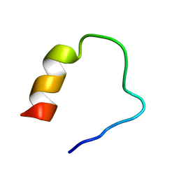
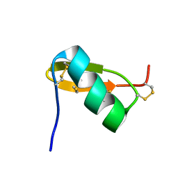
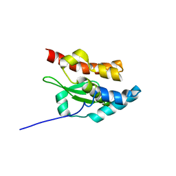
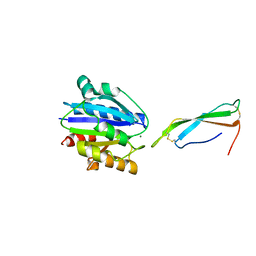
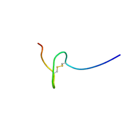
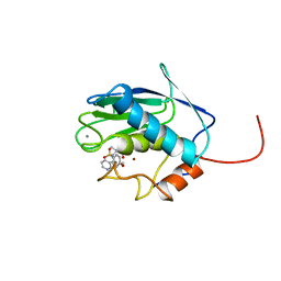
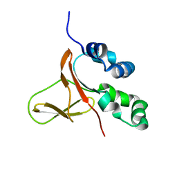
Loading
PDBj
MenuPDBj@FacebookPDBj@TwitterPDBj@YouTubewwPDB FoundationwwPDB
RCSB PDBPDBeBMRBAdv. SearchSearch help

6MI9
DownloadVisualize
BU of 6mi9 by Molmil
The peptide PaAMP1B3 is an analog derived from the PaAMP1. The sequence of the ribosomal protein of Pyrobaculum aerophylum was used as template for rational design, using the Joker algorithm.
Descriptor: PRO-MET-ALA-ARG-ASN-LYS-ILE-LEU-GLY-LYS-ILE-LEU-ARG-LYS-ILE-ALA-ALA-PHE-LYS
Authors:Mundim, H.M, Alves, E.S.F, Rodrigues, L.V, Liao, L.M.
Deposit date:2018-09-19
Release date:2019-09-25
Method:SOLUTION NMR
Cite:NMR SOLUTION STRUCTURE OF THE ANTIMICROBIAL PEPTIDE PAAMP1B3.
To Be Published
5WT7
DownloadVisualize
BU of 5wt7 by Molmil
FAS1-IV domain of Human Periostin
Descriptor: Periostin
Authors:Yun, H, Lee, C.W.
Deposit date:2016-12-09
Release date:2017-12-13
Last modified:2024-05-15
Method:SOLUTION NMR
Cite:1H,13C, and 15N resonance assignments of FAS1-IV domain of human periostin, a component of extracellular matrix proteins.
Biomol NMR Assign, 12, 2018
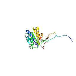
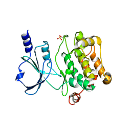
6OBI
DownloadVisualize
BU of 6obi by Molmil
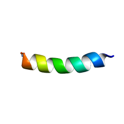
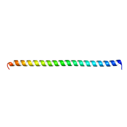
Remarkable rigidity of the single alpha-helical domain of myosin-VI revealed by NMR spectroscopy
Descriptor: Myosin-VI
Authors:Barnes, A, Shen, Y, Ying, J, Takagi, Y, Torchia, D.A, Sellers, J, Bax, A.
Deposit date:2019-03-20
Release date:2019-06-12
Last modified:2024-05-15
Method:SOLUTION NMR
Cite:Remarkable Rigidity of the Single alpha-Helical Domain of Myosin-VI As Revealed by NMR Spectroscopy.
J.Am.Chem.Soc., 141, 2019
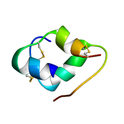
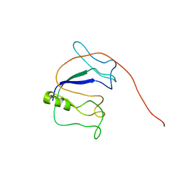
6E4J
DownloadVisualize
BU of 6e4j by Molmil
Solution NMR Structure of Protein PF2048.1
Descriptor: Uncharacterized protein PF2048.1
Authors:Daigham, N.S, Liu, G, Swapna, G.V.T, Cole, C, Valafar, H, Montelione, G.T.
Deposit date:2018-07-17
Release date:2018-08-15
Last modified:2024-05-15
Method:SOLUTION NMR
Cite:REDCRAFT: A Computational Platform Using Residual Dipolar Coupling NMR Data for Determining Structures of Perdeuterated Proteins Without NOEs
To Be Published
6J12
DownloadVisualize
BU of 6j12 by Molmil
Three-dimensional Solution NMR Structure of KYE28-PEG48 in Microgel
Descriptor: Heparin cofactor 2
Authors:Ilyas, H, Bhunia, A.
Deposit date:2018-12-27
Release date:2019-01-16
Last modified:2024-05-01
Method:SOLUTION NMR
Cite:Three-dimensional Solution NMR Structure of KYE28-PEG48 in Microgel.
Colloids Surf A Physicochem Eng Asp, 565, 2019
8VOI
DownloadVisualize
BU of 8voi by Molmil
HADDOCK models of active human alphaM I-domain bound to the the C-terminal domain of the cytokine pleiotrophin
Descriptor: Integrin alpha-M, MAGNESIUM ION, Pleiotrophin
Authors:Wang, X, Nguyen, H.
Deposit date:2024-01-15
Release date:2024-05-15
Method:SOLUTION NMR
Cite:NMR structure of alphaM I-domain of integrin Mac-1 in complex with the Cytokine Pleiotrophin
To Be Published
8VOH
DownloadVisualize
BU of 8voh by Molmil
HADDOCK models of human alphaM I-domain bound to the the N-terminal domain of the cytokine pleiotrophin
Descriptor: Integrin alpha-M, Pleiotrophin
Authors:Wang, X, Nguyen, H.
Deposit date:2024-01-15
Release date:2024-05-15
Method:SOLUTION NMR
Cite:NMR structure of alphaM I-domain of integrin Mac-1 in complex with the Cytokine Pleiotrophin
To Be Published
6K59
DownloadVisualize
BU of 6k59 by Molmil
Structure of Glargine insulin in 20% acetic acid-d4 (pH 1.9)
Descriptor: Glargine Insulin Chain-A, Glargine insulin Chain-B
Authors:Ratha, B.N, Kar, R.K, Bhunia, A.
Deposit date:2019-05-28
Release date:2020-05-06
Method:SOLUTION NMR
Cite:Molecular Details of a Salt Bridge and Its Role in Insulin Fibrillation by NMR and Raman Spectroscopic Analysis.
J.Phys.Chem.B, 124, 2020
5JPX
DownloadVisualize
BU of 5jpx by Molmil
Solution structure of the TRIM21 B-box2 (B2)
Descriptor: E3 ubiquitin-protein ligase TRIM21, ZINC ION
Authors:Wallenhammar, A, Anandapadamanaban, M, Sunnerhagen, M.
Deposit date:2016-05-04
Release date:2017-08-09
Last modified:2024-06-19
Method:SOLUTION NMR
Cite:Solution NMR structure of the TRIM21 B-box2 and identification of residues involved in its interaction with the RING domain.
PLoS ONE, 12, 2017
3BBG
DownloadVisualize
BU of 3bbg by Molmil
MULTI-CONFORMER STRUCTURE OF RAGWEED POLLEN ALLERGEN FROM AMBROSIA TRIFIDA V, NMR, 2 STRUCTURES
Descriptor: POLLEN ALLERGEN 5
Authors:Warren, G.L, Tuner, C.J, Petsko, G.A, Brunger, A.T.
Deposit date:1998-04-24
Release date:1998-06-17
Last modified:2022-03-16
Method:SOLUTION NMR
Cite:A Highly Precise Solution 1H NMR Structure of Ragweed Allergen Amb. T. V
To be Published
6G81
DownloadVisualize
BU of 6g81 by Molmil
Solution structure of the Ni metallochaperone HypA from Helicobacter pylori
Descriptor: Hydrogenase maturation factor HypA, ZINC ION
Authors:Spronk, C.A.E.M, Zerko, S, Gorka, M, Kozminski, W, Bardiaux, B, Zambelli, B, Musiani, F, Piccioli, M, Hu, H, Maroney, M, Ciurli, S.
Deposit date:2018-04-07
Release date:2018-10-10
Last modified:2024-06-19
Method:SOLUTION NMR
Cite:Structure and dynamics of Helicobacter pylori nickel-chaperone HypA: an integrated approach using NMR spectroscopy, functional assays and computational tools.
J. Biol. Inorg. Chem., 23, 2018
1VTP
DownloadVisualize
BU of 1vtp by Molmil
VACUOLAR TARGETING PEPTIDE FROM NA-PROPI
Descriptor: VACUOLAR TARGETING PEPTIDE
Authors:Nielsen, K.J, Hill, J.M, Anderson, M.A, Craik, D.J.
Deposit date:1995-11-14
Release date:1996-03-08
Last modified:2024-05-22
Method:SOLUTION NMR
Cite:Synthesis and structure determination by NMR of a putative vacuolar targeting peptide and model of a proteinase inhibitor from Nicotiana alata.
Biochemistry, 35, 1996
6MM4
DownloadVisualize
BU of 6mm4 by Molmil
Solution NMR Structure of Engineered Cystine Knot Protein 2.5F
Descriptor: Engineered Cystine Knot Protein 2.5F
Authors:Cochran, F.V, Cochran, J.R.
Deposit date:2018-09-29
Release date:2019-10-02
Last modified:2023-06-14
Method:SOLUTION NMR
Cite:Solution NMR Structure of Engineered Cystine Knot Protein 2.5F
To be published
1QKY
DownloadVisualize
BU of 1qky by Molmil
Solution structure of PI7, a non toxic peptide isolated from the scorpion Pandinus Imperator.
Descriptor: TOXIN 7 FROM PANDINUS IMPERATOR
Authors:Delepierre, M, Prochnicka-Chalufour, A, Boisbouvier, J, Possani, L.D.
Deposit date:1999-08-17
Release date:2000-02-03
Last modified:2020-01-15
Method:SOLUTION NMR
Cite:Pi7, an orphan peptide from the scorpion Pandinus imperator: a 1H-NMR analysis using a nano-NMR Probe.
Biochemistry, 38, 1999
6EE9
DownloadVisualize
BU of 6ee9 by Molmil
Cytokine-like Peptide Stress-response Peptide-1 from Manduca Sexta
Descriptor: Stress-response Peptide-1
Authors:Schrag, L.
Deposit date:2018-08-13
Release date:2019-08-21
Last modified:2023-06-14
Method:SOLUTION NMR
Cite:NMR Solution Structure and Expression Profile of Stress Response Peptide-1: A Cytokine from Manduca sexta
To be published
4L67
DownloadVisualize
BU of 4l67 by Molmil
Crystal Structure of Catalytic Domain of PAK4
Descriptor: Serine/threonine-protein kinase PAK 4
Authors:Wang, W, Song, J.
Deposit date:2013-06-12
Release date:2013-08-14
Last modified:2023-11-08
Method:X-RAY DIFFRACTION (2.8 Å)
Cite:NMR binding and crystal structure reveal that intrinsically-unstructured regulatory domain auto-inhibits PAK4 by a mechanism different for that of PAK1
Biochem.Biophys.Res.Commun., 438, 2013
5YQ3
DownloadVisualize
BU of 5yq3 by Molmil
Solution NMR Structure and Backbone Dynamics of the Partially Disordered Arabidopsis thaliana Phloem Protein 16-1, A Putative mRNA Transporter
Descriptor: At3g55470
Authors:Bhuyan, A.K, Sashi, P.
Deposit date:2017-11-04
Release date:2018-04-18
Last modified:2024-05-15
Method:SOLUTION NMR
Cite:Solution NMR Structure and Backbone Dynamics of Partially Disordered Arabidopsis thaliana Phloem Protein 16-1, a Putative mRNA Transporter.
Biochemistry, 57, 2018
2VIK
DownloadVisualize
BU of 2vik by Molmil
REFINED STRUCTURE OF THE ACTIN-SEVERING DOMAIN VILLIN 14T, DETERMINED BY SOLUTION NMR, MINIMIZED AVERAGE STRUCTURE
Descriptor: VILLIN 14T
Authors:Markus, M.A, Matsudaira, P, Wagner, G.
Deposit date:1997-01-16
Release date:1997-04-01
Last modified:2024-05-01
Method:SOLUTION NMR
Cite:Refined structure of villin 14T and a detailed comparison with other actin-severing domains.
Protein Sci., 6, 1997
2VIL
DownloadVisualize
BU of 2vil by Molmil
REFINED STRUCTURE OF THE ACTIN-SEVERING DOMAIN VILLIN 14T, DETERMINED BY SOLUTION NMR, 11 STRUCTURES
Descriptor: VILLIN 14T
Authors:Markus, M.A, Matsudaira, P, Wagner, G.
Deposit date:1997-01-16
Release date:1997-04-01
Last modified:2024-05-01
Method:SOLUTION NMR
Cite:Refined structure of villin 14T and a detailed comparison with other actin-severing domains.
Protein Sci., 6, 1997
6YJL
DownloadVisualize
BU of 6yjl by Molmil
Solution NMR structure of the C-terminal arm of RSV nucleoprotein
Descriptor: Nucleoprotein
Authors:Cardone, C, Eleouet, J.-F, Galloux, M, Sizun, C.
Deposit date:2020-04-03
Release date:2021-04-14
Last modified:2024-06-19
Method:SOLUTION NMR
Cite:Solution NMR structure of the C-terminal arm of RSV nucleoprotein
To Be Published
1BC4
DownloadVisualize
BU of 1bc4 by Molmil
THE SOLUTION STRUCTURE OF A CYTOTOXIC RIBONUCLEASE FROM THE OOCYTES OF RANA CATESBEIANA (BULLFROG), NMR, 15 STRUCTURES
Descriptor: RIBONUCLEASE
Authors:Chang, C.-F, Chen, C, Chen, Y.-C, Hom, K, Huang, R.-F, Huang, T.
Deposit date:1998-05-05
Release date:1998-10-14
Last modified:2019-12-25
Method:SOLUTION NMR
Cite:The solution structure of a cytotoxic ribonuclease from the oocytes of Rana catesbeiana (bullfrog).
J.Mol.Biol., 283, 1998
5JDP
DownloadVisualize
BU of 5jdp by Molmil
E73V mutant of the human voltage-dependent anion channel
Descriptor: Voltage-dependent anion-selective channel protein 1
Authors:Jaremko, M, Jaremko, L, Villinger, S, Schmidt, C, Giller, K, Griesinger, C, Becker, S, Zweckstetter, M.
Deposit date:2016-04-17
Release date:2016-08-10
Last modified:2024-06-19
Method:SOLUTION NMR
Cite:High-Resolution NMR Determination of the Dynamic Structure of Membrane Proteins.
Angew.Chem.Int.Ed.Engl., 55, 2016
1NI7
DownloadVisualize
BU of 1ni7 by Molmil
NORTHEAST STRUCTURAL GENOMIC CONSORTIUM TARGET ER75
Descriptor: Hypothetical protein ygdK
Authors:Liu, G, Chiang, Y, Acton, T, Montelione, G.T, Szyperski, T, Northeast Structural Genomics Consortium (NESG)
Deposit date:2002-12-21
Release date:2003-06-03
Last modified:2024-05-22
Method:SOLUTION NMR
Cite:High-quality homology models derived from NMR and X-ray structures of E. coli proteins YgdK and Suf E suggest that all members of the YgdK/Suf E protein family are enhancers of cysteine desulfurases.
Protein Sci., 14, 2005
1BM6
DownloadVisualize
BU of 1bm6 by Molmil
SOLUTION STRUCTURE OF THE CATALYTIC DOMAIN OF HUMAN STROMELYSIN-1 COMPLEXED TO A POTENT NON-PEPTIDIC INHIBITOR, NMR, 20 STRUCTURES
Descriptor: 1-METHYLOXY-4-SULFONE-BENZENE, 3-METHYLPYRIDINE, CALCIUM ION, ...
Authors:Li, Y, Zhang, X, Melton, R, Ganu, V, Gonnella, N.C.
Deposit date:1998-07-29
Release date:1999-07-29
Last modified:2024-05-22
Method:SOLUTION NMR
Cite:Solution structure of the catalytic domain of human stromelysin-1 complexed to a potent, nonpeptidic inhibitor.
Biochemistry, 37, 1998
1AYG
DownloadVisualize
BU of 1ayg by Molmil
SOLUTION STRUCTURE OF CYTOCHROME C-552, NMR, 20 STRUCTURES
Descriptor: CYTOCHROME C-552, HEME C
Authors:Hasegawa, J, Yoshida, T, Yamazaki, T, Sambongi, Y, Yu, Y, Igarashi, Y, Kodama, T, Yamazaki, K, Hakusui, H, Kyogoku, Y, Kobayashi, Y.
Deposit date:1997-11-04
Release date:1998-11-25
Last modified:2022-02-16
Method:SOLUTION NMR
Cite:Solution structure of thermostable cytochrome c-552 from Hydrogenobacter thermophilus determined by 1H-NMR spectroscopy.
Biochemistry, 37, 1998

222415

數據於2024-07-10公開中

PDB statisticsPDBj update infoContact PDBjnumon