Loading
PDBj
MenuPDBj@FacebookPDBj@TwitterPDBj@YouTubewwPDB FoundationwwPDB
RCSB PDBPDBeBMRBAdv. SearchSearch help

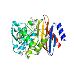
4DE2
DownloadVisualize
BU of 4de2 by Molmil
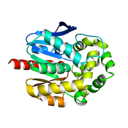
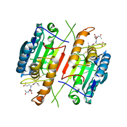
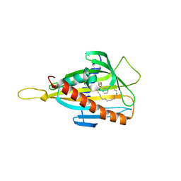
CTX-M-9 class A beta-lactamase complexed with compound 12
Descriptor: 3-[(dimethylamino)methyl]-N-[3-(1H-tetrazol-5-yl)phenyl]benzamide, Beta-lactamase, DIMETHYL SULFOXIDE
Authors:Nichols, D.A, Chen, Y.
Deposit date:2012-01-19
Release date:2012-03-28
Last modified:2023-09-13
Method:X-RAY DIFFRACTION (1.4 Å)
Cite:Structure-Based Design of Potent and Ligand-Efficient Inhibitors of CTX-M Class A Beta-Lactamase
J.Med.Chem., 55, 2012
2O2I
DownloadVisualize
BU of 2o2i by Molmil
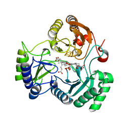
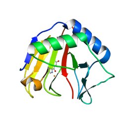
Crystal structure of haloalkane dehalogenase Rv2579 from Mycobacterium tuberculosis complexed with 1,3-propandiol
Descriptor: 1,3-PROPANDIOL, BROMIDE ION, Haloalkane dehalogenase 3
Authors:Mazumdar, P.A, Hulecki, J, Cherney, M.M, Garen, C.R, James, M.N.G, TB Structural Genomics Consortium (TBSGC)
Deposit date:2006-11-29
Release date:2007-11-13
Last modified:2023-08-30
Method:X-RAY DIFFRACTION (1.5 Å)
Cite:Crystal structure of haloalkane dehalogenase Rv2579 from Mycobacterium tuberculosis complexed with 1,3-propandiol
To be Published
3SWK
DownloadVisualize
BU of 3swk by Molmil
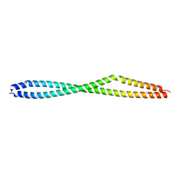
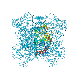
Crystal structure of vimentin coil1B fragment
Descriptor: Vimentin
Authors:Chernyatina, A.A, Strelkov, S.V.
Deposit date:2011-07-14
Release date:2012-08-15
Last modified:2024-02-28
Method:X-RAY DIFFRACTION (1.7 Å)
Cite:Atomic structure of the vimentin central alpha-helical domain and its implications for intermediate filament assembly.
Proc.Natl.Acad.Sci.USA, 109, 2012
3GZ4
DownloadVisualize
BU of 3gz4 by Molmil
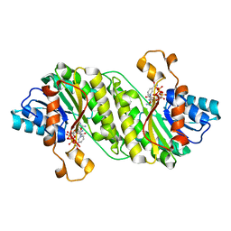
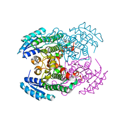
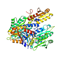
Crystal structure of putative short chain dehydrogenase FROM ESCHERICHIA COLI CFT073 complexed with NADPH
Descriptor: Hypothetical oxidoreductase yciK, NADPH DIHYDRO-NICOTINAMIDE-ADENINE-DINUCLEOTIDE PHOSPHATE
Authors:Malashkevich, V.N, Toro, R, Morano, C, Sauder, J.M, Burley, S.K, Almo, S.C, New York SGX Research Center for Structural Genomics (NYSGXRC)
Deposit date:2009-04-06
Release date:2009-04-14
Last modified:2024-02-21
Method:X-RAY DIFFRACTION (2.1 Å)
Cite:Crystal structure of putative short chain dehydrogenase FROM ESCHERICHIA COLI CFT073 complexed with NADPH
To be Published
4DR5
DownloadVisualize
BU of 4dr5 by Molmil
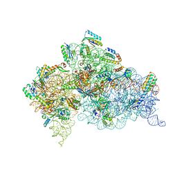
Crystal structure of the Thermus thermophilus (HB8) 30S ribosomal subunit with codon, crystallographically disordered cognate transfer RNA anticodon stem-loop and streptomycin bound
Descriptor: 16S rRNA, 30S ribosomal protein S10, 30S ribosomal protein S11, ...
Authors:Demirci, H, Murphy IV, F, Murphy, E, Gregory, S.T, Dahlberg, A.E, Jogl, G.
Deposit date:2012-02-16
Release date:2012-11-14
Last modified:2013-01-30
Method:X-RAY DIFFRACTION (3.45 Å)
Cite:A structural basis for streptomycin-induced misreading of the genetic code.
Nat Commun, 4, 2013
4HR2
DownloadVisualize
BU of 4hr2 by Molmil
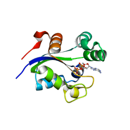
Structure of nucleoside diphosphate kinase (NDK) from Burkholderia thailandensis bound to ADP
Descriptor: ADENOSINE-5'-DIPHOSPHATE, Nucleoside diphosphate kinase
Authors:Seattle Structural Genomics Center for Infectious Disease (SSGCID)
Deposit date:2012-10-26
Release date:2012-11-07
Last modified:2023-09-20
Method:X-RAY DIFFRACTION (1.95 Å)
Cite:Structure of nucleoside diphosphate kinase (NDK) from Burkholderia thailandensis bound to ADP
To be Published
3GTY
DownloadVisualize
BU of 3gty by Molmil
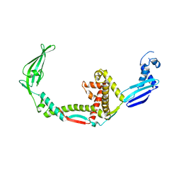
Promiscuous Substrate Recognition in Folding and Assembly Activities of the Trigger Factor Chaperone
Descriptor: 30S ribosomal protein S7, Trigger factor
Authors:Martinez-Hackert, E, Hendrickson, W.A.
Deposit date:2009-03-28
Release date:2009-12-22
Last modified:2024-02-21
Method:X-RAY DIFFRACTION (3.4 Å)
Cite:Promiscuous substrate recognition in folding and assembly activities of the trigger factor chaperone
Cell(Cambridge,Mass.), 138, 2009
3SVG
DownloadVisualize
BU of 3svg by Molmil
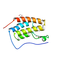
Crystal Structure of the first bromodomain of human BRD4 in complex with a 3,5-dimethylisoxazol ligand
Descriptor: (1R)-1-[3-(3,5-dimethyl-1,2-oxazol-4-yl)-5-ethoxyphenyl]ethanol, 1,2-ETHANEDIOL, Bromodomain-containing protein 4
Authors:Filippakopoulos, P, Picaud, S, Felletar, I, Hewings, S.D, von Delft, F, Arrowsmith, C.H, Edwards, A.M, Weigelt, J, Bountra, C, Conway, S.J, Knapp, S, Structural Genomics Consortium (SGC)
Deposit date:2011-07-12
Release date:2011-08-10
Last modified:2023-09-13
Method:X-RAY DIFFRACTION (1.68 Å)
Cite:Crystal Structure of the first bromodomain of human BRD4 in complex with a 3,5-dimethylisoxazol ligand
TO BE PUBLISHED
2O4L
DownloadVisualize
BU of 2o4l by Molmil
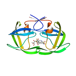
Crystal Structure of HIV-1 Protease (Q7K, I50V) in Complex with Tipranavir
Descriptor: CHLORIDE ION, GLYCEROL, N-(3-{(1R)-1-[(6R)-4-HYDROXY-2-OXO-6-PHENETHYL-6-PROPYL-5,6-DIHYDRO-2H-PYRAN-3-YL]PROPYL}PHENYL)-5-(TRIFLUOROMETHYL)-2-PYRIDINESULFONAMIDE, ...
Authors:Armstrong, A.A, Muzammil, S, Jakalian, A, Bonneau, P.R, Schmelmer, V, Freire, E, Amzel, L.M.
Deposit date:2006-12-04
Release date:2006-12-12
Last modified:2023-08-30
Method:X-RAY DIFFRACTION (1.33 Å)
Cite:Unique thermodynamic response of tipranavir to human immunodeficiency virus type 1 protease drug resistance mutations.
J.Virol., 81, 2007
4DGC
DownloadVisualize
BU of 4dgc by Molmil
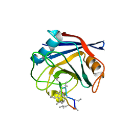
TRIMCyp cyclophilin domain from Macaca mulatta: cyclosporin A complex
Descriptor: TRIMCyp, cyclosporin A
Authors:Caines, M.E.C, Bichel, K, Price, A.J, McEwan, W.A, James, L.C.
Deposit date:2012-01-25
Release date:2012-02-08
Last modified:2023-12-06
Method:X-RAY DIFFRACTION (2.65 Å)
Cite:Diverse HIV viruses are targeted by a conformationally dynamic antiviral.
Nat.Struct.Mol.Biol., 19, 2012
3SVT
DownloadVisualize
BU of 3svt by Molmil
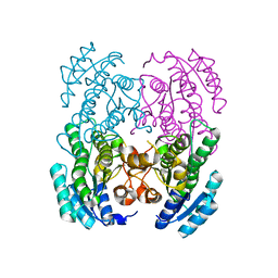
Structure of a short-chain type dehydrogenase/reductase from Mycobacterium ulcerans
Descriptor: CHLORIDE ION, SODIUM ION, Short-chain type dehydrogenase/reductase
Authors:Seattle Structural Genomics Center for Infectious Disease (SSGCID)
Deposit date:2011-07-12
Release date:2011-07-20
Last modified:2023-09-13
Method:X-RAY DIFFRACTION (2 Å)
Cite:Increasing the structural coverage of tuberculosis drug targets.
Tuberculosis (Edinb), 95, 2015
2I51
DownloadVisualize
BU of 2i51 by Molmil
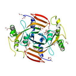
CRYSTAL STRUCTURE OF a pyridoxamine 5'-phosphate oxidase-related, FMN binding protein (NPUN_F5749) FROM NOSTOC PUNCTIFORME PCC 73102 AT 1.40 A RESOLUTION
Descriptor: 1,2-ETHANEDIOL, FLAVIN MONONUCLEOTIDE, GLYCEROL, ...
Authors:Joint Center for Structural Genomics (JCSG)
Deposit date:2006-08-23
Release date:2006-09-05
Last modified:2017-10-25
Method:X-RAY DIFFRACTION (1.4 Å)
Cite:Crystal structure of Uncharacterized conserved protein of COG5135 (ZP_00109616.1) from Nostoc punctiforme PCC 73102 at 1.40 A resolution
To be published
3GVC
DownloadVisualize
BU of 3gvc by Molmil
Crystal structure of probable short-chain dehydrogenase-reductase from Mycobacterium tuberculosis
Descriptor: Probable short-chain type dehydrogenase/reductase
Authors:Seattle Structural Genomics Center for Infectious Disease (SSGCID)
Deposit date:2009-03-30
Release date:2009-04-07
Last modified:2023-09-06
Method:X-RAY DIFFRACTION (2.45 Å)
Cite:Increasing the structural coverage of tuberculosis drug targets.
Tuberculosis (Edinb), 95, 2015
3H1P
DownloadVisualize
BU of 3h1p by Molmil
Mature Caspase-7 I213A with DEVD-CHO inhibitor bound to active site
Descriptor: Caspase-7, N-ACETYL-L-ALPHA-ASPARTYL-L-ALPHA-GLUTAMYL-N-[(2S)-1-CARBOXY-3-HYDROXYPROPAN-2-YL]-L-VALINAMIDE
Authors:Witkowski, W.A, Hardy, J.A.
Deposit date:2009-04-13
Release date:2009-06-30
Last modified:2023-11-22
Method:X-RAY DIFFRACTION (2.61 Å)
Cite:L2' loop is critical for caspase-7 active site formation.
Protein Sci., 18, 2009
4HTR
DownloadVisualize
BU of 4htr by Molmil
N149W variant of SiRHP bound to sulfite
Descriptor: IRON/SULFUR CLUSTER, SIROHEME, SODIUM ION, ...
Authors:Smith, K.W, Stroupe, M.E.
Deposit date:2012-11-01
Release date:2013-01-16
Last modified:2023-12-27
Method:X-RAY DIFFRACTION (1.6 Å)
Cite:Mutational analysis of sulfite reductase hemoprotein reveals the mechanism for coordinated electron and proton transfer.
Biochemistry, 51, 2012
4HU9
DownloadVisualize
BU of 4hu9 by Molmil
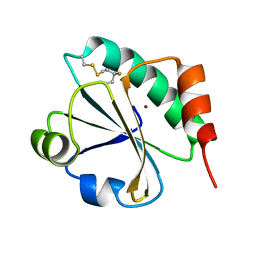
E. coli thioredoxin variant with (4S)-FluoroPro76 as single proline residue
Descriptor: COPPER (II) ION, Thioredoxin-1
Authors:Scharer, M.A, Rubini, M, Capitani, G, Glockshuber, R.
Deposit date:2012-11-02
Release date:2013-05-29
Last modified:2017-09-20
Method:X-RAY DIFFRACTION (1.55 Å)
Cite:(4R)- and (4S)-Fluoroproline in the Conserved cis-Prolyl Peptide Bond of the Thioredoxin Fold: Tertiary Structure Context Dictates Ring Puckering.
Chembiochem, 14, 2013
4DJY
DownloadVisualize
BU of 4djy by Molmil
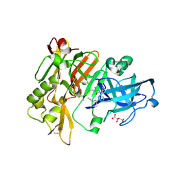
Structure of BACE Bound to (R)-5-cyclopropyl-2-imino-3-methyl-5-(3-(5-(prop-1-yn-1-yl)pyridin-3-yl)phenyl)imidazolidin-4-one
Descriptor: (2E,5R)-5-cyclopropyl-2-imino-3-methyl-5-{3-[5-(prop-1-yn-1-yl)pyridin-3-yl]phenyl}imidazolidin-4-one, Beta-secretase 1, L(+)-TARTARIC ACID
Authors:Strickland, C, Cumming, J.
Deposit date:2012-02-02
Release date:2012-03-21
Last modified:2012-04-04
Method:X-RAY DIFFRACTION (1.86 Å)
Cite:Structure based design of iminohydantoin BACE1 inhibitors: Identification of an orally available, centrally active BACE1 inhibitor.
Bioorg.Med.Chem.Lett., 22, 2012
3H3S
DownloadVisualize
BU of 3h3s by Molmil
Crystal structure of the CERT START domain in complex with HPA-15
Descriptor: Goodpasture antigen binding protein, N-[(1R,3R)-3-hydroxy-1-(hydroxymethyl)-3-phenylpropyl]pentadecanamide
Authors:Kudo, N, Wakatsuki, S, Kato, R.
Deposit date:2009-04-17
Release date:2010-03-02
Last modified:2024-03-20
Method:X-RAY DIFFRACTION (1.66 Å)
Cite:Crystal structures of the CERT START domain with inhibitors provide insights into the mechanism of ceramide transfer.
J.Mol.Biol., 396, 2010
3T3Y
DownloadVisualize
BU of 3t3y by Molmil
Crystal structure of AlkB in complex with Fe(III) and 2-(3-hydroxypicolinomido)acetic acid
Descriptor: Alpha-ketoglutarate-dependent dioxygenase AlkB, FE (III) ION, N-[(3-hydroxypyridin-2-yl)carbonyl]glycine
Authors:Aik, W.S, McDonough, M.A, Schofield, C.J.
Deposit date:2011-07-25
Release date:2012-03-07
Last modified:2023-09-13
Method:X-RAY DIFFRACTION (2.001 Å)
Cite:Dynamic combinatorial mass spectrometry leads to inhibitors of a 2-oxoglutarate-dependent nucleic Acid demethylase.
J.Med.Chem., 55, 2012
4HWG
DownloadVisualize
BU of 4hwg by Molmil
Structure of UDP-N-acetylglucosamine 2-epimerase from Rickettsia bellii
Descriptor: PHOSPHATE ION, UDP-N-acetylglucosamine 2-epimerase
Authors:Seattle Structural Genomics Center for Infectious Disease (SSGCID)
Deposit date:2012-11-07
Release date:2012-11-28
Last modified:2023-09-20
Method:X-RAY DIFFRACTION (2 Å)
Cite:Structure of UDP-N-acetylglucosamine 2-epimerase from Rickettsia bellii
TO BE PUBLISHED
4DLZ
DownloadVisualize
BU of 4dlz by Molmil
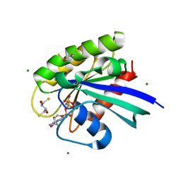
H-Ras Set 2 Ca(OAc)2/DTE, ordered off
Descriptor: (2R,3S)-1,4-DIMERCAPTOBUTANE-2,3-DIOL, CALCIUM ION, GTPase HRas, ...
Authors:Holzapfel, G, Mattos, C.
Deposit date:2012-02-06
Release date:2012-08-08
Last modified:2024-02-28
Method:X-RAY DIFFRACTION (1.662 Å)
Cite:Shift in the Equilibrium between On and Off States of the Allosteric Switch in Ras-GppNHp Affected by Small Molecules and Bulk Solvent Composition.
Biochemistry, 51, 2012
3H5G
DownloadVisualize
BU of 3h5g by Molmil
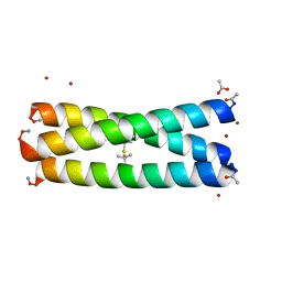
Switching the Chirality of the Metal Environment Alters the Coordination Mode in Designed Peptides.
Descriptor: ACETATE ION, COIL SER L16D-Pen, ZINC ION
Authors:Peacock, A.F.A, Stuckey, J.A, Pecoraro, V.L.
Deposit date:2009-04-22
Release date:2009-07-14
Last modified:2023-11-22
Method:X-RAY DIFFRACTION (1.71 Å)
Cite:Switching the Chirality of the Metal Environment Alters the Coordination Mode in Designed Peptides.
Angew.Chem.Int.Ed.Engl., 48, 2009
4HWP
DownloadVisualize
BU of 4hwp by Molmil
Crystal structure of E. coli Threonyl-tRNA synthetase bound to a novel inhibitor
Descriptor: N-{[3-(4-amino-2-methylquinazolin-7-yl)phenyl]sulfonyl}-L-threoninamide, Threonine--tRNA ligase, ZINC ION
Authors:Hilgers, M.T.
Deposit date:2012-11-08
Release date:2013-09-18
Last modified:2024-02-28
Method:X-RAY DIFFRACTION (1.807 Å)
Cite:Identification of bacteria-selective threonyl-tRNA synthetase substrate inhibitors by structure-based design.
J.Med.Chem., 56, 2013
3T46
DownloadVisualize
BU of 3t46 by Molmil
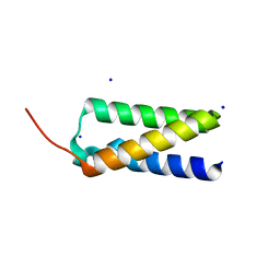
Crystal structure of Staphylococcal Complement Inhibitor D (SCIN-D) at 1.5 Angstrom
Descriptor: SCIN-D, SODIUM ION
Authors:Garcia, B.L, Geisbrecht, B.V, Summers, B.J.
Deposit date:2011-07-25
Release date:2011-11-23
Last modified:2023-09-13
Method:X-RAY DIFFRACTION (1.5 Å)
Cite:Diversity in the C3b Convertase Contact Residues and Tertiary Structures of the Staphylococcal Complement Inhibitor (SCIN) Protein Family.
J.Biol.Chem., 287, 2012
3T49
DownloadVisualize
BU of 3t49 by Molmil
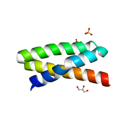
Crystal structure of truncated form of Staphylococcal Complement Inhibitor B (SCIN-B) at 1.5 Angstrom
Descriptor: Fibrinogen-binding protein, GLYCEROL, SULFATE ION
Authors:Garcia, B.L, Geisbrecht, B.V, Summers, B.J.
Deposit date:2011-07-25
Release date:2011-11-23
Last modified:2023-09-13
Method:X-RAY DIFFRACTION (1.45 Å)
Cite:Diversity in the C3b Convertase Contact Residues and Tertiary Structures of the Staphylococcal Complement Inhibitor (SCIN) Protein Family.
J.Biol.Chem., 287, 2012

223790

數據於2024-08-14公開中

PDB statisticsPDBj update infoContact PDBjnumon