Loading
PDBj
MenuPDBj@FacebookPDBj@TwitterPDBj@YouTubewwPDB FoundationwwPDB
RCSB PDBPDBeBMRBAdv. SearchSearch help

3DQO
DownloadVisualize
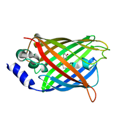
BU of 3dqo by Molmil
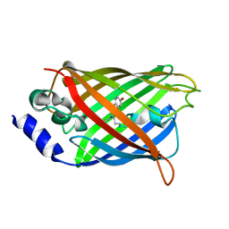
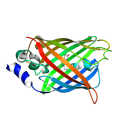
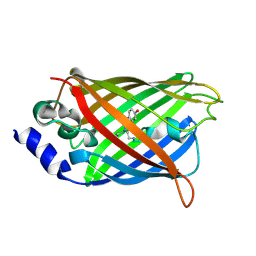
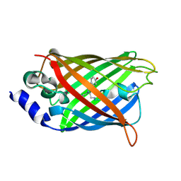
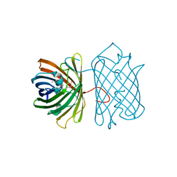
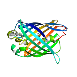
Structure of the Yellow Fluorescent Protein Citrine Frozen at 1 Atmosphere Number 2: Structure 2 in a Series of 26 High Pressure Structures
Descriptor: Green fluorescent protein
Authors:Barstow, B, Kim, C.U.
Deposit date:2008-07-09
Release date:2008-09-23
Last modified:2023-11-15
Method:X-RAY DIFFRACTION (1.5 Å)
Cite:Alteration of citrine structure by hydrostatic pressure explains the accompanying spectral shift.
Proc.Natl.Acad.Sci.Usa, 105, 2008
3DPX
DownloadVisualize
BU of 3dpx by Molmil
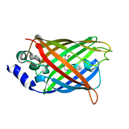
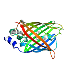
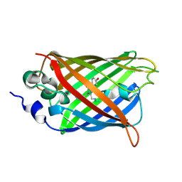
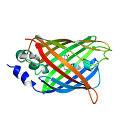
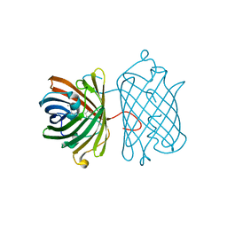
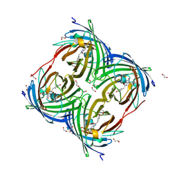
Structure of the Yellow Fluorescent Protein Citrine Frozen at 5000 Atmospheres: Structure 26 in a Series of 26 High Pressure Structures
Descriptor: Green fluorescent protein
Authors:Barstow, B, Kim, C.U.
Deposit date:2008-07-09
Release date:2008-09-23
Last modified:2023-11-15
Method:X-RAY DIFFRACTION (1.5 Å)
Cite:Alteration of citrine structure by hydrostatic pressure explains the accompanying spectral shift.
Proc.Natl.Acad.Sci.Usa, 105, 2008
3DQ5
DownloadVisualize
BU of 3dq5 by Molmil
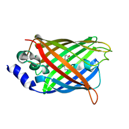
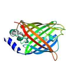
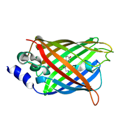
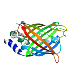
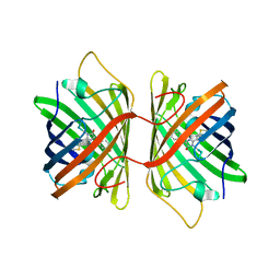
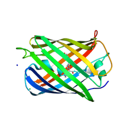
Structure of the Yellow Fluorescent Protein Citrine Frozen at 1960 Atmospheres: Structure 19 in a Series of 26 High Pressure Structures
Descriptor: Green fluorescent protein
Authors:Barstow, B, Kim, C.U.
Deposit date:2008-07-09
Release date:2008-09-23
Last modified:2023-11-15
Method:X-RAY DIFFRACTION (1.5 Å)
Cite:Alteration of citrine structure by hydrostatic pressure explains the accompanying spectral shift.
Proc.Natl.Acad.Sci.Usa, 105, 2008
3DQE
DownloadVisualize
BU of 3dqe by Molmil
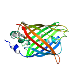
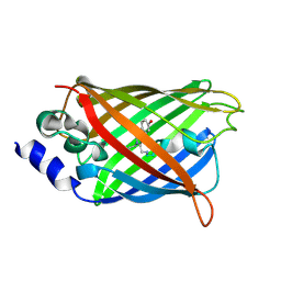
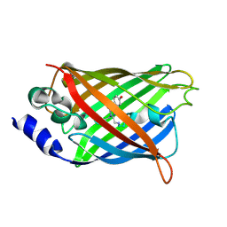
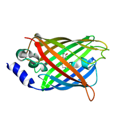
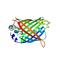
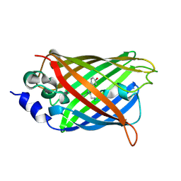
Structure of the Yellow Fluorescent Protein Citrine Frozen at 1250 Atmospheres Number 1: Structure 11 in a Series of 26 High Pressure Structures
Descriptor: Green fluorescent protein
Authors:Barstow, B, Kim, C.U.
Deposit date:2008-07-09
Release date:2008-09-23
Last modified:2023-11-15
Method:X-RAY DIFFRACTION (1.43 Å)
Cite:Alteration of citrine structure by hydrostatic pressure explains the accompanying spectral shift.
Proc.Natl.Acad.Sci.Usa, 105, 2008
3DQI
DownloadVisualize
BU of 3dqi by Molmil
Structure of the Yellow Fluorescent Protein Citrine Frozen at 1000 Atmospheres Number 4: Structure 8 in a Series of 26 High Pressure Structures
Descriptor: Green fluorescent protein
Authors:Barstow, B, Kim, C.U.
Deposit date:2008-07-09
Release date:2008-09-23
Last modified:2023-11-15
Method:X-RAY DIFFRACTION (1.42 Å)
Cite:Alteration of citrine structure by hydrostatic pressure explains the accompanying spectral shift.
Proc.Natl.Acad.Sci.Usa, 105, 2008
3DQU
DownloadVisualize
BU of 3dqu by Molmil
Structure of the Yellow Fluorescent Protein Citrine Frozen at 2000 Atmospheres Number 1: Structure 20 in a Series of 26 High Pressure Structures
Descriptor: Green fluorescent protein
Authors:Barstow, B, Kim, C.U.
Deposit date:2008-07-09
Release date:2008-09-23
Last modified:2023-11-15
Method:X-RAY DIFFRACTION (1.42 Å)
Cite:Alteration of citrine structure by hydrostatic pressure explains the accompanying spectral shift.
Proc.Natl.Acad.Sci.Usa, 105, 2008
3DQJ
DownloadVisualize
BU of 3dqj by Molmil
Structure of the Yellow Fluorescent Protein Citrine Frozen at 1000 Atmospheres Number 3: Structure 7 in a Series of 26 High Pressure Structures
Descriptor: Green fluorescent protein
Authors:Barstow, B, Kim, C.U.
Deposit date:2008-07-09
Release date:2008-09-23
Last modified:2023-11-15
Method:X-RAY DIFFRACTION (1.51 Å)
Cite:Alteration of citrine structure by hydrostatic pressure explains the accompanying spectral shift.
Proc.Natl.Acad.Sci.Usa, 105, 2008
3GJ1
DownloadVisualize
BU of 3gj1 by Molmil
Non photoactivated state of PA-GFP
Descriptor: CHLORIDE ION, Green fluorescent protein, SULFATE ION
Authors:Henderson, J.N, Gepshtein, R, Heenan, J.R, Kallio, K, Huppert, D, Remington, S.J.
Deposit date:2009-03-07
Release date:2009-03-24
Last modified:2023-11-22
Method:X-RAY DIFFRACTION (1.8 Å)
Cite:Structure and mechanism of the photoactivatable green fluorescent protein.
J.Am.Chem.Soc., 131, 2009
3DQ9
DownloadVisualize
BU of 3dq9 by Molmil
Structure of the Yellow Fluorescent Protein Citrine Frozen at 1500 Atmospheres Number 1: Structure 15 in a Series of 26 High Pressure Structures
Descriptor: Green fluorescent protein
Authors:Barstow, B, Kim, C.U.
Deposit date:2008-07-09
Release date:2008-09-23
Last modified:2023-11-15
Method:X-RAY DIFFRACTION (1.4 Å)
Cite:Alteration of citrine structure by hydrostatic pressure explains the accompanying spectral shift.
Proc.Natl.Acad.Sci.Usa, 105, 2008
3DQM
DownloadVisualize
BU of 3dqm by Molmil
Structure of the Yellow Fluorescent Protein Citrine Frozen at 750 Atmospheres: Structure 4 in a Series of 26 High Pressure Structures
Descriptor: Green fluorescent protein
Authors:Barstow, B, Kim, C.U.
Deposit date:2008-07-09
Release date:2008-09-23
Last modified:2023-11-15
Method:X-RAY DIFFRACTION (1.44 Å)
Cite:Alteration of citrine structure by hydrostatic pressure explains the accompanying spectral shift.
Proc.Natl.Acad.Sci.Usa, 105, 2008
3DQH
DownloadVisualize
BU of 3dqh by Molmil
Structure of the Yellow Fluorescent Protein Citrine Frozen at 1000 Atmospheres Number 5: Structure 9 in a Series of 26 High Pressure Structures
Descriptor: Green fluorescent protein
Authors:Barstow, B, Kim, C.U.
Deposit date:2008-07-09
Release date:2008-09-23
Last modified:2023-11-15
Method:X-RAY DIFFRACTION (1.45 Å)
Cite:Alteration of citrine structure by hydrostatic pressure explains the accompanying spectral shift.
Proc.Natl.Acad.Sci.Usa, 105, 2008
3DQ2
DownloadVisualize
BU of 3dq2 by Molmil
Structure of the Yellow Fluorescent Protein Citrine Frozen at 4000 Atmospheres Number 1: Structure 23 in a Series of 26 High Pressure Structures
Descriptor: Green fluorescent protein
Authors:Barstow, B, Kim, C.U.
Deposit date:2008-07-09
Release date:2008-09-23
Last modified:2023-11-15
Method:X-RAY DIFFRACTION (1.6 Å)
Cite:Alteration of citrine structure by hydrostatic pressure explains the accompanying spectral shift.
Proc.Natl.Acad.Sci.Usa, 105, 2008
3DQC
DownloadVisualize
BU of 3dqc by Molmil
Structure of the Yellow Fluorescent Protein Citrine Frozen at 1250 Atmospheres Number 3: Structure 13 in a Series of 26 High Pressure Structures
Descriptor: Green fluorescent protein
Authors:Barstow, B, Kim, C.U.
Deposit date:2008-07-09
Release date:2008-09-23
Last modified:2023-11-15
Method:X-RAY DIFFRACTION (1.49 Å)
Cite:Alteration of citrine structure by hydrostatic pressure explains the accompanying spectral shift.
Proc.Natl.Acad.Sci.Usa, 105, 2008
3G9A
DownloadVisualize
BU of 3g9a by Molmil
Green fluorescent protein bound to minimizer nanobody
Descriptor: Green fluorescent protein, Minimizer
Authors:Kirchhofer, A, Helma, J, Schmidthals, K, Frauer, C, Cui, S, Karcher, A, Pellis, M, Muyldermans, S, Delucci, C.C, Cardoso, M.C, Leonhardt, H, Hopfner, K.-P, Rothbauer, U.
Deposit date:2009-02-13
Release date:2009-12-08
Last modified:2023-11-15
Method:X-RAY DIFFRACTION (1.614 Å)
Cite:Modulation of protein properties in living cells using nanobodies
Nat.Struct.Mol.Biol., 17, 2010
3GB3
DownloadVisualize
BU of 3gb3 by Molmil
X-ray structure of genetically encoded photosensitizer KillerRed in native form
Descriptor: KillerRed, SULFATE ION
Authors:Pletnev, S, Pletneva, N.V, Pletnev, V.Z.
Deposit date:2009-02-18
Release date:2009-09-08
Last modified:2023-11-15
Method:X-RAY DIFFRACTION (1.75 Å)
Cite:Structural basis for phototoxicity of the genetically encoded photosensitizer KillerRed.
J.Biol.Chem., 284, 2009
3DQ7
DownloadVisualize
BU of 3dq7 by Molmil
Structure of the Yellow Fluorescent Protein Citrine Frozen at 1920 Atmospheres Number 1: Structure 17 in a Series of 26 High Pressure Structures
Descriptor: Green fluorescent protein
Authors:Barstow, B, Kim, C.U.
Deposit date:2008-07-09
Release date:2008-09-23
Last modified:2023-11-15
Method:X-RAY DIFFRACTION (1.23 Å)
Cite:Alteration of citrine structure by hydrostatic pressure explains the accompanying spectral shift.
Proc.Natl.Acad.Sci.Usa, 105, 2008
3DQK
DownloadVisualize
BU of 3dqk by Molmil
Structure of the Yellow Fluorescent Protein Citrine Frozen at 1000 Atmospheres Number 2: Structure 6 in a Series of 26 High Pressure Structures
Descriptor: Green fluorescent protein
Authors:Barstow, B, Kim, C.U.
Deposit date:2008-07-09
Release date:2008-09-23
Last modified:2023-11-15
Method:X-RAY DIFFRACTION (1.4 Å)
Cite:Alteration of citrine structure by hydrostatic pressure explains the accompanying spectral shift.
Proc.Natl.Acad.Sci.Usa, 105, 2008
3DPZ
DownloadVisualize
BU of 3dpz by Molmil
Structure of the Yellow Fluorescent Protein Citrine Frozen at 4000 Atmospheres Number 3: Structure 25 in a Series of 26 High Pressure Structures
Descriptor: Green fluorescent protein
Authors:Barstow, B, Kim, C.U.
Deposit date:2008-07-09
Release date:2008-09-23
Last modified:2023-11-15
Method:X-RAY DIFFRACTION (1.7 Å)
Cite:Alteration of citrine structure by hydrostatic pressure explains the accompanying spectral shift.
Proc.Natl.Acad.Sci.Usa, 105, 2008
3DQ8
DownloadVisualize
BU of 3dq8 by Molmil
Structure of the Yellow Fluorescent Protein Citrine Frozen at 1500 Atmospheres Number 2: Structure 16 in a Series of 26 High Pressure Structures
Descriptor: Green fluorescent protein
Authors:Barstow, B, Kim, C.U.
Deposit date:2008-07-09
Release date:2008-09-23
Last modified:2023-11-15
Method:X-RAY DIFFRACTION (1.51 Å)
Cite:Alteration of citrine structure by hydrostatic pressure explains the accompanying spectral shift.
Proc.Natl.Acad.Sci.Usa, 105, 2008
3GEX
DownloadVisualize
BU of 3gex by Molmil
1.6 angstrom crystal structure of fluorescent protein Cypet
Descriptor: Green fluorescent protein
Authors:Hu, X, Liu, R.
Deposit date:2009-02-26
Release date:2010-03-09
Last modified:2023-11-22
Method:X-RAY DIFFRACTION (1.6 Å)
Cite:1.6 angstrom crystal structure of fluorescent protein Cypet
To be Published
7A7N
DownloadVisualize
BU of 7a7n by Molmil
rsGreen0.7b in the green-off state
Descriptor: Green fluorescent protein
Authors:De Zitter, E, Dedecker, P, Van Meervelt, L.
Deposit date:2020-08-30
Release date:2021-02-17
Last modified:2023-11-15
Method:X-RAY DIFFRACTION (1.2 Å)
Cite:Structure-Function Dataset Reveals Environment Effects within a Fluorescent Protein Model System*.
Angew.Chem.Int.Ed.Engl., 60, 2021
7A84
DownloadVisualize
BU of 7a84 by Molmil
rsGreen0.7-K206A-F145H partially in the green-off state
Descriptor: Green fluorescent protein
Authors:De Zitter, E, Dedecker, P, Van Meervelt, L.
Deposit date:2020-08-30
Release date:2021-02-17
Last modified:2021-05-05
Method:X-RAY DIFFRACTION (2.1 Å)
Cite:Structure-Function Dataset Reveals Environment Effects within a Fluorescent Protein Model System*.
Angew.Chem.Int.Ed.Engl., 60, 2021
7A8C
DownloadVisualize
BU of 7a8c by Molmil
rsGreen0.7-K206A-F145S in the green-off state
Descriptor: Green fluorescent protein, TRIETHYLENE GLYCOL
Authors:De Zitter, E, Dedecker, P, Van Meervelt, L.
Deposit date:2020-08-30
Release date:2021-02-17
Last modified:2021-05-05
Method:X-RAY DIFFRACTION (2.13 Å)
Cite:Structure-Function Dataset Reveals Environment Effects within a Fluorescent Protein Model System*.
Angew.Chem.Int.Ed.Engl., 60, 2021
7A7L
DownloadVisualize
BU of 7a7l by Molmil
rsEGFP in the green-off state
Descriptor: Green fluorescent protein, TETRAETHYLENE GLYCOL
Authors:De Zitter, E, Dedecker, P, Van Meervelt, L.
Deposit date:2020-08-30
Release date:2021-02-17
Last modified:2021-05-05
Method:X-RAY DIFFRACTION (1.3 Å)
Cite:Structure-Function Dataset Reveals Environment Effects within a Fluorescent Protein Model System*.
Angew.Chem.Int.Ed.Engl., 60, 2021
7A81
DownloadVisualize
BU of 7a81 by Molmil
rsGreen0.7-K206A-F145A partially in the green-on state
Descriptor: Green fluorescent protein
Authors:De Zitter, E, Dedecker, P, Van Meervelt, L.
Deposit date:2020-08-30
Release date:2021-02-17
Last modified:2021-05-05
Method:X-RAY DIFFRACTION (1.7 Å)
Cite:Structure-Function Dataset Reveals Environment Effects within a Fluorescent Protein Model System*.
Angew.Chem.Int.Ed.Engl., 60, 2021

221051

數據於2024-06-12公開中

PDB statisticsPDBj update infoContact PDBjnumon