Loading
PDBj
MenuPDBj@FacebookPDBj@X(formerly Twitter)PDBj@BlueSkyPDBj@YouTubewwPDB FoundationwwPDB
RCSB PDBPDBeBMRBAdv. SearchSearch help
PDB: 227344 results

1HUA
DownloadVisualize
BU of 1hua by Molmil
THE SOLUTION CONFORMATION OF HYALURONAN: A COMBINED NMR AND MOLECULAR DYNAMICS STUDY
Descriptor: beta-D-glucopyranuronic acid-(1-3)-2-acetamido-2-deoxy-beta-D-galactopyranose-(1-4)-2,6-anhydro-L-gulonic acid
Authors:Holmbeck, S.M.A, Petillo, P.A, Lerner, L.E.
Deposit date:1994-01-31
Release date:1994-11-30
Last modified:2024-05-01
Method:SOLUTION NMR
Cite:The solution conformation of hyaluronan: a combined NMR and molecular dynamics study.
Biochemistry, 33, 1994
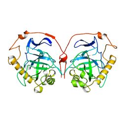
1HUC
DownloadVisualize
BU of 1huc by Molmil
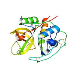
THE REFINED 2.15 ANGSTROMS X-RAY CRYSTAL STRUCTURE OF HUMAN LIVER CATHEPSIN B: THE STRUCTURAL BASIS FOR ITS SPECIFICITY
Descriptor: CATHEPSIN B
Authors:Musil, D, Bode, W.
Deposit date:1993-04-21
Release date:1995-01-26
Last modified:2024-10-23
Method:X-RAY DIFFRACTION (2.1 Å)
Cite:The refined 2.15 A X-ray crystal structure of human liver cathepsin B: the structural basis for its specificity.
EMBO J., 10, 1991
1HUE
DownloadVisualize
BU of 1hue by Molmil
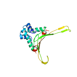
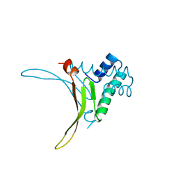
HISTONE-LIKE PROTEIN
Descriptor: HU PROTEIN
Authors:Vis, H, Mariani, M, Vorgias, C.E, Wilson, K.S, Kaptein, R, Boelens, R.
Deposit date:1995-05-26
Release date:1995-10-15
Last modified:2024-05-22
Method:SOLUTION NMR
Cite:Solution structure of the HU protein from Bacillus stearothermophilus.
J.Mol.Biol., 254, 1995
1HUF
DownloadVisualize
BU of 1huf by Molmil
CRYSTAL STRUCTURE OF THE N-TERMINAL DOMAIN OF THE TYROSINE PHOSPHATASE YOPH FROM YERSINIA PESTIS.
Descriptor: TYROSINE PHOSPHATASE YOPH
Authors:Evdokimov, A.G, Waugh, D.S.
Deposit date:2001-01-04
Release date:2001-05-23
Last modified:2024-02-07
Method:X-RAY DIFFRACTION (2 Å)
Cite:Structure of the N-terminal domain of Yersinia pestis YopH at 2.0 A resolution.
Acta Crystallogr.,Sect.D, 57, 2001
1HUG
DownloadVisualize
BU of 1hug by Molmil
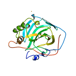
Differences in anionic inhibition of Human Carbonic Anhydrase I revealed from the structures of iodide and gold cyanide inhibitor complexes
Descriptor: CARBONIC ANHYDRASE I, GOLD (I) CYANIDE ION, ZINC ION
Authors:Kumar, V, Kannan, K.K.
Deposit date:1993-10-28
Release date:1994-04-30
Last modified:2024-02-07
Method:X-RAY DIFFRACTION (2 Å)
Cite:Differences in anionic inhibition of human carbonic anhydrase I revealed from the structures of iodide and gold cyanide inhibitor complexes.
Acta Crystallogr.,Sect.D, 50, 1994
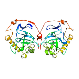
1HUH
DownloadVisualize
BU of 1huh by Molmil
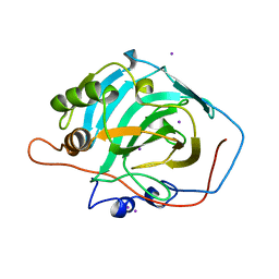
DIFFERENCES IN ANIONIC INHIBITION OF HUMAN CARBONIC ANHYDRASE I REVEALED FROM THE STRUCTURES OF IODIDE AND GOLD CYANIDE INHIBITOR COMPLEXES
Descriptor: CARBONIC ANHYDRASE I, IODIDE ION, ZINC ION
Authors:Kumar, V, Kannan, K.K.
Deposit date:1993-10-28
Release date:1994-04-30
Last modified:2024-02-07
Method:X-RAY DIFFRACTION (2.2 Å)
Cite:Differences in anionic inhibition of human carbonic anhydrase I revealed from the structures of iodide and gold cyanide inhibitor complexes.
Acta Crystallogr.,Sect.D, 50, 1994
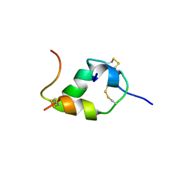
1HUI
DownloadVisualize
BU of 1hui by Molmil
INSULIN MUTANT (B1, B10, B16, B27)GLU, DES-B30, NMR, 25 STRUCTURES
Descriptor: INSULIN
Authors:Olsen, H.B, Ludvigsen, S, Kaarsholm, N.C.
Deposit date:1996-03-29
Release date:1997-03-12
Last modified:2024-10-30
Method:SOLUTION NMR
Cite:Solution structure of an engineered insulin monomer at neutral pH.
Biochemistry, 35, 1996
1HUJ
DownloadVisualize
BU of 1huj by Molmil
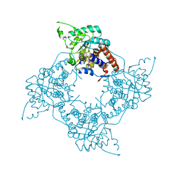
REFINED STRUCTURE OF YEAST INORGANIC PYROPHOSPHATASE AND ITS K61R MUTANT
Descriptor: INORGANIC PYROPHOSPHATASE, MAGNESIUM ION
Authors:Swaminathan, K, Cooperman, B.S, Lahti, R, Voet, D.
Deposit date:1997-12-26
Release date:1998-04-08
Last modified:2024-05-22
Method:X-RAY DIFFRACTION (2.1 Å)
Cite:Refined Structure of Yeast Inorganic Pyrophosphatase and its K61R Mutant
To be Published
1HUK
DownloadVisualize
BU of 1huk by Molmil
REFINED STRUCTURE OF YEAST INORGANIC PYROPHOSPHATASE AND ITS K61R MUTANT
Descriptor: INORGANIC PYROPHOSPHATASE, MAGNESIUM ION
Authors:Swaminathan, K, Cooperman, B.S, Lahti, R, Voet, D.
Deposit date:1997-12-26
Release date:1998-04-08
Last modified:2024-05-22
Method:X-RAY DIFFRACTION (2.2 Å)
Cite:Refined Structure of Yeast Inorganic Pyrophosphatase and its K61R Mutant
To be Published
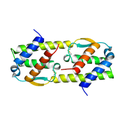
1HUL
DownloadVisualize
BU of 1hul by Molmil
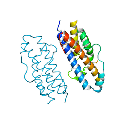
A NOVEL DIMER CONFIGURATION REVEALED BY THE CRYSTAL STRUCTURE AT 2.4 ANGSTROMS RESOLUTION OF HUMAN INTERLEUKIN-5
Descriptor: INTERLEUKIN-5
Authors:Milburn, M.V.
Deposit date:1995-03-17
Release date:1995-12-07
Last modified:2024-10-09
Method:X-RAY DIFFRACTION (2.4 Å)
Cite:A novel dimer configuration revealed by the crystal structure at 2.4 A resolution of human interleukin-5.
Nature, 363, 1993
1HUM
DownloadVisualize
BU of 1hum by Molmil
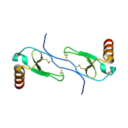
SOLUTION STRUCTURE OF THE CHEMOKINE HMIP-1BETA(SLASH)ACT-2 BY MULTI-DIMENSIONAL NMR: A NOVEL CHEMOKINE DIMER
Descriptor: HUMAN MACROPHAGE INFLAMMATORY PROTEIN 1 BETA
Authors:Clore, G.M, Lodi, P.J, Garrett, D.S, Gronenborn, A.M.
Deposit date:1994-01-31
Release date:1994-04-30
Last modified:2024-10-23
Method:SOLUTION NMR
Cite:High-resolution solution structure of the beta chemokine hMIP-1 beta by multidimensional NMR.
Science, 263, 1994
1HUN
DownloadVisualize
BU of 1hun by Molmil
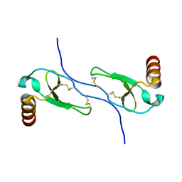
SOLUTION STRUCTURE OF THE CHEMOKINE HMIP-1BETA(SLASH)ACT-2 BY MULTI-DIMENSIONAL NMR: A NOVEL CHEMOKINE DIMER
Descriptor: HUMAN MACROPHAGE INFLAMMATORY PROTEIN 1 BETA
Authors:Clore, G.M, Lodi, P.J, Garrett, D.S, Gronenborn, A.M.
Deposit date:1994-01-31
Release date:1994-04-30
Last modified:2022-02-23
Method:SOLUTION NMR
Cite:High-resolution solution structure of the beta chemokine hMIP-1 beta by multidimensional NMR.
Science, 263, 1994
1HUO
DownloadVisualize
BU of 1huo by Molmil
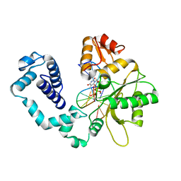
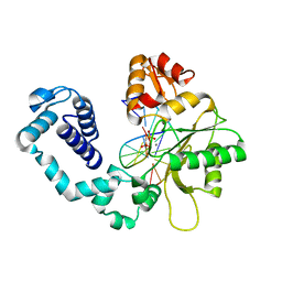
CRYSTAL STRUCTURE OF DNA POLYMERASE BETA COMPLEXED WITH DNA AND CR-TMPPCP
Descriptor: 5'-D(*AP*AP*TP*AP*GP*GP*CP*GP*TP*CP*G)-3', 5'-D(P*CP*GP*AP*CP*GP*CP*C)-3', CHROMIUM ION, ...
Authors:Arndt, J.W, Gong, W, Zhong, X, Showalter, A.K, Liu, J, Lin, Z, Paxson, C, Tsai, M.-D, Chan, M.K.
Deposit date:2001-01-04
Release date:2001-04-23
Last modified:2024-02-07
Method:X-RAY DIFFRACTION (2.6 Å)
Cite:Insight into the catalytic mechanism of DNA polymerase beta: structures of intermediate complexes.
Biochemistry, 40, 2001
1HUP
DownloadVisualize
BU of 1hup by Molmil
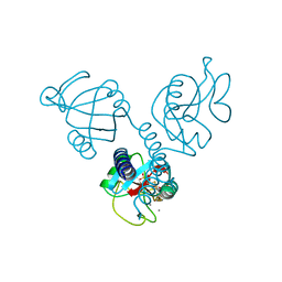
HUMAN MANNOSE BINDING PROTEIN CARBOHYDRATE RECOGNITION DOMAIN TRIMERIZES THROUGH A TRIPLE ALPHA-HELICAL COILED-COIL
Descriptor: CALCIUM ION, MANNOSE-BINDING PROTEIN, SULFATE ION
Authors:Sheriff, S.
Deposit date:1994-09-21
Release date:1995-10-15
Last modified:2024-11-06
Method:X-RAY DIFFRACTION (2.5 Å)
Cite:Human mannose-binding protein carbohydrate recognition domain trimerizes through a triple alpha-helical coiled-coil.
Nat.Struct.Biol., 1, 1994
1HUQ
DownloadVisualize
BU of 1huq by Molmil
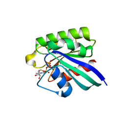
1.8A CRYSTAL STRUCTURE OF THE MONOMERIC GTPASE RAB5C (MOUSE)
Descriptor: MAGNESIUM ION, PHOSPHOAMINOPHOSPHONIC ACID-GUANYLATE ESTER, RAB5C
Authors:Merithew, E, Hatherly, S, Dumas, J.J, Lawe, D.C, Heller-Harrison, R, Lambright, D.G.
Deposit date:2001-01-04
Release date:2001-02-07
Last modified:2023-08-09
Method:X-RAY DIFFRACTION (1.8 Å)
Cite:Structural plasticity of an invariant hydrophobic triad in the switch regions of Rab GTPases is a determinant of effector recognition.
J.Biol.Chem., 276, 2001
1HUR
DownloadVisualize
BU of 1hur by Molmil
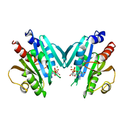
HUMAN ADP-RIBOSYLATION FACTOR 1 COMPLEXED WITH GDP, FULL LENGTH NON-MYRISTOYLATED
Descriptor: GUANOSINE-5'-DIPHOSPHATE, HUMAN ADP-RIBOSYLATION FACTOR 1, MAGNESIUM ION
Authors:Amor, J.C, Harrison, D.H, Kahn, R.A, Ringe, D.
Deposit date:1995-04-19
Release date:1995-07-10
Last modified:2024-02-07
Method:X-RAY DIFFRACTION (2 Å)
Cite:Structure of the human ADP-ribosylation factor 1 complexed with GDP.
Nature, 372, 1994
1HUS
DownloadVisualize
BU of 1hus by Molmil
RIBOSOMAL PROTEIN S7
Descriptor: RIBOSOMAL PROTEIN S7
Authors:Hosaka, H, Nakagawa, A, Tanaka, I.
Deposit date:1997-08-08
Release date:1998-01-28
Last modified:2024-10-16
Method:X-RAY DIFFRACTION (2.5 Å)
Cite:Ribosomal protein S7: a new RNA-binding motif with structural similarities to a DNA architectural factor.
Structure, 5, 1997
1HUT
DownloadVisualize
BU of 1hut by Molmil
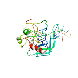
THE STRUCTURE OF ALPHA-THROMBIN INHIBITED BY A 15-MER SINGLE-STRANDED DNA APTAMER
Descriptor: ALPHA-Thrombin heavy chain, ALPHA-Thrombin light chain, D-phenylalanyl-N-[(3S)-6-carbamimidamido-1-chloro-2-oxohexan-3-yl]-L-prolinamide, ...
Authors:Padmanabhan, K, Padmanabhan, K.P, Ferrara, J.D, Sadler, J.E, Tulinsky, A.
Deposit date:1993-05-27
Release date:1994-06-22
Last modified:2024-10-16
Method:X-RAY DIFFRACTION (2.9 Å)
Cite:The structure of alpha-thrombin inhibited by a 15-mer single-stranded DNA aptamer.
J.Biol.Chem., 268, 1993
1HUU
DownloadVisualize
BU of 1huu by Molmil
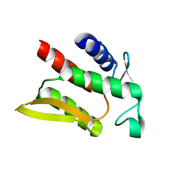
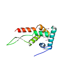
DNA-BINDING PROTEIN HU FROM BACILLUS STEAROTHERMOPHILUS
Descriptor: PROTEIN HU
Authors:White, S.W, Tanaka, I, Appelt, K, Wilson, K.S.
Deposit date:1998-07-06
Release date:1999-01-13
Last modified:2024-02-07
Method:X-RAY DIFFRACTION (2 Å)
Cite:A protein structural motif that bends DNA.
Proteins, 5, 1989
1HUV
DownloadVisualize
BU of 1huv by Molmil
CRYSTAL STRUCTURE OF A SOLUBLE MUTANT OF THE MEMBRANE-ASSOCIATED (S)-MANDELATE DEHYDROGENASE FROM PSEUDOMONAS PUTIDA AT 2.15A RESOLUTION
Descriptor: 2-(N-MORPHOLINO)-ETHANESULFONIC ACID, FLAVIN MONONUCLEOTIDE, L(+)-MANDELATE DEHYDROGENASE, ...
Authors:Mathews, F.S, Sukumar, N.
Deposit date:2001-01-04
Release date:2001-09-19
Last modified:2023-08-09
Method:X-RAY DIFFRACTION (2.15 Å)
Cite:Structure of an active soluble mutant of the membrane-associated (S)-mandelate dehydrogenase.
Biochemistry, 40, 2001
1HUW
DownloadVisualize
BU of 1huw by Molmil
THE CRYSTAL STRUCTURE OF AFFINITY-MATURED HUMAN GROWTH HORMONE AT 2 ANGSTROMS RESOLUTION
Descriptor: HUMAN GROWTH HORMONE
Authors:Ultsch, M.H, Somers, W.S, Kossiakoff, A.A, De Vos, A.M.
Deposit date:1993-09-22
Release date:1994-01-31
Last modified:2024-10-23
Method:X-RAY DIFFRACTION (2 Å)
Cite:The crystal structure of affinity-matured human growth hormone at 2 A resolution.
J.Mol.Biol., 236, 1994
1HUX
DownloadVisualize
BU of 1hux by Molmil
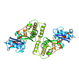
CRYSTAL STRUCTURE OF THE ACIDAMINOCOCCUS FERMENTANS (R)-2-HYDROXYGLUTARYL-COA DEHYDRATASE COMPONENT A
Descriptor: ACTIVATOR OF (R)-2-HYDROXYGLUTARYL-COA DEHYDRATASE, ADENOSINE-5'-DIPHOSPHATE, IRON/SULFUR CLUSTER
Authors:Locher, K.P, Hans, M, Yeh, A.P, Schmid, B, Buckel, W, Rees, D.C.
Deposit date:2001-01-04
Release date:2001-03-21
Last modified:2024-02-07
Method:X-RAY DIFFRACTION (3 Å)
Cite:Crystal structure of the Acidaminococcus fermentans 2-hydroxyglutaryl-CoA dehydratase component A.
J.Mol.Biol., 307, 2001
1HUY
DownloadVisualize
BU of 1huy by Molmil
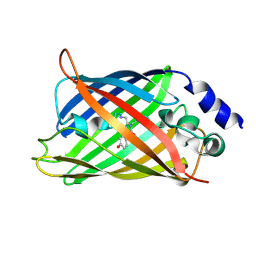
CRYSTAL STRUCTURE OF CITRINE, AN IMPROVED YELLOW VARIANT OF GREEN FLUORESCENT PROTEIN
Descriptor: GREEN FLUORESCENT PROTEIN
Authors:Griesbeck, O, Baird, G.S, Campbell, R.E, Zacharias, D.A, Tsien, R.Y.
Deposit date:2001-01-04
Release date:2001-07-04
Last modified:2023-11-15
Method:X-RAY DIFFRACTION (2.2 Å)
Cite:Reducing the environmental sensitivity of yellow fluorescent protein. Mechanism and applications
J.Biol.Chem., 276, 2001
1HUZ
DownloadVisualize
BU of 1huz by Molmil
CRYSTAL STRUCTURE OF DNA POLYMERASE COMPLEXED WITH DNA AND CR-PCP
Descriptor: 5'-D(*AP*AP*TP*AP*GP*GP*CP*GP*TP*CP*G)-3', 5'-D(P*CP*GP*AP*CP*GP*CP*CP*T)-3', CHROMIUM ION, ...
Authors:Arndt, J.W, Gong, W, Zhong, X, Showalter, A.K, Liu, J, Lin, Z, Paxson, C, Tsai, M.-D, Chan, M.K.
Deposit date:2001-01-04
Release date:2001-04-23
Last modified:2024-02-07
Method:X-RAY DIFFRACTION (2.6 Å)
Cite:Insight into the catalytic mechanism of DNA polymerase beta: structures of intermediate complexes.
Biochemistry, 40, 2001
1HV0
DownloadVisualize
BU of 1hv0 by Molmil
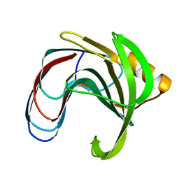
DISSECTING ELECTROSTATIC INTERACTIONS AND THE PH-DEPENDENT ACTIVITY OF A FAMILY 11 GLYCOSIDASE
Descriptor: ENDO-1,4-BETA-XYLANASE
Authors:Joshi, M.D, Sidhu, G, Nielsen, J.E, Brayer, G.D, Withers, S.G, McIntosh, L.P.
Deposit date:2001-01-05
Release date:2001-09-14
Last modified:2023-08-09
Method:X-RAY DIFFRACTION (1.6 Å)
Cite:Dissecting the electrostatic interactions and pH-dependent activity of a family 11 glycosidase.
Biochemistry, 40, 2001

227344

數據於2024-11-13公開中

PDB statisticsPDBj update infoContact PDBjnumon