Loading
PDBj
MenuPDBj@FacebookPDBj@TwitterPDBj@YouTubewwPDB FoundationwwPDB
RCSB PDBPDBeBMRBAdv. SearchSearch help

3FJJ
DownloadVisualize
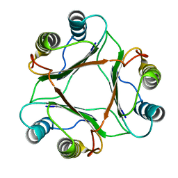
BU of 3fjj by Molmil
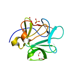
Crystal structure of C83V mutant of Human acidic fibroblast growth factor
Descriptor: FORMIC ACID, Heparin-binding growth factor 1, SULFATE ION
Authors:Blaber, M, Lee, J.
Deposit date:2008-12-14
Release date:2009-10-06
Last modified:2023-09-06
Method:X-RAY DIFFRACTION (1.9 Å)
Cite:Structural basis of conserved cysteine in the fibroblast growth factor family: evidence for a vestigial half-cystine.
J.Mol.Biol., 393, 2009
3UR1
DownloadVisualize
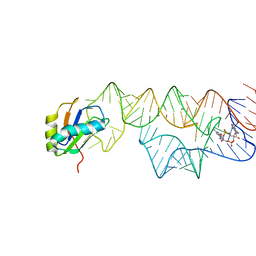
BU of 3ur1 by Molmil
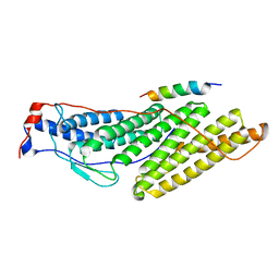
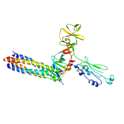
The structure of a ternary complex between CheA domains P4 and P5 with CheW and with a truncated fragment of TM14, a chemoreceptor analog from Thermotoga maritima.
Descriptor: Chemotaxis protein CheA, Chemotaxis protein CheW, Methyl-accepting chemotaxis protein
Authors:Li, X, Crane, B.R, Bilwes, A.M.
Deposit date:2011-11-21
Release date:2012-03-07
Last modified:2024-02-28
Method:X-RAY DIFFRACTION (4.5 Å)
Cite:The structure of native bacterial chemoreceptor arrays
Proc.Natl.Acad.Sci.USA, 2012
3UM3
DownloadVisualize
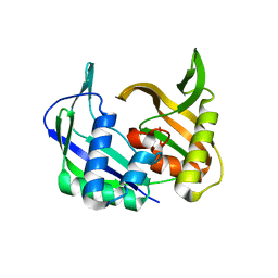
BU of 3um3 by Molmil
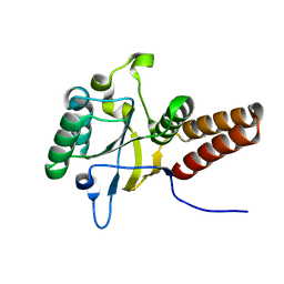
Crystal structure of the Brox Bro1 domain in complex with the C-terminal tail of CHMP4B
Descriptor: BRO1 domain-containing protein BROX, Charged multivesicular body protein 4b
Authors:Jiang, J.S, Mu, R.L, Xiao, T.
Deposit date:2011-11-12
Release date:2012-04-18
Last modified:2023-09-13
Method:X-RAY DIFFRACTION (3.8 Å)
Cite:Two Distinct Binding Modes Define the Interaction of Brox with the C-Terminal Tails of CHMP5 and CHMP4B.
Structure, 20, 2012
3FCY
DownloadVisualize
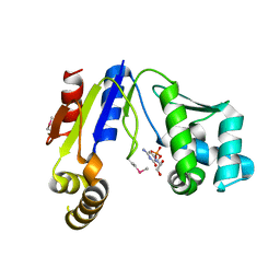
BU of 3fcy by Molmil
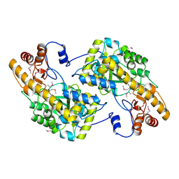
Crystal Structure of Acetyl Xylan Esterase 1 from Thermoanaerobacterium sp. JW/SL YS485
Descriptor: CALCIUM ION, Xylan esterase 1
Authors:Krastanova, I, Cassetta, A, Lamba, D.
Deposit date:2008-11-24
Release date:2009-11-17
Last modified:2023-09-06
Method:X-RAY DIFFRACTION (2.1 Å)
Cite:Crystal Structure Analysis of Acetyl Xylan Esterase 1 from Thermoanaerobacterium sp. JW/SL YS485
To be Published
3VH4
DownloadVisualize
BU of 3vh4 by Molmil
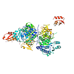
Crystal structure of Atg7CTD-Atg8-MgATP complex
Descriptor: ADENOSINE-5'-TRIPHOSPHATE, Autophagy-related protein 8, MAGNESIUM ION, ...
Authors:Noda, N.N, Satoo, K, Inagaki, F.
Deposit date:2011-08-23
Release date:2011-09-21
Last modified:2023-11-08
Method:X-RAY DIFFRACTION (2.65 Å)
Cite:Structural basis of Atg8 activation by a homodimeric E1, Atg7.
Mol.Cell, 44, 2011
3G7S
DownloadVisualize
BU of 3g7s by Molmil
Crystal structure of a long-chain-fatty-acid-CoA ligase (FadD1) from Archaeoglobus fulgidus
Descriptor: Long-chain-fatty-acid--CoA ligase (FadD-1)
Authors:Palani, K, Kumaran, D, Burley, S.K, Swaminathan, S, New York SGX Research Center for Structural Genomics (NYSGXRC)
Deposit date:2009-02-10
Release date:2009-03-03
Last modified:2021-02-10
Method:X-RAY DIFFRACTION (2.15 Å)
Cite:Crystal structure of a long-chain-fatty-acid-CoA ligase (FadD1) from Archaeoglobus fulgidus
To be Published
3GDF
DownloadVisualize
BU of 3gdf by Molmil
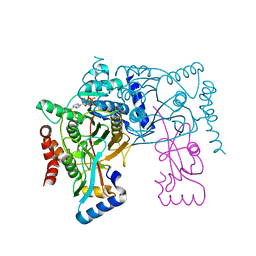
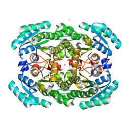
Crystal structure of the NADP-dependent mannitol dehydrogenase from Cladosporium herbarum.
Descriptor: Probable NADP-dependent mannitol dehydrogenase, ZINC ION
Authors:Nuess, D, Goettig, P, Magler, I, Denk, U, Breitenbach, M, Schneider, P.B, Brandstetter, H, Simon-Nobbe, B.
Deposit date:2009-02-24
Release date:2010-05-26
Last modified:2023-09-06
Method:X-RAY DIFFRACTION (2.5 Å)
Cite:Crystal structure of the NADP-dependent mannitol dehydrogenase from Cladosporium herbarum: Implications for oligomerisation and catalysis.
Biochimie, 92, 2010
3VZ6
DownloadVisualize
BU of 3vz6 by Molmil
Crystal Structure Analysis of the Mini-chaperonines, variant with Gly 184 replaced with Ile and Leu 185 replaced Val and Val 186 replaced with Leu.
Descriptor: 60 kDa chaperonin
Authors:Saijo, S, Sato, T.
Deposit date:2012-10-09
Release date:2013-11-13
Last modified:2023-11-08
Method:X-RAY DIFFRACTION (1.5 Å)
Cite:Binding Energy(BE) from Crystal Packing of Mini-chaperones (mcpn) Provided Insights into the Allosteric Interaction of GroEL/ES Complex
To be Published
3W1K
DownloadVisualize
BU of 3w1k by Molmil
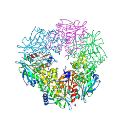
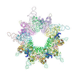
Crystal structure of the selenocysteine synthase SelA and tRNASec complex
Descriptor: L-seryl-tRNA(Sec) selenium transferase, selenocysteine tRNA
Authors:Itoh, Y, Sekine, S, Yokoyama, S.
Deposit date:2012-11-15
Release date:2013-06-05
Last modified:2023-12-06
Method:X-RAY DIFFRACTION (7.5 Å)
Cite:Decameric SelA-tRNA(Sec) ring structure reveals mechanism of bacterial selenocysteine formation
Science, 340, 2013
3GES
DownloadVisualize
BU of 3ges by Molmil
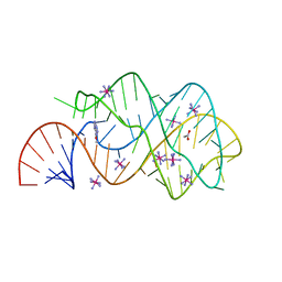
Crystal structure of the guanine riboswitch C74U mutant bound to 6-O-methylguanine
Descriptor: 6-O-methylguanine, ACETATE ION, COBALT HEXAMMINE(III), ...
Authors:Gilbert, S.D, Batey, R.T.
Deposit date:2009-02-26
Release date:2009-06-23
Last modified:2023-09-06
Method:X-RAY DIFFRACTION (2.15 Å)
Cite:Adaptive ligand binding by the purine riboswitch in the recognition of Guanine and adenine analogs
Structure, 17, 2009
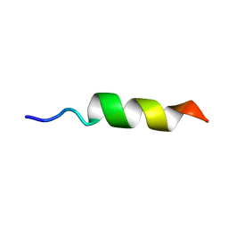
1OMQ
DownloadVisualize
BU of 1omq by Molmil
Structure of penetratin in bicellar solution
Descriptor: Homeotic antennapedia protein
Authors:Lindberg, M, Biverstahl, H, Graslund, A, Maler, L.
Deposit date:2003-02-26
Release date:2003-07-29
Last modified:2024-05-22
Method:SOLUTION NMR
Cite:Structure and positioning comparison of two variants of penetratin in two different membrane mimicking systems by NMR
Eur.J.Biochem., 270, 2003
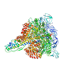
3W3A
DownloadVisualize
BU of 3w3a by Molmil
Crystal structure of V1-ATPase at 3.9 angstrom resolution
Descriptor: ADENOSINE-5'-DIPHOSPHATE, V-type ATP synthase alpha chain, V-type ATP synthase beta chain, ...
Authors:Nagamatsu, Y, Takeda, K, Kuranaga, T, Numoto, N, Miki, K.
Deposit date:2012-12-14
Release date:2013-05-15
Last modified:2024-03-20
Method:X-RAY DIFFRACTION (3.9 Å)
Cite:Origin of Asymmetry at the Intersubunit Interfaces of V1-ATPase from Thermusthermophilus
J.Mol.Biol., 425, 2013
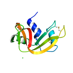
3I7W
DownloadVisualize
BU of 3i7w by Molmil
High pressure structure of wild-type RNase A (0.67 GPa)
Descriptor: CHLORIDE ION, Ribonuclease pancreatic
Authors:Lewinski, K, Kurpiewska, K, Dziubek, K, Katrusiak, A, Font, J, Ribo, M, Vilanova, M.
Deposit date:2009-07-09
Release date:2009-08-04
Last modified:2023-11-01
Method:X-RAY DIFFRACTION (2.35 Å)
Cite:Structural investigation of ribonuclease A conformational preferences using high pressure protein crystallography
Chem.Phys., 468, 2016
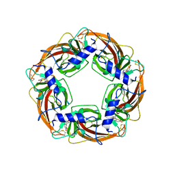
3WTM
DownloadVisualize
BU of 3wtm by Molmil
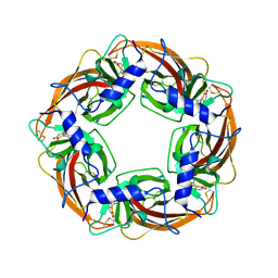
Crystal Structure of Lymnaea stagnalis Acetylcholine-Binding Protein Q55R Mutant Complexed with Nitromethylene Analogue of Imidacloprid
Descriptor: 2-chloro-5-{[(2E)-2-(nitromethylidene)imidazolidin-1-yl]methyl}pyridine, Acetylcholine-binding protein
Authors:Okajima, T, Ihara, M, Yamashita, A, Oda, T, Matsuda, K.
Deposit date:2014-04-11
Release date:2015-02-04
Method:X-RAY DIFFRACTION (2.48 Å)
Cite:Studies on an acetylcholine binding protein identify a basic residue in loop G on the beta 1 strand as a new structural determinant of neonicotinoid actions
Mol.Pharmacol., 86, 2014
1P1G
DownloadVisualize
BU of 1p1g by Molmil
MACROPHAGE MIGRATION INHIBITORY FACTOR (MIF) WITH PRO-1 MUTATED TO GLY-1
Descriptor: PROTEIN (MACROPHAGE MIGRATION INHIBITORY FACTOR)
Authors:Lubetsky, J.B, Lolis, E.
Deposit date:1999-03-24
Release date:1999-06-07
Last modified:2023-12-27
Method:X-RAY DIFFRACTION (2.5 Å)
Cite:Pro-1 of macrophage migration inhibitory factor functions as a catalytic base in the phenylpyruvate tautomerase activity.
Biochemistry, 38, 1999
3IWN
DownloadVisualize
BU of 3iwn by Molmil
Co-crystal structure of a bacterial c-di-GMP riboswitch
Descriptor: 9,9'-[(2R,3R,3aS,5S,7aR,9R,10R,10aS,12S,14aR)-3,5,10,12-tetrahydroxy-5,12-dioxidooctahydro-2H,7H-difuro[3,2-d:3',2'-j][1,3,7,9,2,8]tetraoxadiphosphacyclododecine-2,9-diyl]bis(2-amino-1,9-dihydro-6H-purin-6-one), C-di-GMP riboswitch, U1 small nuclear ribonucleoprotein A
Authors:Kulshina, N, Baird, N.J, Ferre-D'Amare, A.R.
Deposit date:2009-09-02
Release date:2009-11-10
Last modified:2024-02-21
Method:X-RAY DIFFRACTION (3.2 Å)
Cite:Recognition of the bacterial second messenger cyclic diguanylate by its cognate riboswitch.
Nat.Struct.Mol.Biol., 16, 2009
3UFC
DownloadVisualize
BU of 3ufc by Molmil
Characterization of a Cas6-related gene from Pyrococcus furiosus
Descriptor: Putative uncharacterized protein
Authors:Park, H.M, Kim, J.S.
Deposit date:2011-11-01
Release date:2012-03-21
Last modified:2024-03-20
Method:X-RAY DIFFRACTION (2.03 Å)
Cite:Crystal structure of a Cas6 paralogous protein from Pyrococcus furiosus
Proteins, 80, 2012
3W8N
DownloadVisualize
BU of 3w8n by Molmil
Open form structure of CMP kinase in complex with CMP from Thermus thermophilus HB8
Descriptor: CYTIDINE-5'-MONOPHOSPHATE, Cytidylate kinase
Authors:Nakagawa, N, Mega, R, Masui, R, Kuramitsu, S, RIKEN Structural Genomics/Proteomics Initiative (RSGI)
Deposit date:2013-03-19
Release date:2013-09-25
Last modified:2017-11-22
Method:X-RAY DIFFRACTION (2.2 Å)
Cite:Crystal structure of CMP kinase: insights into initial substrate recognition and reaction mechanisms
To be Published
3UAK
DownloadVisualize
BU of 3uak by Molmil
Crystal Structure of De Novo designed cysteine esterase ECH14, Northeast Structural Genomics Consortium Target OR54
Descriptor: De Novo designed cysteine esterase ECH14
Authors:Kuzin, A, Su, M, Seetharaman, J, Sahdev, S, Xiao, R, Ciccosanti, C, Richter, F, Everett, J.K, Nair, R, Acton, T.B, Rost, B, Baker, D, Montelione, G.T, Hunt, J.F, Tong, L, Northeast Structural Genomics Consortium (NESG)
Deposit date:2011-10-21
Release date:2011-12-07
Last modified:2023-12-06
Method:X-RAY DIFFRACTION (3.232 Å)
Cite:Computational design of catalytic dyads and oxyanion holes for ester hydrolysis.
J.Am.Chem.Soc., 134, 2012
3UB3
DownloadVisualize
BU of 3ub3 by Molmil
D96N variant of TIR domain of Mal/TIRAP
Descriptor: 2,3-DIHYDROXY-1,4-DITHIOBUTANE, Toll/interleukin-1 receptor domain-containing adapter protein
Authors:Shen, Y, Lin, Z.
Deposit date:2011-10-23
Release date:2012-05-16
Last modified:2023-11-01
Method:X-RAY DIFFRACTION (2.75 Å)
Cite:Structural Insights into TIR Domain Specificity of the Bridging Adaptor Mal in TLR4 Signaling
Plos One, 7, 2012
3WOY
DownloadVisualize
BU of 3woy by Molmil
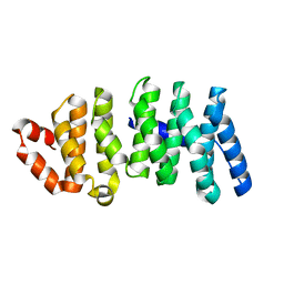
Crystal structure of CLASP2 TOG domain (TOG2)
Descriptor: CLIP-associating protein 2
Authors:Hayashi, I, Maki, T.
Deposit date:2014-01-06
Release date:2015-05-27
Last modified:2024-03-20
Method:X-RAY DIFFRACTION (2.1 Å)
Cite:CLASP2 Has Two Distinct TOG Domains That Contribute Differently to Microtubule Dynamics
J. Mol. Biol., 427, 2015
3WTL
DownloadVisualize
BU of 3wtl by Molmil
Crystal Structure of Lymnaea stagnalis Acetylcholine Binding Protein Complexed with Nitromethylene Analogue of Imidacloprid
Descriptor: 2-chloro-5-{[(2E)-2-(nitromethylidene)imidazolidin-1-yl]methyl}pyridine, Acetylcholine-binding protein
Authors:Okajima, T, Ihara, M, Yamashita, A, Oda, T, Matsuda, K.
Deposit date:2014-04-11
Release date:2015-02-04
Last modified:2023-11-08
Method:X-RAY DIFFRACTION (2.3 Å)
Cite:Studies on an acetylcholine binding protein identify a basic residue in loop G on the beta 1 strand as a new structural determinant of neonicotinoid actions
Mol.Pharmacol., 86, 2014
3WW3
DownloadVisualize
BU of 3ww3 by Molmil
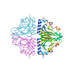
X-ray structures of Cellulomonas parahominis L-ribose isomerase with no ligand
Descriptor: L-ribose isomerase, MANGANESE (II) ION
Authors:Terami, Y, Yoshida, H, Takata, G, Kamitori, S.
Deposit date:2014-06-13
Release date:2015-04-29
Last modified:2024-05-29
Method:X-RAY DIFFRACTION (1.9 Å)
Cite:Essentiality of tetramer formation of Cellulomonas parahominis L-ribose isomerase involved in novel L-ribose metabolic pathway.
Appl.Microbiol.Biotechnol., 99, 2015
3W59
DownloadVisualize
BU of 3w59 by Molmil
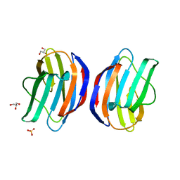
Crystal structure of Galectin-1 in the lactose-unbound state(P212121)
Descriptor: BETA-MERCAPTOETHANOL, GLYCEROL, Galectin-1, ...
Authors:Saburi, H, Tanaka, T, Kunishima, N.
Deposit date:2013-01-26
Release date:2014-01-29
Last modified:2023-11-08
Method:X-RAY DIFFRACTION (2.1 Å)
Cite:Crystal structure of Galectin-1 in the lactose-unbound state
To be Published
3W90
DownloadVisualize
BU of 3w90 by Molmil
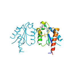
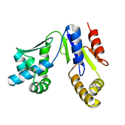
Crystal structure of CMP kinase from Thermus thermophilus HB8
Descriptor: Cytidylate kinase
Authors:Nakagawa, N, Mega, R, Masui, R, Kuramitsu, S, RIKEN Structural Genomics/Proteomics Initiative (RSGI)
Deposit date:2013-03-22
Release date:2013-09-25
Last modified:2023-11-08
Method:X-RAY DIFFRACTION (1.65 Å)
Cite:Crystal structure of CMP kinase: insights into initial substrate recognition and reaction mechanisms
To be Published

224931

数据于2024-09-11公开中

PDB statisticsPDBj update infoContact PDBjnumon