Loading
PDBj
MenuPDBj@FacebookPDBj@TwitterPDBj@YouTubewwPDB FoundationwwPDB
RCSB PDBPDBeBMRBAdv. SearchSearch help

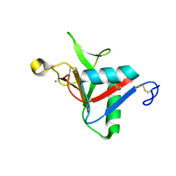
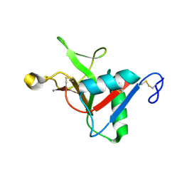
4J6K
DownloadVisualize
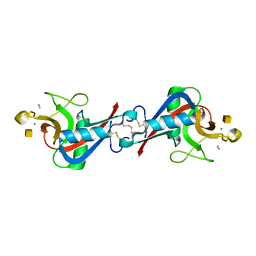
BU of 4j6k by Molmil
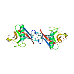
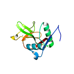
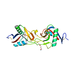
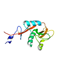
Crystal structure of calcium2+-free wild-type CD23 lectin domain (crystal form B)
Descriptor: Low affinity immunoglobulin epsilon Fc receptor, SULFATE ION
Authors:Dhaliwal, B, Pang, M.O.Y, Sutton, B.J.
Deposit date:2013-02-11
Release date:2013-08-28
Method:X-RAY DIFFRACTION (2.3 Å)
Cite:Conformational plasticity at the IgE-binding site of the B-cell receptor CD23.
Mol.Immunol., 56, 2013
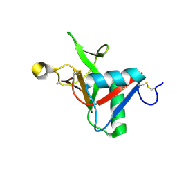
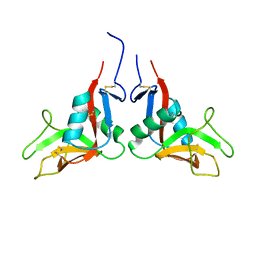
4J6Q
DownloadVisualize
BU of 4j6q by Molmil
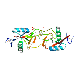
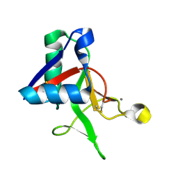
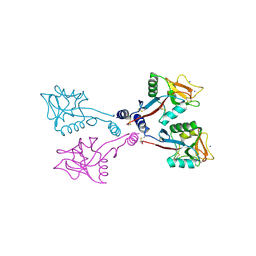
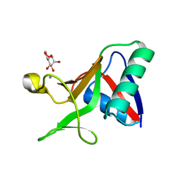
Crystal structure of calcium2+-free wild-type CD23 lectin domain (crystal form G)
Descriptor: Low affinity immunoglobulin epsilon Fc receptor
Authors:Dhaliwal, B, Pang, M.O.Y, Sutton, B.J.
Deposit date:2013-02-11
Release date:2013-08-28
Last modified:2023-09-20
Method:X-RAY DIFFRACTION (2.539 Å)
Cite:Conformational plasticity at the IgE-binding site of the B-cell receptor CD23.
Mol.Immunol., 56, 2013
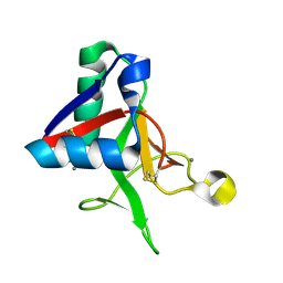
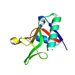
4J6N
DownloadVisualize
BU of 4j6n by Molmil
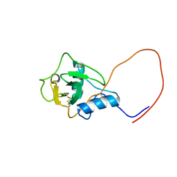
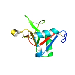
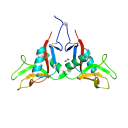
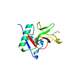
Crystal structure of calcium2+-free wild-type CD23 lectin domain (crystal form E)
Descriptor: Low affinity immunoglobulin epsilon Fc receptor
Authors:Dhaliwal, B, Pang, M.O.Y, Sutton, B.J.
Deposit date:2013-02-11
Release date:2013-08-28
Last modified:2023-09-20
Method:X-RAY DIFFRACTION (2.85 Å)
Cite:Conformational plasticity at the IgE-binding site of the B-cell receptor CD23.
Mol.Immunol., 56, 2013
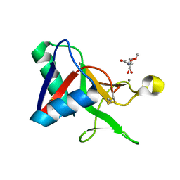
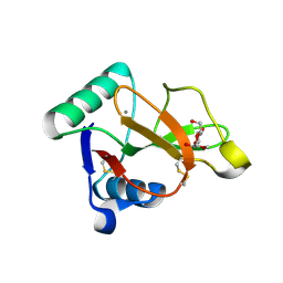
4J6J
DownloadVisualize
BU of 4j6j by Molmil
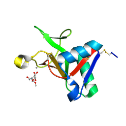
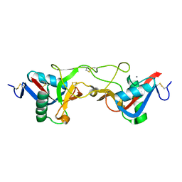
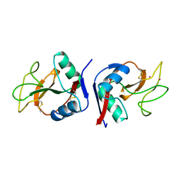
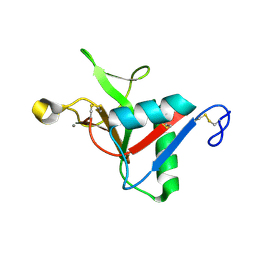
Crystal structure of calcium2+-free wild-type CD23 lectin domain (crystal form A)
Descriptor: Low affinity immunoglobulin epsilon Fc receptor, SULFATE ION
Authors:Dhaliwal, B, Yahya, N, Sutton, B.J.
Deposit date:2013-02-11
Release date:2013-08-28
Last modified:2023-09-20
Method:X-RAY DIFFRACTION (1.9 Å)
Cite:Conformational plasticity at the IgE-binding site of the B-cell receptor CD23.
Mol.Immunol., 56, 2013
4J6L
DownloadVisualize
BU of 4j6l by Molmil
Crystal structure of calcium2+-free wild-type CD23 lectin domain (crystal form C)
Descriptor: Low affinity immunoglobulin epsilon Fc receptor
Authors:Dhaliwal, B, Yuan, D, Sutton, B.J.
Deposit date:2013-02-11
Release date:2013-08-28
Last modified:2023-09-20
Method:X-RAY DIFFRACTION (3.15 Å)
Cite:Conformational plasticity at the IgE-binding site of the B-cell receptor CD23.
Mol.Immunol., 56, 2013
4KZW
DownloadVisualize
BU of 4kzw by Molmil
Structure of the carbohydrate-recognition domain of the C-type lectin mincle
Descriptor: C-TYPE LECTIN MINCLE, CALCIUM ION, CITRATE ANION, ...
Authors:Feinberg, H, Jegouzo, S.A.F, Rowntree, T.J.W, Guan, Y, Brash, M.A, Taylor, M.E, Weis, W.I, Drickamer, K.
Deposit date:2013-05-30
Release date:2013-08-28
Last modified:2017-11-15
Method:X-RAY DIFFRACTION (1.85 Å)
Cite:Mechanism for Recognition of an Unusual Mycobacterial Glycolipid by the Macrophage Receptor Mincle.
J.Biol.Chem., 288, 2013
4N34
DownloadVisualize
BU of 4n34 by Molmil
Structure of langerin CRD I313 with alpha-MeGlcNAc
Descriptor: C-type lectin domain family 4 member K, CALCIUM ION, methyl 2-acetamido-2-deoxy-alpha-D-glucopyranoside
Authors:Feinberg, H, Rowntree, T.J.W, Tan, S.L.W, Drickamer, K, Weis, W.I, Taylor, M.E.
Deposit date:2013-10-06
Release date:2013-11-20
Last modified:2023-09-20
Method:X-RAY DIFFRACTION (1.75 Å)
Cite:Common polymorphisms in human langerin change specificity for glycan ligands.
J.Biol.Chem., 288, 2013
4KMB
DownloadVisualize
BU of 4kmb by Molmil
COMPLEX OF 4'-SULFO-LEWIS-X WITH A SELECTIN-LIKE MUTANT OF MANNOSE-BINDING PROTEIN A
Descriptor: CALCIUM ION, CHLORIDE ION, MANNOSE-BINDING PROTEIN-A, ...
Authors:Ng, K.K.-S, Weis, W.I.
Deposit date:1996-11-07
Release date:1997-02-12
Last modified:2023-08-09
Method:X-RAY DIFFRACTION (2 Å)
Cite:Structure of a selectin-like mutant of mannose-binding protein complexed with sialylated and sulfated Lewis(x) oligosaccharides.
Biochemistry, 36, 1997
4N37
DownloadVisualize
BU of 4n37 by Molmil
Structure of langerin CRD I313 D288 complexed with Me-Man
Descriptor: C-type lectin domain family 4 member K, CALCIUM ION, methyl alpha-D-mannopyranoside
Authors:Feinberg, H, Rowntree, T.J.W, Tan, S.L.W, Drickamer, K, Weis, W.I, Taylor, M.E.
Deposit date:2013-10-06
Release date:2013-11-20
Last modified:2023-09-20
Method:X-RAY DIFFRACTION (2 Å)
Cite:Common polymorphisms in human langerin change specificity for glycan ligands.
J.Biol.Chem., 288, 2013
4MTH
DownloadVisualize
BU of 4mth by Molmil
Crystal structure of mature human RegIIIalpha
Descriptor: Regenerating islet-derived protein 3-alpha 15 kDa form, ZINC ION
Authors:Derebe, M.G.
Deposit date:2013-09-19
Release date:2013-11-27
Last modified:2017-11-15
Method:X-RAY DIFFRACTION (1.47 Å)
Cite:Antibacterial membrane attack by a pore-forming intestinal C-type lectin.
Nature, 505, 2013
4N33
DownloadVisualize
BU of 4n33 by Molmil
Structure of langerin CRD complexed with GlcNAc-beta1-3Gal-beta1-4Glc-beta-CH2CH2N3
Descriptor: 2-acetamido-2-deoxy-beta-D-glucopyranose-(1-3)-beta-D-galactopyranose, 2-acetamido-2-deoxy-beta-D-glucopyranose-(1-3)-beta-D-galactopyranose-(1-4)-beta-D-glucopyranose, C-type lectin domain family 4 member K, ...
Authors:Feinberg, H, Rowntree, T.J.W, Tan, S.L.W, Drickamer, K, Weis, W.I, Taylor, M.E.
Deposit date:2013-10-06
Release date:2013-11-20
Last modified:2023-09-20
Method:X-RAY DIFFRACTION (1.85 Å)
Cite:Common polymorphisms in human langerin change specificity for glycan ligands.
J.Biol.Chem., 288, 2013
1UV0
DownloadVisualize
BU of 1uv0 by Molmil
Pancreatitis-associated protein 1 from human
Descriptor: PANCREATITIS-ASSOCIATED PROTEIN 1, ZINC ION
Authors:Abergel, C, Shepard, W, Christal, L.
Deposit date:2004-01-12
Release date:2004-01-14
Last modified:2023-12-13
Method:X-RAY DIFFRACTION (1.78 Å)
Cite:Crystallization and preliminary crystallographic study of HIP/PAP, a human C-lectin overexpressed in primary liver cancers.
Acta Crystallogr.,Sect.D, 55, 1999
1WMZ
DownloadVisualize
BU of 1wmz by Molmil
Crystal Structure of C-type Lectin CEL-I complexed with N-acetyl-D-galactosamine
Descriptor: 2-acetamido-2-deoxy-alpha-D-galactopyranose, 2-acetamido-2-deoxy-beta-D-galactopyranose, CALCIUM ION, ...
Authors:Sugawara, H, Kusunoki, M, Kurisu, G, Fujimoto, T, Aoyagi, H, Hatakeyama, T.
Deposit date:2004-07-22
Release date:2004-09-07
Last modified:2020-07-29
Method:X-RAY DIFFRACTION (1.7 Å)
Cite:Characteristic Recognition of N-Acetylgalactosamine by an Invertebrate C-type Lectin, CEL-I, Revealed by X-ray Crystallographic Analysis
J.Biol.Chem., 279, 2004
1WMY
DownloadVisualize
BU of 1wmy by Molmil
Crystal Structure of C-type Lectin CEL-I from Cucumaria echinata
Descriptor: (4S)-2-METHYL-2,4-PENTANEDIOL, CALCIUM ION, lectin CEL-I, ...
Authors:Sugawara, H, Kusunoki, M, Kurisu, G, Fujimoto, T, Aoyagi, H, Hatakeyama, T.
Deposit date:2004-07-22
Release date:2004-09-07
Last modified:2023-10-25
Method:X-RAY DIFFRACTION (2 Å)
Cite:Characteristic Recognition of N-Acetylgalactosamine by an Invertebrate C-type Lectin, CEL-I, Revealed by X-ray Crystallographic Analysis
J.Biol.Chem., 279, 2004
1X2W
DownloadVisualize
BU of 1x2w by Molmil
Crystal Structure of Apo-Habu IX-bp at pH 4.6
Descriptor: CHLORIDE ION, Coagulation factor IX/X-binding protein A chain, Coagulation factor IX/factor X-binding protein B chain, ...
Authors:Suzuki, N, Fujimoto, Z, Morita, T, Fukamizu, A, Mizuno, H.
Deposit date:2005-04-26
Release date:2005-10-04
Last modified:2023-10-25
Method:X-RAY DIFFRACTION (2.29 Å)
Cite:pH-Dependent Structural Changes at Ca(2+)-binding sites of Coagulation Factor IX-binding Protein
J.Mol.Biol., 353, 2005
1WK1
DownloadVisualize
BU of 1wk1 by Molmil
Solution structure of Lectin C-type domain derived from a hypothetical protein from C. elegans
Descriptor: Hypothetical protein yk1067a12
Authors:Kobayashi, N, Koshiba, S, Inoue, M, Tochio, N, Kigawa, T, Yokoyama, S, RIKEN Structural Genomics/Proteomics Initiative (RSGI)
Deposit date:2004-05-29
Release date:2004-11-29
Last modified:2022-03-02
Method:SOLUTION NMR
Cite:Solution structure of Lectin C-type domain derived from a hypothetical protein from C. elegans
To be Published
1WT9
DownloadVisualize
BU of 1wt9 by Molmil
crystal structure of Aa-X-bp-I, a snake venom protein with the activity of binding to coagulation factor X from Agkistrodon acutus
Descriptor: CALCIUM ION, agkisacutacin A chain, anticoagulant protein-B
Authors:Zhu, Z, Liu, S, Mo, X, Yu, X, Liang, Z, Zang, J, Zhao, W, Teng, M, Niu, L.
Deposit date:2004-11-18
Release date:2006-03-07
Last modified:2011-07-13
Method:X-RAY DIFFRACTION (2.01 Å)
Cite:Characterizations and Crystal structures of two snake venom proteins with the activity of binding coagulation factor X from Agkistrodon acutus
To be Published
1X2T
DownloadVisualize
BU of 1x2t by Molmil
Crystal Structure of Habu IX-bp at pH 6.5
Descriptor: CALCIUM ION, Coagulation factor IX/X-binding protein A chain, Coagulation factor IX/factor X-binding protein B chain, ...
Authors:Suzuki, N, Fujimoto, Z, Morita, T, Fukamizu, A, Mizuno, H.
Deposit date:2005-04-26
Release date:2005-10-04
Last modified:2023-10-25
Method:X-RAY DIFFRACTION (1.72 Å)
Cite:pH-Dependent Structural Changes at Ca(2+)-binding sites of Coagulation Factor IX-binding Protein
J.Mol.Biol., 353, 2005
1XAR
DownloadVisualize
BU of 1xar by Molmil
Crystal Structure of a fragment of DC-SIGNR (containing the carbohydrate recognition domain and two repeats of the neck).
Descriptor: CD209 antigen-like protein 1, SODIUM ION
Authors:Feinberg, H, Guo, Y, Mitchell, D.A, Drickamer, K, Weis, W.I.
Deposit date:2004-08-26
Release date:2004-11-16
Last modified:2011-07-13
Method:X-RAY DIFFRACTION (2.25 Å)
Cite:Extended Neck Regions Stabilize Tetramers of the Receptors DC-SIGN and DC-SIGNR
J.Biol.Chem., 280, 2005
1XPH
DownloadVisualize
BU of 1xph by Molmil
Structure of DC-SIGNR and a portion of repeat domain 8
Descriptor: CALCIUM ION, CD209 antigen-like protein 1
Authors:Snyder, G.A, Colonna, M, Sun, P.D.
Deposit date:2004-10-08
Release date:2005-04-19
Last modified:2023-08-23
Method:X-RAY DIFFRACTION (1.41 Å)
Cite:The structure of DC-SIGNR with a portion of its repeat domain lends insights to modeling of the receptor tetramer.
J.Mol.Biol., 347, 2005
1UMR
DownloadVisualize
BU of 1umr by Molmil
Crystal structure of the platelet activator convulxin, a disulfide linked a4b4 cyclic tetramer from the venom of Crotalus durissus terrificus
Descriptor: CONVULXIN ALPHA, CONVULXIN BETA
Authors:Murakami, M.T, Zela, S.P, Gava, L.M, Michelan-Duarte, S, Cintra, A.C.O, Arni, R.K.
Deposit date:2003-08-28
Release date:2003-11-21
Last modified:2023-12-13
Method:X-RAY DIFFRACTION (2.4 Å)
Cite:Crystal Structure of the Platelet Activator Convulxin, a Disulfide Linked A4B4 Cyclic Tetramer from the Venom of Crotalus Durissus Terrificus
Biochem.Biophys.Res.Commun., 310, 2003
1YPU
DownloadVisualize
BU of 1ypu by Molmil
Human Oxidized Low Density Lipoprotein Receptor LOX-1 C2 Space Group
Descriptor: oxidised low density lipoprotein (lectin-like) receptor 1
Authors:Park, H, Adsit, F.G, Boyington, J.C.
Deposit date:2005-01-31
Release date:2005-02-08
Last modified:2023-08-23
Method:X-RAY DIFFRACTION (2.05 Å)
Cite:The 1.4 angstrom crystal structure of the human oxidized low density lipoprotein receptor lox-1.
J.Biol.Chem., 280, 2005
1YPO
DownloadVisualize
BU of 1ypo by Molmil
Human Oxidized Low Density Lipoprotein Receptor LOX-1 P3 1 21 Space Group
Descriptor: oxidised low density lipoprotein (lectin-like) receptor 1
Authors:Park, H, Adsit, F.G, Boyington, J.C.
Deposit date:2005-01-31
Release date:2005-02-08
Last modified:2023-08-23
Method:X-RAY DIFFRACTION (3 Å)
Cite:The 1.4 angstrom crystal structure of the human oxidized low density lipoprotein receptor lox-1.
J.Biol.Chem., 280, 2005
1YPQ
DownloadVisualize
BU of 1ypq by Molmil
Human Oxidized Low Density Lipoprotein Receptor LOX-1 Dioxane Complex
Descriptor: 1,4-DIETHYLENE DIOXIDE, oxidised low density lipoprotein (lectin-like) receptor 1
Authors:Park, H, Adsit, F.G, Boyington, J.C.
Deposit date:2005-01-31
Release date:2005-02-08
Last modified:2023-08-23
Method:X-RAY DIFFRACTION (1.4 Å)
Cite:The 1.4 angstrom crystal structure of the human oxidized low density lipoprotein receptor lox-1.
J.Biol.Chem., 280, 2005
1Y17
DownloadVisualize
BU of 1y17 by Molmil
crystal structure of Aa-X-bp-II, a snake venom protein with the activity of binding to coagulation factor X from Agkistrodon acutus
Descriptor: CALCIUM ION, anticoagulant protein A, anticoagulant protein-B
Authors:Zhu, Z, Liu, S, Mo, X, Yu, X, Liang, Z, Zang, J, Zhao, W, Teng, M, Niu, L.
Deposit date:2004-11-17
Release date:2006-03-07
Last modified:2011-07-13
Method:X-RAY DIFFRACTION (2.4 Å)
Cite:Characterizations and Crystal structures of two snake venom proteins with the activity of binding coagulation factor X from Agkistrodon acutus
To be Published

221371

数据于2024-06-19公开中

PDB statisticsPDBj update infoContact PDBjnumon