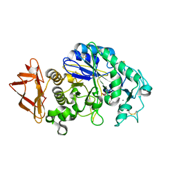
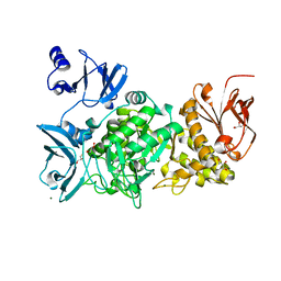
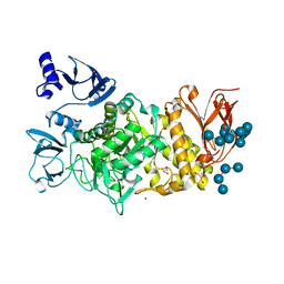
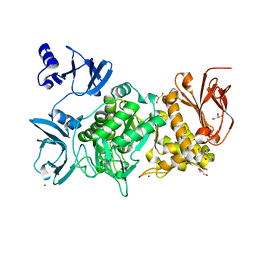
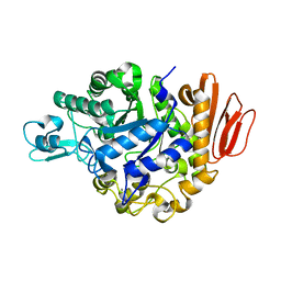
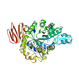
Loading
PDBj
MenuPDBj@FacebookPDBj@TwitterPDBj@YouTubewwPDB FoundationwwPDB
RCSB PDBPDBeBMRBAdv. SearchSearch help
PDB: 575 results

5E6Y
DownloadVisualize
BU of 5e6y by Molmil
Crystal structure of E.Coli branching enzyme in complex with alpha cyclodextrin
Descriptor: 1,4-alpha-glucan branching enzyme GlgB, Cyclohexakis-(1-4)-(alpha-D-glucopyranose), GLYCEROL
Authors:Feng, L, Nosrati, M, Geiger, J.H.
Deposit date:2015-10-11
Release date:2015-12-16
Last modified:2023-09-27
Method:X-RAY DIFFRACTION (2.6 Å)
Cite:Crystal structures of Escherichia coli branching enzyme in complex with cyclodextrins.
Acta Crystallogr D Struct Biol, 72, 2016
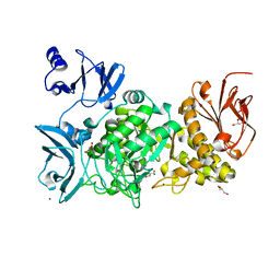
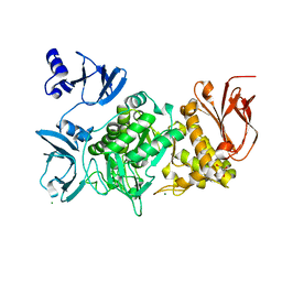
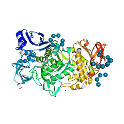
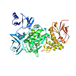
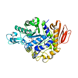
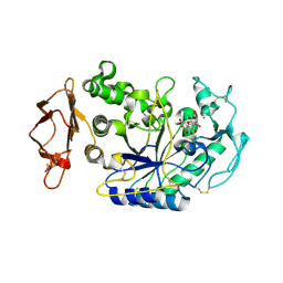
1DED
DownloadVisualize
BU of 1ded by Molmil
CRYSTAL STRUCTURE OF ALKALOPHILIC ASPARAGINE 233-REPLACED CYCLODEXTRIN GLUCANOTRANSFERASE COMPLEXED WITH AN INHIBITOR, ACARBOSE, AT 2.0 A RESOLUTION
Descriptor: 4,6-dideoxy-4-{[(1S,4R,5S,6S)-4,5,6-trihydroxy-3-(hydroxymethyl)cyclohex-2-en-1-yl]amino}-alpha-D-glucopyranose-(1-4)-alpha-D-glucopyranose-(1-4)-beta-D-glucopyranose, CALCIUM ION, CYCLODEXTRIN GLUCANOTRANSFERASE
Authors:Ishii, N, Haga, K, Yamane, K, Harata, K.
Deposit date:1999-11-14
Release date:2000-04-07
Last modified:2021-11-03
Method:X-RAY DIFFRACTION (2 Å)
Cite:Crystal structure of alkalophilic asparagine 233-replaced cyclodextrin glucanotransferase complexed with an inhibitor, acarbose, at 2.0 A resolution.
J.Biochem.(Tokyo), 127, 2000
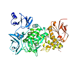
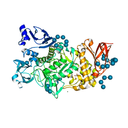
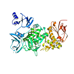
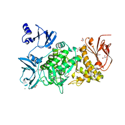
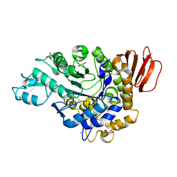
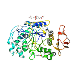
5EMY
DownloadVisualize
BU of 5emy by Molmil
Human Pancreatic Alpha-Amylase in complex with the mechanism based inactivator glucosyl epi-cyclophellitol
Descriptor: (1R,2R,3S,5R,6S)-2,3,5-trihydroxy-6-(hydroxymethyl)cyclohexyl alpha-D-glucopyranoside, CALCIUM ION, CHLORIDE ION, ...
Authors:Caner, S, Brayer, G.D.
Deposit date:2015-11-06
Release date:2016-07-06
Last modified:2023-09-27
Method:X-RAY DIFFRACTION (1.231 Å)
Cite:Glucosyl epi-cyclophellitol allows mechanism-based inactivation and structural analysis of human pancreatic alpha-amylase.
Febs Lett., 590, 2016
5GR0
DownloadVisualize
BU of 5gr0 by Molmil
Crystal structure of branching enzyme D501A mutant from Cyanothece sp. ATCC 51142
Descriptor: 1,4-alpha-glucan branching enzyme GlgB, GLYCEROL, MAGNESIUM ION
Authors:Suzuki, R, Suzuki, E.
Deposit date:2016-08-08
Release date:2017-08-16
Last modified:2023-11-08
Method:X-RAY DIFFRACTION (1.95 Å)
Cite:Structural basis for substrate binding and catalysis of branching enzyme from Cyanothece sp. ATCC 51142
To be published
5GTW
DownloadVisualize
BU of 5gtw by Molmil
The N253R mutant structures of trehalose synthase from Deinococcus radiodurans display two different active-site conformations
Descriptor: 2-AMINO-2-HYDROXYMETHYL-PROPANE-1,3-DIOL, CALCIUM ION, MAGNESIUM ION, ...
Authors:Chow, S.Y, Wei, Y.J, Liaw, S.H.
Deposit date:2016-08-23
Release date:2017-08-30
Last modified:2023-11-08
Method:X-RAY DIFFRACTION (2.93 Å)
Cite:The N253R mutant structures of trehalose synthase from Deinococcus radiodurans display two different active-site conformations
To Be Published
5GQU
DownloadVisualize
BU of 5gqu by Molmil
Crystal structure of branching enzyme from Cyanothece sp. ATCC 51142
Descriptor: 1,4-alpha-glucan branching enzyme GlgB, GLYCEROL, MAGNESIUM ION
Authors:Suzuki, R, Suzuki, E.
Deposit date:2016-08-08
Release date:2017-02-22
Last modified:2023-11-08
Method:X-RAY DIFFRACTION (1.85 Å)
Cite:Bound Substrate in the Structure of Cyanobacterial Branching Enzyme Supports a New Mechanistic Model
J. Biol. Chem., 292, 2017
5GR6
DownloadVisualize
BU of 5gr6 by Molmil
Crystal structure of branching enzyme Y500A/D501A double mutant from Cyanothece sp. ATCC 51142
Descriptor: 1,4-alpha-glucan branching enzyme GlgB, GLYCEROL, MAGNESIUM ION
Authors:Suzuki, R, Suzuki, E.
Deposit date:2016-08-08
Release date:2017-08-16
Last modified:2023-11-08
Method:X-RAY DIFFRACTION (2 Å)
Cite:Structural basis for substrate binding and catalysis of branching enzyme from Cyanothece sp. ATCC 51142
To be published
5GR3
DownloadVisualize
BU of 5gr3 by Molmil
Crystal structure of branching enzyme L541A/W655A mutant from Cyanothece sp. ATCC 51142
Descriptor: 1,4-alpha-glucan branching enzyme GlgB, GLYCEROL, MAGNESIUM ION
Authors:Suzuki, R, Suzuki, E.
Deposit date:2016-08-08
Release date:2017-08-16
Last modified:2023-11-08
Method:X-RAY DIFFRACTION (2.6 Å)
Cite:Structural basis for substrate binding and catalysis of branching enzyme from Cyanothece sp. ATCC 51142
To be published
5GQV
DownloadVisualize
BU of 5gqv by Molmil
Crystal structure of branching enzyme from Cyanothece sp. ATCC 51142 in complex with maltohexaose
Descriptor: 1,4-alpha-glucan branching enzyme GlgB, GLYCEROL, MAGNESIUM ION, ...
Authors:Suzuki, R, Suzuki, E.
Deposit date:2016-08-08
Release date:2017-02-22
Last modified:2023-11-08
Method:X-RAY DIFFRACTION (3 Å)
Cite:Bound Substrate in the Structure of Cyanobacterial Branching Enzyme Supports a New Mechanistic Model
J. Biol. Chem., 292, 2017
5GR4
DownloadVisualize
BU of 5gr4 by Molmil
Crystal structure of branching enzyme L541A mutant from Cyanothece sp. ATCC 51142 in complex with maltoheptaose
Descriptor: 1,4-alpha-glucan branching enzyme GlgB, GLYCEROL, MAGNESIUM ION, ...
Authors:Suzuki, R, Suzuki, E.
Deposit date:2016-08-08
Release date:2017-08-16
Last modified:2024-03-20
Method:X-RAY DIFFRACTION (2 Å)
Cite:Structural basis for substrate binding and catalysis of branching enzyme from Cyanothece sp. ATCC 51142
To be published
5GR1
DownloadVisualize
BU of 5gr1 by Molmil
Crystal structure of branching enzyme Y500A/D501A mutant from Cyanothece sp. ATCC 51142 in complex with maltoheptaose
Descriptor: 1,4-alpha-glucan branching enzyme GlgB, GLYCEROL, MAGNESIUM ION, ...
Authors:Suzuki, R, Suzuki, E.
Deposit date:2016-08-08
Release date:2017-08-16
Last modified:2024-03-20
Method:X-RAY DIFFRACTION (2.5 Å)
Cite:Structural basis for substrate binding and catalysis of branching enzyme from Cyanothece sp. ATCC 51142
To be published
5GQY
DownloadVisualize
BU of 5gqy by Molmil
Crystal structure of branching enzyme from Cyanothece sp. ATCC 51142 in complex with maltoheptaose
Descriptor: 1,4-alpha-glucan branching enzyme GlgB, GLYCEROL, MAGNESIUM ION, ...
Authors:Suzuki, R, Suzuki, E.
Deposit date:2016-08-08
Release date:2017-08-16
Last modified:2023-11-08
Method:X-RAY DIFFRACTION (2 Å)
Cite:Structural basis for substrate binding and catalysis of branching enzyme from Cyanothece sp. ATCC 51142
To be published
5GR2
DownloadVisualize
BU of 5gr2 by Molmil
Crystal structure of branching enzyme L541A mutant from Cyanothece sp. ATCC 51142
Descriptor: 1,4-alpha-glucan branching enzyme GlgB, GLYCEROL, MAGNESIUM ION
Authors:Suzuki, R, Suzuki, E.
Deposit date:2016-08-08
Release date:2017-08-16
Last modified:2023-11-08
Method:X-RAY DIFFRACTION (1.95 Å)
Cite:Structural basis for substrate binding and catalysis of branching enzyme from Cyanothece sp. ATCC 51142
To be published
5GQX
DownloadVisualize
BU of 5gqx by Molmil
Crystal structure of branching enzyme W610N mutant from Cyanothece sp. ATCC 51142 in complex with maltoheptaose
Descriptor: 1,4-alpha-glucan branching enzyme GlgB, GLYCEROL, MAGNESIUM ION, ...
Authors:Suzuki, R, Suzuki, E.
Deposit date:2016-08-08
Release date:2017-02-22
Last modified:2023-11-08
Method:X-RAY DIFFRACTION (2.3 Å)
Cite:Bound Substrate in the Structure of Cyanobacterial Branching Enzyme Supports a New Mechanistic Model
J. Biol. Chem., 292, 2017
5GR5
DownloadVisualize
BU of 5gr5 by Molmil
Crystal structure of branching enzyme W610A mutant from Cyanothece sp. ATCC 51142
Descriptor: 1,4-alpha-glucan branching enzyme GlgB, GLYCEROL, MAGNESIUM ION
Authors:Suzuki, R, Suzuki, E.
Deposit date:2016-08-08
Release date:2017-08-16
Last modified:2023-11-08
Method:X-RAY DIFFRACTION (2.4 Å)
Cite:Structural basis for substrate binding and catalysis of branching enzyme from Cyanothece sp. ATCC 51142
To be published
5GQW
DownloadVisualize
BU of 5gqw by Molmil
Crystal structure of branching enzyme W610N mutant from Cyanothece sp. ATCC 51142
Descriptor: 1,4-alpha-glucan branching enzyme GlgB, GLYCEROL, MAGNESIUM ION
Authors:Suzuki, R, Suzuki, E.
Deposit date:2016-08-08
Release date:2017-02-22
Last modified:2023-11-08
Method:X-RAY DIFFRACTION (1.8 Å)
Cite:Bound Substrate in the Structure of Cyanobacterial Branching Enzyme Supports a New Mechanistic Model
J. Biol. Chem., 292, 2017
5H2T
DownloadVisualize
BU of 5h2t by Molmil
Structure of trehalose synthase
Descriptor: Trehalose synthase
Authors:Wang, D, Huang, H, Zhou, J, Jiang, L.
Deposit date:2016-10-18
Release date:2017-10-18
Last modified:2024-03-20
Method:X-RAY DIFFRACTION (2.796 Å)
Cite:Structure of trehalose synthase
To Be Published
5GQZ
DownloadVisualize
BU of 5gqz by Molmil
Crystal structure of branching enzyme Y500A mutant from Cyanothece sp. ATCC 51142
Descriptor: 1,4-alpha-glucan branching enzyme GlgB, GLYCEROL, MAGNESIUM ION
Authors:Suzuki, R, Suzuki, E.
Deposit date:2016-08-08
Release date:2017-08-16
Last modified:2023-11-08
Method:X-RAY DIFFRACTION (1.85 Å)
Cite:Structural basis for substrate binding and catalysis of branching enzyme from Cyanothece sp. ATCC 51142
To be published
4GI9
DownloadVisualize
BU of 4gi9 by Molmil
Crystal structure of the MUTB F164L mutant from crystals soaked with Trehalulose
Descriptor: 2-AMINO-2-HYDROXYMETHYL-PROPANE-1,3-DIOL, CALCIUM ION, GLYCEROL, ...
Authors:Lipski, A, Haser, R, Aghajari, N.
Deposit date:2012-08-08
Release date:2013-02-13
Last modified:2023-11-08
Method:X-RAY DIFFRACTION (2.15 Å)
Cite:Mutations inducing an active-site aperture in Rhizobium sp. sucrose isomerase confer hydrolytic activity
Acta Crystallogr.,Sect.D, 69, 2013
4GI8
DownloadVisualize
BU of 4gi8 by Molmil
Crystal structure of the MUTB F164L mutant from crystals soaked with the substrate sucrose
Descriptor: 2-AMINO-2-HYDROXYMETHYL-PROPANE-1,3-DIOL, CALCIUM ION, GLYCEROL, ...
Authors:Lipski, A, Haser, R, Aghajari, N.
Deposit date:2012-08-08
Release date:2013-02-13
Last modified:2023-11-08
Method:X-RAY DIFFRACTION (1.95 Å)
Cite:Mutations inducing an active-site aperture in Rhizobium sp. sucrose isomerase confer hydrolytic activity
Acta Crystallogr.,Sect.D, 69, 2013
4GIN
DownloadVisualize
BU of 4gin by Molmil
Crystal structure of the MUTB R284C mutant from crystals soaked with the inhibitor deoxynojirimycin
Descriptor: CALCIUM ION, GLYCEROL, Sucrose isomerase
Authors:Lipski, A, Ravaud, S, Robert, X, Haser, R, Aghajari, N.
Deposit date:2012-08-08
Release date:2013-02-13
Last modified:2023-11-08
Method:X-RAY DIFFRACTION (1.9 Å)
Cite:Mutations inducing an active-site aperture in Rhizobium sp. sucrose isomerase confer hydrolytic activity
Acta Crystallogr.,Sect.D, 69, 2013
4GI6
DownloadVisualize
BU of 4gi6 by Molmil
Crystal structure of the MUTB F164L mutant in complex with glucose
Descriptor: 2-AMINO-2-HYDROXYMETHYL-PROPANE-1,3-DIOL, CALCIUM ION, GLYCEROL, ...
Authors:Lipski, A, Haser, R, Aghajari, N.
Deposit date:2012-08-08
Release date:2013-02-13
Last modified:2023-11-08
Method:X-RAY DIFFRACTION (2.15 Å)
Cite:Mutations inducing an active-site aperture in Rhizobium sp. sucrose isomerase confer hydrolytic activity
Acta Crystallogr.,Sect.D, 69, 2013
4GO9
DownloadVisualize
BU of 4go9 by Molmil
CRYSTAL STRUCTURE of the TREHALULOSE SYNTHASE MUTANT, MUTB D415N, in COMPLEX with TRIS
Descriptor: 2-AMINO-2-HYDROXYMETHYL-PROPANE-1,3-DIOL, CALCIUM ION, Sucrose isomerase
Authors:Lipski, A, Ravaud, S, Robert, X, Haser, R, Aghajari, N.
Deposit date:2012-08-19
Release date:2013-08-21
Last modified:2023-11-08
Method:X-RAY DIFFRACTION (2.2 Å)
Cite:CRYSTAL STRUCTURE of the TREHALULOSE SYNTHASE MUTB, MUTANT D415N, in COMPLEX with TRIS
To be Published, 2013
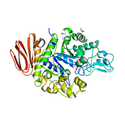
4GQR
DownloadVisualize
BU of 4gqr by Molmil
Human Pancreatic alpha-amylase in complex with myricetin
Descriptor: 2-acetamido-2-deoxy-beta-D-glucopyranose, 3,5,7-TRIHYDROXY-2-(3,4,5-TRIHYDROXYPHENYL)-4H-CHROMEN-4-ONE, CALCIUM ION, ...
Authors:Williams, L.K, Brayer, G.D.
Deposit date:2012-08-23
Release date:2012-10-24
Last modified:2020-07-29
Method:X-RAY DIFFRACTION (1.2 Å)
Cite:Order and disorder: differential structural impacts of myricetin and ethyl caffeate on human amylase, an antidiabetic target.
J.Med.Chem., 55, 2012
4GQQ
DownloadVisualize
BU of 4gqq by Molmil
Human pancreatic alpha-amylase with bound ethyl caffeate
Descriptor: 2-acetamido-2-deoxy-beta-D-glucopyranose, CALCIUM ION, CHLORIDE ION, ...
Authors:Williams, L.K, Brayer, G.D.
Deposit date:2012-08-23
Release date:2012-10-24
Last modified:2020-07-29
Method:X-RAY DIFFRACTION (1.35 Å)
Cite:Order and disorder: differential structural impacts of myricetin and ethyl caffeate on human amylase, an antidiabetic target.
J.Med.Chem., 55, 2012

220472

건을2024-05-29부터공개중

PDB statisticsPDBj update infoContact PDBjnumon