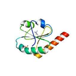
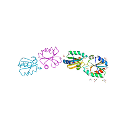
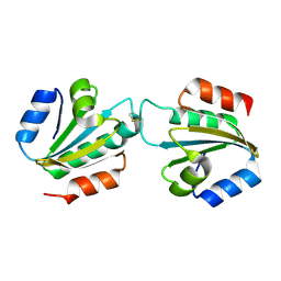
3UEM
 
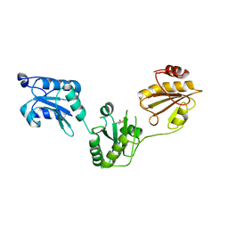 | | Crystal structure of human PDI bb'a' domains | | 分子名称: | (4S,5S)-1,2-DITHIANE-4,5-DIOL, Protein disulfide-isomerase | | 著者 | Yu, J, Wang, C, Huo, L, Feng, W, Wang, C.-C. | | 登録日 | 2011-10-30 | | 公開日 | 2011-11-23 | | 最終更新日 | 2024-03-20 | | 実験手法 | X-RAY DIFFRACTION (2.29 Å) | | 主引用文献 | Human protein-disulfide isomerase is a redox-regulated chaperone activated by oxidation of domain a'
J.Biol.Chem., 287, 2012
|
|
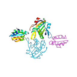
3UJ1
 | |
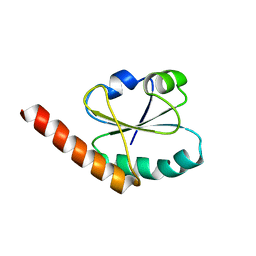
3UL3
 | |
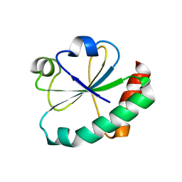
3UVT
 | |
3VFI
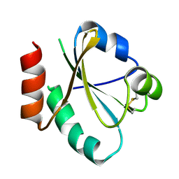
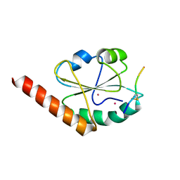
 | | Crystal Structure of a Metagenomic Thioredoxin | | 分子名称: | thioredoxin | | 著者 | Craig, T.K, Gardberg, A, Lorimer, D.D, Burgin Jr, A.B, Segall, A, Rohwer, F. | | 登録日 | 2012-01-09 | | 公開日 | 2013-01-23 | | 実験手法 | X-RAY DIFFRACTION (1.75 Å) | | 主引用文献 | Structure of a Metagenomic Thioredoxin
To be Published
|
|
3VWW
 | |
3W8J
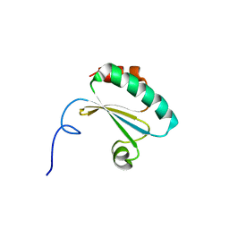
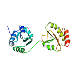
 | | Crystal structure of P5 a0 in a complex with Prx4 c-term | | 分子名称: | C-terminal peptide from Peroxiredoxin-4, GLYCEROL, POTASSIUM ION, ... | | 著者 | Inaba, K, Suzuki, M, Kojima, R. | | 登録日 | 2013-03-13 | | 公開日 | 2013-09-04 | | 実験手法 | X-RAY DIFFRACTION (2.1 Å) | | 主引用文献 | Synergistic cooperation of PDI family members in peroxiredoxin 4-driven oxidative protein folding
Sci Rep, 3, 2013
|
|
3WGD
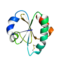
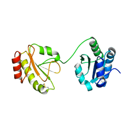
 | | Crystal structure of ERp46 Trx1 | | 分子名称: | GLYCEROL, PHOSPHATE ION, POTASSIUM ION, ... | | 著者 | Inaba, K, Suzuki, M, Kojima, R. | | 登録日 | 2013-08-04 | | 公開日 | 2014-06-25 | | 実験手法 | X-RAY DIFFRACTION (2.5 Å) | | 主引用文献 | Radically different thioredoxin domain arrangement of ERp46, an efficient disulfide bond introducer of the mammalian PDI family
Structure, 22, 2014
|
|
3WGE
 | | Crystal structure of ERp46 Trx2 | | 分子名称: | GLYCEROL, Thioredoxin domain-containing protein 5 | | 著者 | Inaba, K, Suzuki, M, Kojima, R. | | 登録日 | 2013-08-04 | | 公開日 | 2014-06-25 | | 実験手法 | X-RAY DIFFRACTION (0.95 Å) | | 主引用文献 | Radically different thioredoxin domain arrangement of ERp46, an efficient disulfide bond introducer of the mammalian PDI family
Structure, 22, 2014
|
|
3WGX
 | | Crystal structure of ERp46 Trx2 in a complex with Prx4 C-term | | 分子名称: | GLYCEROL, Peroxiredoxin-4, Thioredoxin domain-containing protein 5 | | 著者 | Inaba, K, Suzuki, M, Kojima, R. | | 登録日 | 2013-08-13 | | 公開日 | 2014-06-25 | | 実験手法 | X-RAY DIFFRACTION (0.92 Å) | | 主引用文献 | Radically different thioredoxin domain arrangement of ERp46, an efficient disulfide bond introducer of the mammalian PDI family
Structure, 22, 2014
|
|
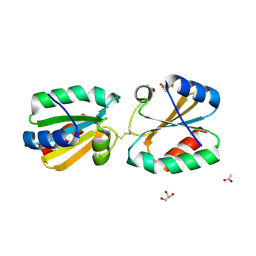
3WT1
 | | Crystal structure of the b'-a' domain of thermophilic fungal protein disulfide isomerase (reduced form) | | 分子名称: | GLYCEROL, Protein disulfide-isomerase | | 著者 | Inagaki, K, Satoh, T, Itoh, S.G, Okumura, H, Kato, K. | | 登録日 | 2014-04-02 | | 公開日 | 2014-11-26 | | 最終更新日 | 2023-11-08 | | 実験手法 | X-RAY DIFFRACTION (1.85 Å) | | 主引用文献 | Redox-dependent conformational transition of catalytic domain of protein disulfide isomerase indicated by crystal structure-based molecular dynamics simulation
Chem.Phys.Lett., 618, 2015
|
|
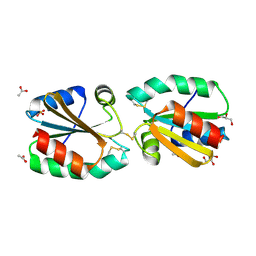
3WT2
 | | Crystal structure of the b'-a' domain of thermophilic fungal protein disulfide isomerase (oxidized form) | | 分子名称: | Protein disulfide-isomerase | | 著者 | Inagaki, K, Satoh, T, Itoh, S.G, Okumura, H, Kato, K. | | 登録日 | 2014-04-02 | | 公開日 | 2014-11-26 | | 最終更新日 | 2023-11-08 | | 実験手法 | X-RAY DIFFRACTION (3.3 Å) | | 主引用文献 | Redox-dependent conformational transition of catalytic domain of protein disulfide isomerase indicated by crystal structure-based molecular dynamics simulation
Chem.Phys.Lett., 618, 2015
|
|
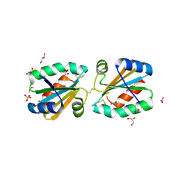
3ZZX
 | | Crystallographic structure of thioredoxin from Litopenaeus vannamei | | 分子名称: | (2R,3S)-1,4-DIMERCAPTOBUTANE-2,3-DIOL, ACETATE ION, GLYCEROL, ... | | 著者 | Campos-Acevedo, A.A, Sotelo-Mundo, R.R, Rudino-Pinera, E. | | 登録日 | 2011-09-05 | | 公開日 | 2012-09-26 | | 最終更新日 | 2023-12-20 | | 実験手法 | X-RAY DIFFRACTION (1.88 Å) | | 主引用文献 | Expression, Purification, Crystallization and X-Ray Crystallographic Studies of Different Redox States of the Active Site of Thioredoxin 1 from the Whiteleg Shrimp Litopenaeus Vannamei
Acta Crystallogr.,Sect.F, 69, 2013
|
|
4AJ6
 | | Crystallographic structure of thioredoxin from Litopenaeus vannamei (reduced form). | | 分子名称: | 2,3-DIHYDROXY-1,4-DITHIOBUTANE, ACETATE ION, GLYCEROL, ... | | 著者 | Campos-Acevedo, A.A, Sotelo-Mundo, R.R, Rudino-Pinera, E. | | 登録日 | 2012-02-15 | | 公開日 | 2013-03-06 | | 最終更新日 | 2023-12-20 | | 実験手法 | X-RAY DIFFRACTION (2 Å) | | 主引用文献 | Expression, Purification, Crystallization and X-Ray Crystallographic Studies of Different Redox States of the Active Site of Thioredoxin 1 from the Whiteleg Shrimp Litopenaeus Vannamei
Acta Crystallogr.,Sect.F, 69, 2013
|
|
4AJ7
 | | Crystallographic structure of thioredoxin from Litopenaeus vannamei (oxidized form). | | 分子名称: | ACETATE ION, GLYCEROL, SULFATE ION, ... | | 著者 | Campos-Acevedo, A.A, Sotelo-Mundo, R.R, Rudino-Pinera, E. | | 登録日 | 2012-02-15 | | 公開日 | 2013-03-06 | | 最終更新日 | 2023-12-20 | | 実験手法 | X-RAY DIFFRACTION (2.035 Å) | | 主引用文献 | Expression, Purification, Crystallization and X-Ray Crystallographic Studies of Different Redox States of the Active Site of Thioredoxin 1 from the Whiteleg Shrimp Litopenaeus Vannamei
Acta Crystallogr.,Sect.F, 69, 2013
|
|
4AJ8
 | | Crystallographic structure of thioredoxin from Litopenaeus vannamei (partially reduced). | | 分子名称: | ACETATE ION, GLYCEROL, SULFATE ION, ... | | 著者 | Campos-Acevedo, A.A, Sotelo-Mundo, R.R, Rudino-Pinera, E. | | 登録日 | 2012-02-16 | | 公開日 | 2013-03-06 | | 最終更新日 | 2023-12-20 | | 実験手法 | X-RAY DIFFRACTION (1.54 Å) | | 主引用文献 | Expression, Purification, Crystallization and X-Ray Crystallographic Studies of Different Redox States of the Active Site of Thioredoxin 1 from the Whiteleg Shrimp Litopenaeus Vannamei
Acta Crystallogr.,Sect.F, 69, 2013
|
|
4CW9
 | | Entamoeba histolytica thiredoxin C34S mutant | | 分子名称: | THIOREDOXIN | | 著者 | Parsonage, D, Kells, P.M, Vieira, D.F, Hirata, K, Debnath, A, Poole, L.B, McKerrow, J.H, Reed, S.L, Podust, L.M. | | 登録日 | 2014-04-01 | | 公開日 | 2015-05-13 | | 最終更新日 | 2023-12-20 | | 実験手法 | X-RAY DIFFRACTION (1.84 Å) | | 主引用文献 | X-Ray Structures of Thioredoxin and Thioredoxin Reductase from Entamoeba Histolytica and Prevailing Hypothesis of the Mechanism of Auranofin Action.
J.Struct.Biol., 194, 2016
|
|
4DSS
 | | Crystal structure of peroxiredoxin Ahp1 from Saccharomyces cerevisiae in complex with thioredoxin Trx2 | | 分子名称: | Peroxiredoxin type-2, Thioredoxin-2 | | 著者 | Lian, F.M, Yu, J, Ma, X.X, Yu, X.J, Chen, Y, Zhou, C.Z. | | 登録日 | 2012-02-19 | | 公開日 | 2012-04-11 | | 最終更新日 | 2023-11-08 | | 実験手法 | X-RAY DIFFRACTION (2.1 Å) | | 主引用文献 | Structural Snapshots of Yeast Alkyl Hydroperoxide Reductase Ahp1 Peroxiredoxin Reveal a Novel Two-cysteine Mechanism of Electron Transfer to Eliminate Reactive Oxygen Species.
J.Biol.Chem., 287, 2012
|
|
4EF0
 | |
4EKZ
 | | Crystal structure of reduced hPDI (abb'xa') | | 分子名称: | Protein disulfide-isomerase | | 著者 | Wang, C, Li, W, Ren, J, Ke, H, Gong, W, Feng, W, Wang, C.-C. | | 登録日 | 2012-04-10 | | 公開日 | 2013-04-10 | | 最終更新日 | 2023-11-08 | | 実験手法 | X-RAY DIFFRACTION (2.51 Å) | | 主引用文献 | Structural insights into the redox-regulated dynamic conformations of human protein disulfide isomerase
Antioxid Redox Signal, 19, 2013
|
|
4EL1
 | | Crystal structure of oxidized hPDI (abb'xa') | | 分子名称: | Protein disulfide-isomerase | | 著者 | Wang, C, Li, W, Ren, J, Ke, H, Gong, W, Feng, W, Wang, C.-C. | | 登録日 | 2012-04-10 | | 公開日 | 2013-04-10 | | 最終更新日 | 2023-11-08 | | 実験手法 | X-RAY DIFFRACTION (2.883 Å) | | 主引用文献 | Structural insights into the redox-regulated dynamic conformations of human protein disulfide isomerase
Antioxid Redox Signal, 19, 2013
|
|
4GWR
 | |
4HU7
 | | E. coli thioredoxin variant with Pro76 as single proline residue | | 分子名称: | COPPER (II) ION, SODIUM ION, Thioredoxin-1 | | 著者 | Glockshuber, R, Scharer, M.A, Capitani, G, Rubini, M. | | 登録日 | 2012-11-02 | | 公開日 | 2013-05-29 | | 最終更新日 | 2023-09-20 | | 実験手法 | X-RAY DIFFRACTION (1.4 Å) | | 主引用文献 | (4R)- and (4S)-Fluoroproline in the Conserved cis-Prolyl Peptide Bond of the Thioredoxin Fold: Tertiary Structure Context Dictates Ring Puckering.
Chembiochem, 14, 2013
|
|
4HU9
 | | E. coli thioredoxin variant with (4S)-FluoroPro76 as single proline residue | | 分子名称: | COPPER (II) ION, Thioredoxin-1 | | 著者 | Scharer, M.A, Rubini, M, Capitani, G, Glockshuber, R. | | 登録日 | 2012-11-02 | | 公開日 | 2013-05-29 | | 最終更新日 | 2017-09-20 | | 実験手法 | X-RAY DIFFRACTION (1.55 Å) | | 主引用文献 | (4R)- and (4S)-Fluoroproline in the Conserved cis-Prolyl Peptide Bond of the Thioredoxin Fold: Tertiary Structure Context Dictates Ring Puckering.
Chembiochem, 14, 2013
|
|
4HUA
 | | E. coli thioredoxin variant with (4R)-FluoroPro76 as single proline residue | | 分子名称: | COPPER (II) ION, Thioredoxin-1 | | 著者 | Scharer, M.A, Rubini, M, Capitani, G, Glockshuber, R. | | 登録日 | 2012-11-02 | | 公開日 | 2013-05-29 | | 最終更新日 | 2023-12-06 | | 実験手法 | X-RAY DIFFRACTION (1.1 Å) | | 主引用文献 | (4R)- and (4S)-Fluoroproline in the Conserved cis-Prolyl Peptide Bond of the Thioredoxin Fold: Tertiary Structure Context Dictates Ring Puckering.
Chembiochem, 14, 2013
|
|


