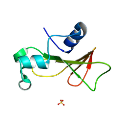
3P9N
 
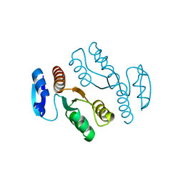
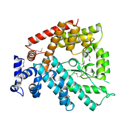
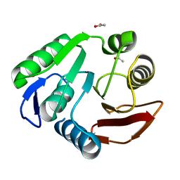 | | Rv2966c of M. tuberculosis is a RsmD-like methyltransferase | | 分子名称: | ACETATE ION, POSSIBLE METHYLTRANSFERASE (METHYLASE) | | 著者 | Kumar, A, Malhotra, K, Saigal, K, Sinha, K.M, Taneja, B. | | 登録日 | 2010-10-18 | | 公開日 | 2011-04-06 | | 最終更新日 | 2023-11-01 | | 実験手法 | X-RAY DIFFRACTION (1.9 Å) | | 主引用文献 | Structural and functional characterization of Rv2966c protein reveals an RsmD-like methyltransferase from Mycobacterium tuberculosis and the role of its N-terminal domain in target recognition
J.Biol.Chem., 286, 2011
|
|
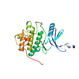
3OPT
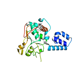
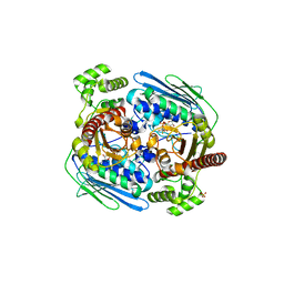
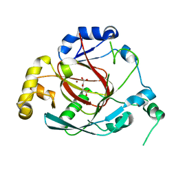 | | Crystal structure of the Rph1 catalytic core with a-ketoglutarate | | 分子名称: | 2-OXOGLUTARIC ACID, DNA damage-responsive transcriptional repressor RPH1, NICKEL (II) ION | | 著者 | Chang, Y, Wu, J, Tong, X, Zhou, J, Ding, J. | | 登録日 | 2010-09-02 | | 公開日 | 2010-12-22 | | 最終更新日 | 2024-03-20 | | 実験手法 | X-RAY DIFFRACTION (2.2 Å) | | 主引用文献 | Crystal structure of the catalytic core of Saccharomyces cerevesiae histone demethylase Rph1: insights into the substrate specificity and catalytic mechanism
Biochem.J., 433, 2011
|
|
1J25
 | | Crystal structure of archaeal XPF/Mus81 homolog, Hef from Pyrococcus furiosus, nuclease domain, Mn cocrystal | | 分子名称: | ATP-dependent RNA helicase, putative, MANGANESE (II) ION | | 著者 | Nishino, T, Komori, K, Ishino, Y, Morikawa, K. | | 登録日 | 2002-12-25 | | 公開日 | 2003-04-22 | | 最終更新日 | 2024-04-03 | | 実験手法 | X-RAY DIFFRACTION (1.78 Å) | | 主引用文献 | X-Ray and Biochemical Anatomy of an Archaeal XPF/Rad1/Mus81 Family Nuclease. Similarity between Its Endonuclease Domain and Restriction Enzymes
Structure, 11, 2003
|
|
1J24
 | | Crystal structure of archaeal XPF/Mus81 homolog, Hef from Pyrococcus furiosus, nuclease domain, Ca cocrystal | | 分子名称: | ATP-dependent RNA helicase, putative, CALCIUM ION | | 著者 | Nishino, T, Komori, K, Ishino, Y, Morikawa, K. | | 登録日 | 2002-12-25 | | 公開日 | 2003-04-22 | | 最終更新日 | 2024-04-03 | | 実験手法 | X-RAY DIFFRACTION (1.78 Å) | | 主引用文献 | X-Ray and Biochemical Anatomy of an Archaeal XPF/Rad1/Mus81 Family Nuclease. Similarity between Its Endonuclease Domain and Restriction Enzymes
Structure, 11, 2003
|
|
1J22
 | | Crystal structure of archaeal XPF/Mus81 homolog, Hef from Pyrococcus furiosus, nuclease domain, selenomet derivative | | 分子名称: | ATP-dependent RNA helicase, putative | | 著者 | Nishino, T, Komori, K, Ishino, Y, Morikawa, K. | | 登録日 | 2002-12-25 | | 公開日 | 2003-04-22 | | 最終更新日 | 2023-12-27 | | 実験手法 | X-RAY DIFFRACTION (1.8 Å) | | 主引用文献 | X-Ray and Biochemical Anatomy of an Archaeal XPF/Rad1/Mus81 Family Nuclease. Similarity between Its Endonuclease Domain and Restriction Enzymes
Structure, 11, 2003
|
|
1U0J
 | | Crystal Structure of AAV2 Rep40-ADP complex | | 分子名称: | ADENOSINE-5'-DIPHOSPHATE, DNA replication protein | | 著者 | James, J.A, Aggarwal, A.K, Linden, R.M, Escalante, C.R. | | 登録日 | 2004-07-13 | | 公開日 | 2004-08-24 | | 最終更新日 | 2023-08-23 | | 実験手法 | X-RAY DIFFRACTION (2.1 Å) | | 主引用文献 | Structure of adeno-associated virus type 2 Rep40-ADP complex: Insight into
nucleotide recognition and catalysis by superfamily 3 helicases
Proc.Natl.Acad.Sci.USA, 101, 2004
|
|
1K6Y
 | | Crystal Structure of a Two-Domain Fragment of HIV-1 Integrase | | 分子名称: | Integrase, PHOSPHATE ION, POTASSIUM ION, ... | | 著者 | Wang, J, Ling, H, Yang, W, Craigie, R. | | 登録日 | 2001-10-17 | | 公開日 | 2001-12-21 | | 最終更新日 | 2024-02-07 | | 実験手法 | X-RAY DIFFRACTION (2.4 Å) | | 主引用文献 | Structure of a two-domain fragment of HIV-1 integrase: implications for domain organization in the intact protein.
EMBO J., 20, 2001
|
|
1YQA
 | |
1RNB
 | |
1IA8
 | | THE 1.7 A CRYSTAL STRUCTURE OF HUMAN CELL CYCLE CHECKPOINT KINASE CHK1 | | 分子名称: | CHK1 CHECKPOINT KINASE, SULFATE ION | | 著者 | Chen, P, Luo, C, Deng, Y, Ryan, K, Register, J, Margosiak, S, Tempczyk-Russell, A, Nguyen, B, Myers, P, Lundgren, K, Chen Kan, C.-C, O'Connor, P.M. | | 登録日 | 2001-03-22 | | 公開日 | 2001-04-18 | | 最終更新日 | 2024-04-03 | | 実験手法 | X-RAY DIFFRACTION (1.7 Å) | | 主引用文献 | The 1.7 A crystal structure of human cell cycle checkpoint kinase Chk1: implications for Chk1 regulation.
Cell(Cambridge,Mass.), 100, 2000
|
|
1JI4
 | | NAP protein from helicobacter pylori | | 分子名称: | (4S)-2-METHYL-2,4-PENTANEDIOL, FE (III) ION, NEUTROPHIL-ACTIVATING PROTEIN A, ... | | 著者 | Zanotti, G, Papinutto, E, Dundon, W.G, Battistutta, R, Seveso, M, Del Giudice, G, Rappuoli, R, Montecucco, C. | | 登録日 | 2001-06-29 | | 公開日 | 2002-10-09 | | 最終更新日 | 2023-08-16 | | 実験手法 | X-RAY DIFFRACTION (2.52 Å) | | 主引用文献 | Structure of the Neutrophil-activating Protein from Helicobacter pylori
J.Mol.Biol., 323, 2002
|
|
1Z5A
 | | Topoisomerase VI-B, ADP-bound dimer form | | 分子名称: | ADENOSINE-5'-DIPHOSPHATE, MAGNESIUM ION, Type II DNA topoisomerase VI subunit B | | 著者 | Corbett, K.D, Berger, J.M. | | 登録日 | 2005-03-17 | | 公開日 | 2005-06-14 | | 最終更新日 | 2023-08-23 | | 実験手法 | X-RAY DIFFRACTION (2.2 Å) | | 主引用文献 | Structural dissection of ATP turnover in the prototypical GHL ATPase TopoVI.
Structure, 13, 2005
|
|
1T2R
 | | Structural basis for 3' end recognition of nucleic acids by the Drosophila Argonaute 2 PAZ domain | | 分子名称: | 5'-R(*CP*UP*CP*AP*C)-3', Argonaute 2 | | 著者 | Lingel, A, Simon, B, Izaurralde, E, Sattler, M. | | 登録日 | 2004-04-22 | | 公開日 | 2004-06-01 | | 最終更新日 | 2024-05-22 | | 実験手法 | SOLUTION NMR | | 主引用文献 | Nucleic acid 3'-end recognition by the Argonaute2 PAZ domain.
Nat.Struct.Mol.Biol., 11, 2004
|
|
1TPT
 | | THREE-DIMENSIONAL STRUCTURE OF THYMIDINE PHOSPHORYLASE FROM ESCHERICHIA COLI AT 2.8 ANGSTROMS RESOLUTION | | 分子名称: | SULFATE ION, THYMIDINE PHOSPHORYLASE, THYMINE | | 著者 | Walter, M.R, Cook, W.J, Cole, L.B, Short, S.A, Koszalka, G.W, Krenitsky, T.A, Ealick, S.E. | | 登録日 | 1990-06-14 | | 公開日 | 1991-07-15 | | 最終更新日 | 2024-02-14 | | 実験手法 | X-RAY DIFFRACTION (2.8 Å) | | 主引用文献 | Three-dimensional structure of thymidine phosphorylase from Escherichia coli at 2.8 A resolution.
J.Biol.Chem., 265, 1990
|
|
1Z59
 | |
1Z5C
 | |
3RPU
 | |
1V4A
 | | Structure of the N-terminal Domain of Escherichia coli Glutamine Synthetase adenylyltransferase | | 分子名称: | Glutamate-ammonia-ligase adenylyltransferase | | 著者 | Xu, Y, Zhang, R, Joachimiak, A, Carr, P.D, Ollis, D.L, Vasudevan, S.G. | | 登録日 | 2003-11-12 | | 公開日 | 2004-07-27 | | 最終更新日 | 2023-12-27 | | 実験手法 | X-RAY DIFFRACTION (2 Å) | | 主引用文献 | Structure of the n-terminal domain of Escherichia coli glutamine synthetase adenylyltransferase
Structure, 12, 2004
|
|
1Z5B
 | |
3QBZ
 | | Crystal structure of the Rad53-recognition domain of Saccharomyces cerevisiae Dbf4 | | 分子名称: | DDK kinase regulatory subunit DBF4, SULFATE ION | | 著者 | Matthews, L.A, Jones, D.R, Prasad, A.A, Duncker, B.P, Guarne, A. | | 登録日 | 2011-01-14 | | 公開日 | 2011-12-07 | | 最終更新日 | 2023-09-13 | | 実験手法 | X-RAY DIFFRACTION (2.692 Å) | | 主引用文献 | Saccharomyces cerevisiae Dbf4 has unique fold necessary for interaction with Rad53 kinase.
J.Biol.Chem., 287, 2012
|
|
1LTQ
 | | CRYSTAL STRUCTURE OF T4 POLYNUCLEOTIDE KINASE | | 分子名称: | ADENOSINE-5'-DIPHOSPHATE, DIMETHYL SULFOXIDE, POLYNUCLEOTIDE KINASE | | 著者 | Galburt, E.A, Pelletier, J, Wilson, G, Stoddard, B.L. | | 登録日 | 2002-05-20 | | 公開日 | 2002-10-09 | | 最終更新日 | 2024-10-09 | | 実験手法 | X-RAY DIFFRACTION (2.33 Å) | | 主引用文献 | Structure of a tRNA repair enzyme and molecular biology workhorse: T4 polynucleotide kinase.
Structure, 10, 2002
|
|
1QL1
 | |
1QGT
 | |
1VYB
 | | Endonuclease domain of human LINE1 ORF2p | | 分子名称: | GLYCEROL, ORF2 CONTAINS A REVERSE TRANSCRIPTASE DOMAIN, SULFATE ION, ... | | 著者 | Weichenrieder, O, Repanas, K, Perrakis, A. | | 登録日 | 2004-04-25 | | 公開日 | 2004-06-04 | | 最終更新日 | 2023-12-13 | | 実験手法 | X-RAY DIFFRACTION (1.8 Å) | | 主引用文献 | Crystal structure of the targeting endonuclease of the human LINE-1 retrotransposon.
Structure, 12, 2004
|
|
1YBL
 | |


