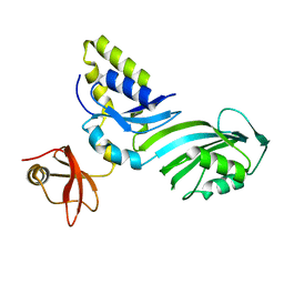
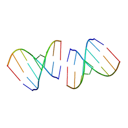
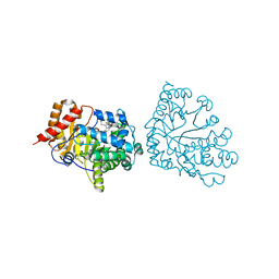
1R3B
 
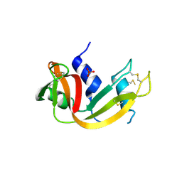
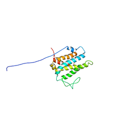 | | Solution structure of xenopus laevis Mob1 | | 分子名称: | MOB1 | | 著者 | Ponchon, L, Dumas, C, Kajava, A.V, Fesquet, D, Padilla, A. | | 登録日 | 2003-10-01 | | 公開日 | 2004-09-28 | | 最終更新日 | 2024-05-22 | | 実験手法 | SOLUTION NMR | | 主引用文献 | NMR solution structure of Mob1, a mitotic exit network protein and its interaction with an NDR kinase peptide
J.Mol.Biol., 337, 2004
|
|
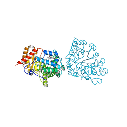
1R3C
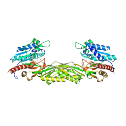
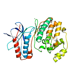 | | THE STRUCTURE OF P38ALPHA C162S MUTANT | | 分子名称: | MAGNESIUM ION, Mitogen-activated protein kinase 14 | | 著者 | Patel, S.B, Cameron, P.M, Frantz-Wattley, B, O'Neill, E, Becker, J.W, Scapin, G. | | 登録日 | 2003-10-01 | | 公開日 | 2004-01-20 | | 最終更新日 | 2023-08-23 | | 実験手法 | X-RAY DIFFRACTION (2 Å) | | 主引用文献 | Lattice stabilization and enhanced diffraction in human p38 alpha crystals by protein engineering.
Biochim.Biophys.Acta, 1696, 2004
|
|
1R3D
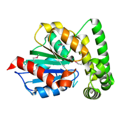 | |
1R3E
 | | Crystal Structure of tRNA Pseudouridine Synthase TruB and Its RNA Complex: RNA-protein Recognition Through a Combination of Rigid Docking and Induced Fit | | 分子名称: | 5'-R(*CP*UP*GP*UP*GP*UP*(FHU)P*CP*GP*AP*UP*CP*CP*AP*CP*AP*G)-3', 5'-R(*CP*UP*GP*UP*GP*UP*UP*CP*GP*AP*UP*CP*CP*AP*CP*AP*G)-3', tRNA pseudouridine synthase B | | 著者 | Pan, H, Agarwalla, S, Moustakas, D.T, Finer-Moore, J, Stroud, R.M. | | 登録日 | 2003-10-01 | | 公開日 | 2003-11-04 | | 最終更新日 | 2018-02-07 | | 実験手法 | X-RAY DIFFRACTION (2.1 Å) | | 主引用文献 | Crystal Structure of tRNA Pseudouridine Synthase TruB and Its RNA Complex: RNA Recognition Through a Combination of Rigid Docking and Induced Fit
Proc.Natl.Acad.Sci.USA, 100, 2003
|
|
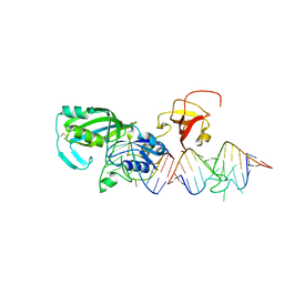
1R3F
 | | Crystal Structure of tRNA Pseudouridine Synthase TruB and Its RNA Complex: RNA-protein Recognition Through a Combination of Rigid Docking and Induced Fit | | 分子名称: | tRNA pseudouridine synthase B | | 著者 | Pan, H, Agarwalla, S, Moustakas, D.T, Finer-Moore, J, Stroud, R.M. | | 登録日 | 2003-10-01 | | 公開日 | 2003-11-04 | | 最終更新日 | 2024-02-14 | | 実験手法 | X-RAY DIFFRACTION (1.85 Å) | | 主引用文献 | Structure of tRNA Pseudouridine Synthase TruB and Its RNA Complex: RNA Recognition Through a Combination of Rigid Docking and Induced Fit
Proc.Natl.Acad.Sci.USA, 100, 2003
|
|
1R3G
 | | 1.16A X-ray structure of the synthetic DNA fragment with the incorporated 2'-O-[(2-Guanidinium)ethyl]-5-methyluridine residues | | 分子名称: | 5'-D(*GP*CP*GP*TP*AP*(GMU)P*AP*CP*GP*C)-3'), MAGNESIUM ION | | 著者 | Prakash, T.P, Puschl, A, Lesnik, E, Tereshko, V, Egli, M, Manoharan, M. | | 登録日 | 2003-10-01 | | 公開日 | 2003-10-21 | | 最終更新日 | 2024-02-14 | | 実験手法 | X-RAY DIFFRACTION (1.16 Å) | | 主引用文献 | 2'-O-[2-(Guanidinium)ethyl]-Modified Oligonucleotides: Stabilizing Effect on Duplex and Triplex Structures.
Org.Lett., 6, 2004
|
|
1R3H
 | | Crystal Structure of T10 | | 分子名称: | Beta-2-microglobulin, MHC H2-TL-T10-129 | | 著者 | Rudolph, M.G, Wilson, I.A. | | 登録日 | 2003-10-02 | | 公開日 | 2004-03-30 | | 最終更新日 | 2023-08-23 | | 実験手法 | X-RAY DIFFRACTION (2.5 Å) | | 主引用文献 | Combined pseudo-merohedral twinning, non-crystallographic symmetry and pseudo-translation in a monoclinic crystal form of the gammadelta T-cell ligand T10.
Acta Crystallogr.,Sect.D, 60, 2004
|
|
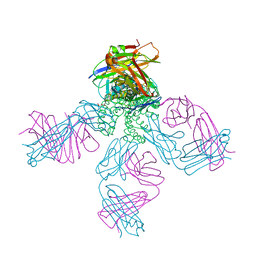
1R3I
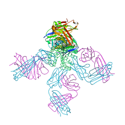 | | potassium channel KcsA-Fab complex in Rb+ | | 分子名称: | Antibody Fab fragment heavy chain, Antibody Fab fragment light chain, DIACYL GLYCEROL, ... | | 著者 | Zhou, Y, MacKinnon, R. | | 登録日 | 2003-10-02 | | 公開日 | 2003-11-25 | | 最終更新日 | 2023-08-23 | | 実験手法 | X-RAY DIFFRACTION (2.4 Å) | | 主引用文献 | The occupancy of ions in the K+ selectivity filter: Charge balance and coupling of ion binding to a protein conformational change underlie high conduction rates
J.Mol.Biol., 333, 2003
|
|
1R3J
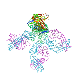 | | potassium channel KcsA-Fab complex in high concentration of Tl+ | | 分子名称: | Antibody Fab fragment heavy chain, Antibody Fab fragment light chain, DIACYL GLYCEROL, ... | | 著者 | Zhou, Y, MacKinnon, R. | | 登録日 | 2003-10-02 | | 公開日 | 2003-11-25 | | 最終更新日 | 2023-08-23 | | 実験手法 | X-RAY DIFFRACTION (1.9 Å) | | 主引用文献 | The occupancy of ions in the K+ selectivity filter: Charge balance and coupling of ion binding to a protein conformational change underlie high conduction rates
J.Mol.Biol., 333, 2003
|
|
1R3K
 | | potassium channel KcsA-Fab complex in low concentration of Tl+ | | 分子名称: | Antibody Fab fragment heavy chain, Antibody Fab fragment light chain, DIACYL GLYCEROL, ... | | 著者 | Zhou, Y, MacKinnon, R. | | 登録日 | 2003-10-02 | | 公開日 | 2003-11-25 | | 最終更新日 | 2023-08-23 | | 実験手法 | X-RAY DIFFRACTION (2.8 Å) | | 主引用文献 | The occupancy of ions in the K+ selectivity filter: Charge balance and coupling of ion binding to a protein conformational change underlie high conduction rates
J.Mol.Biol., 333, 2003
|
|
1R3L
 | | potassium channel KcsA-Fab complex in Cs+ | | 分子名称: | Antibody Fab fragment heavy chain, Antibody Fab fragment light chain, CESIUM ION, ... | | 著者 | Zhou, Y, MacKinnon, R. | | 登録日 | 2003-10-02 | | 公開日 | 2003-11-25 | | 最終更新日 | 2023-08-23 | | 実験手法 | X-RAY DIFFRACTION (2.41 Å) | | 主引用文献 | The occupancy of ions in the K+ selectivity filter: Charge balance and coupling of ion binding to a protein conformational change underlie high conduction rates
J.Mol.Biol., 333, 2003
|
|
1R3M
 | | Crystal structure of the dimeric unswapped form of bovine seminal ribonuclease | | 分子名称: | PHOSPHATE ION, Ribonuclease, seminal | | 著者 | Berisio, R, Sica, F, De Lorenzo, C, Di Fiore, A, Piccoli, R, Zagari, A, Mazzarella, L. | | 登録日 | 2003-10-02 | | 公開日 | 2003-11-18 | | 最終更新日 | 2023-08-23 | | 実験手法 | X-RAY DIFFRACTION (2.2 Å) | | 主引用文献 | Crystal structure of the dimeric unswapped form of bovine seminal ribonuclease
Febs Lett., 554, 2003
|
|
1R3N
 | | Crystal structure of beta-alanine synthase from Saccharomyces kluyveri | | 分子名称: | BETA-AMINO ISOBUTYRATE, ZINC ION, beta-alanine synthase | | 著者 | Lundgren, S, Gojkovic, Z, Piskur, J, Dobritzsch, D. | | 登録日 | 2003-10-02 | | 公開日 | 2003-11-11 | | 最終更新日 | 2024-04-03 | | 実験手法 | X-RAY DIFFRACTION (2.7 Å) | | 主引用文献 | Yeast beta-Alanine Synthase Shares a Structural Scaffold and Origin with Dizinc-dependent Exopeptidases
J.Biol.Chem., 278, 2003
|
|
1R3O
 | | Crystal structure of the first RNA duplex in L-conformation at 1.9A resolution | | 分子名称: | L-RNA | | 著者 | Vallazza, M, Perbandt, M, Klussmann, S, Rypniewski, W, Erdmann, V.A, Betzel, C. | | 登録日 | 2003-10-02 | | 公開日 | 2003-12-23 | | 最終更新日 | 2024-02-14 | | 実験手法 | X-RAY DIFFRACTION (1.9 Å) | | 主引用文献 | First look at RNA in L-configuration.
Acta Crystallogr.,Sect.D, 60, 2004
|
|
1R3Q
 | | Uroporphyrinogen Decarboxylase in complex with coproporphyrinogen-I | | 分子名称: | CARBON DIOXIDE, COPROPORPHYRINOGEN I, Uroporphyrinogen Decarboxylase | | 著者 | Phillips, J.D, Whitby, F.G, Kushner, J.P, Hill, C.P. | | 登録日 | 2003-10-03 | | 公開日 | 2003-12-09 | | 最終更新日 | 2023-08-23 | | 実験手法 | X-RAY DIFFRACTION (1.7 Å) | | 主引用文献 | Structural basis for tetrapyrrole coordination by uroporphyrinogen decarboxylase
Embo J., 22, 2003
|
|
1R3R
 | |
1R3S
 | | Uroporphyrinogen Decarboxylase single mutant D86G in complex with coproporphyrinogen-I | | 分子名称: | COPROPORPHYRINOGEN I, Uroporphyrinogen Decarboxylase | | 著者 | Phillips, J.D, Whitby, F.G, Kushner, J.P, Hill, C.P. | | 登録日 | 2003-10-03 | | 公開日 | 2003-12-09 | | 最終更新日 | 2023-08-23 | | 実験手法 | X-RAY DIFFRACTION (1.65 Å) | | 主引用文献 | Structural basis for tetrapyrrole coordination by uroporphyrinogen decarboxylase
Embo J., 22, 2003
|
|
1R3T
 | | Uroporphyrinogen Decarboxylase single mutant D86G in complex with coproporphyrinogen-III | | 分子名称: | COPROPORPHYRINOGEN III, Uroporphyrinogen Decarboxylase | | 著者 | Phillips, J.D, Whitby, F.G, Kushner, J.P, Hill, C.P. | | 登録日 | 2003-10-03 | | 公開日 | 2003-12-09 | | 最終更新日 | 2023-08-23 | | 実験手法 | X-RAY DIFFRACTION (1.7 Å) | | 主引用文献 | Structural basis for tetrapyrrole coordination by uroporphyrinogen decarboxylase
Embo J., 22, 2003
|
|
1R3U
 | | Crystal Structure of Hypoxanthine-Guanine Phosphoribosyltransferase from Thermoanaerobacter tengcongensis | | 分子名称: | ACETATE ION, Hypoxanthine-guanine phosphoribosyltransferase, MAGNESIUM ION | | 著者 | Chen, Q, Liang, Y.H, Gu, X.C, Luo, M, Su, X.D. | | 登録日 | 2003-10-03 | | 公開日 | 2004-10-19 | | 最終更新日 | 2023-10-25 | | 実験手法 | X-RAY DIFFRACTION (2.5 Å) | | 主引用文献 | Crystal Structure of Hypoxanthine-Guanine Phosphoribosyltransferase from Thermoanaerobacter tengcongensis
To be published
|
|
1R3V
 | | Uroporphyrinogen Decarboxylase single mutant D86E in complex with coproporphyrinogen-I | | 分子名称: | BETA-MERCAPTOETHANOL, COPROPORPHYRINOGEN I, Uroporphyrinogen Decarboxylase | | 著者 | Phillips, J.D, Whitby, F.G, Kushner, J.P, Hill, C.P. | | 登録日 | 2003-10-03 | | 公開日 | 2003-12-09 | | 最終更新日 | 2023-08-23 | | 実験手法 | X-RAY DIFFRACTION (1.9 Å) | | 主引用文献 | Structural basis for tetrapyrrole coordination by uroporphyrinogen decarboxylase
Embo J., 22, 2003
|
|
1R3W
 | | Uroporphyrinogen Decarboxylase Y164F mutant in complex with coproporphyrinogen-III | | 分子名称: | COPROPORPHYRINOGEN III, Uroporphyrinogen Decarboxylase | | 著者 | Phillips, J.D, Whitby, F.G, Kushner, J.P, Hill, C.P. | | 登録日 | 2003-10-03 | | 公開日 | 2003-12-09 | | 最終更新日 | 2023-08-23 | | 実験手法 | X-RAY DIFFRACTION (1.7 Å) | | 主引用文献 | Structural basis for tetrapyrrole coordination by uroporphyrinogen decarboxylase
Embo J., 22, 2003
|
|
1R3X
 | | INTRAMOLECULAR DNA TRIPLEX WITH RNA THIRD STRAND, NMR, 10 STRUCTURES | | 分子名称: | DNA (5'-D(*AP*GP*AP*GP*AP*GP*AP*A)-3'), DNA (5'-D(*TP*TP*CP*TP*CP*TP*CP*T)-3'), RNA (5'-R(*UP*CP*UP*CP*UP*CP*UP*U)-3') | | 著者 | Gotfredsen, C.H, Schultze, P, Feigon, J. | | 登録日 | 1998-02-06 | | 公開日 | 1998-05-20 | | 最終更新日 | 2024-05-22 | | 実験手法 | SOLUTION NMR | | 主引用文献 | Solution Structure of an Intramolecular Pyrimidine-Purine-Pyrimidine Triplex Containing an RNA Third Strand
J.Am.Chem.Soc., 120, 1998
|
|
1R3Y
 | | Uroporphyrinogen Decarboxylase in complex with coproporphyrinogen-III | | 分子名称: | COPROPORPHYRINOGEN III, Uroporphyrinogen Decarboxylase | | 著者 | Phillips, J.D, Whitby, F.G, Kushner, J.P, Hill, C.P. | | 登録日 | 2003-10-03 | | 公開日 | 2003-12-09 | | 最終更新日 | 2023-08-23 | | 実験手法 | X-RAY DIFFRACTION (1.755 Å) | | 主引用文献 | Structural basis for tetrapyrrole coordination by uroporphyrinogen decarboxylase
Embo J., 22, 2003
|
|
1R3Z
 | | Crystal structures of d(Gm5CGm5CGCGC) and d(GCGCGm5CGm5C): Effects of methylation on alternating DNA octamers | | 分子名称: | 5'-D(*GP*(5CM)P*GP*(5CM)P*GP*CP*GP*C)-3' | | 著者 | Shi, K, Pan, B, Tippin, D, Sundaralingam, M. | | 登録日 | 2003-10-03 | | 公開日 | 2003-12-23 | | 最終更新日 | 2024-02-14 | | 実験手法 | X-RAY DIFFRACTION (1.7 Å) | | 主引用文献 | Structures of d(Gm5)CGm5CGCGC) and d(GCGCGm5CGm5C): effects of methylation on alternating DNA octamers.
Acta Crystallogr.,Sect.D, 60, 2004
|
|
1R41
 | | Crystal structures of d(Gm5CGm5CGCGC) and d(GCGCGm5CGm5C): Effects of methylation on alternating DNA octamers | | 分子名称: | 5'-D(*GP*CP*GP*CP*GP*(5CM)P*GP*(5CM))-3' | | 著者 | Shi, K, Pan, B, Tippin, D, Sundaralingam, M. | | 登録日 | 2003-10-03 | | 公開日 | 2003-12-23 | | 最終更新日 | 2024-02-14 | | 実験手法 | X-RAY DIFFRACTION (1.9 Å) | | 主引用文献 | Structures of d(Gm5)CGm5CGCGC) and d(GCGCGm5CGm5C): effects of methylation on alternating DNA octamers.
Acta Crystallogr.,Sect.D, 60, 2004
|
|


