3K4A
 
 | |
3K4W
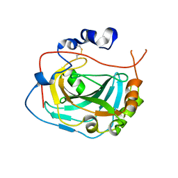
 | | CRYSTAL STRUCTURE OF Uncharacterized Tim-Barrel Protein Bb4693 From Bordetella Bronchiseptica | | 分子名称: | uncharacterized protein Bb4693 | | 著者 | Malashkevich, V.N, Toro, R, Sauder, J.M, Burley, S.K, Almo, S.C, New York SGX Research Center for Structural Genomics (NYSGXRC) | | 登録日 | 2009-10-06 | | 公開日 | 2009-11-24 | | 最終更新日 | 2024-02-21 | | 実験手法 | X-RAY DIFFRACTION (1.92 Å) | | 主引用文献 | CRYSTAL STRUCTURE OF Uncharacterized Tim-Barrel Protein
Bb4693 From Bordetella Bronchiseptica
To be Published
|
|
3SEW
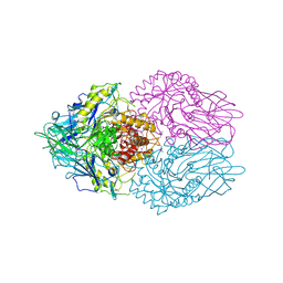
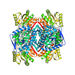
 | | Zn-mediated Polymer of Maltose-binding Protein A216H/K220H by Synthetic Symmetrization (Form I) | | 分子名称: | CHLORIDE ION, GLYCEROL, Maltose-binding periplasmic protein, ... | | 著者 | Zhao, M, Soriaga, A.B, Laganowsky, A, Sawaya, M.R, Cascio, D, Yeates, T.O. | | 登録日 | 2011-06-11 | | 公開日 | 2011-09-21 | | 最終更新日 | 2024-02-28 | | 実験手法 | X-RAY DIFFRACTION (1.55 Å) | | 主引用文献 | An approach to crystallizing proteins by metal-mediated synthetic symmetrization.
Protein Sci., 20, 2011
|
|
4UZ8
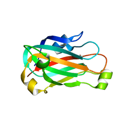
 | | The SeMet structure of the family 46 carbohydrate-binding module (CBM46) of endo-beta-1,4-glucanase B (Cel5B) from Bacillus halodurans | | 分子名称: | ENDO-BETA-1,4-GLUCANASE (CELULASE B), SULFATE ION | | 著者 | Venditto, I, Santos, H, Ferreira, L.M.A, Sakka, K, Fontes, C.M.G.A, Najmudin, S. | | 登録日 | 2014-09-04 | | 公開日 | 2015-02-25 | | 最終更新日 | 2024-11-06 | | 実験手法 | X-RAY DIFFRACTION (2.3 Å) | | 主引用文献 | Family 46 Carbohydrate-Binding Modules Contribute to the Enzymatic Hydrolysis of Xyloglucan and Beta-1,3-1,4-Glucans Through Distinct Mechanisms.
J.Biol.Chem., 290, 2015
|
|
3K77
 | | X-ray crystal structure of XRCC1 | | 分子名称: | DNA repair protein XRCC1 | | 著者 | Cuneo, M.J, London, R.E. | | 登録日 | 2009-10-12 | | 公開日 | 2010-04-28 | | 最終更新日 | 2023-09-06 | | 実験手法 | X-RAY DIFFRACTION (2.597 Å) | | 主引用文献 | Oxidation state of the XRCC1 N-terminal domain regulates DNA polymerase beta binding affinity.
Proc.Natl.Acad.Sci.USA, 107, 2010
|
|
3K9I
 | |
3K9W
 | |
3KA3
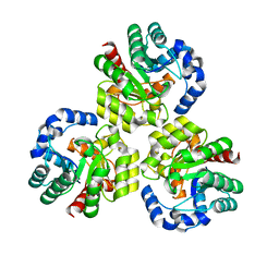
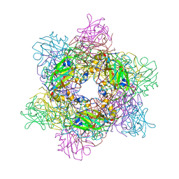
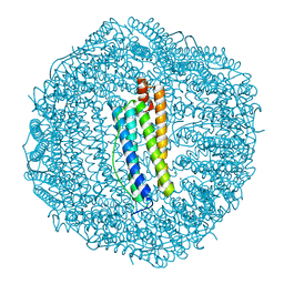 | | Frog M-ferritin with magnesium | | 分子名称: | CHLORIDE ION, Ferritin, middle subunit, ... | | 著者 | Tosha, T, Ng, H.L, Theil, E, Alber, T, Bhattasali, O. | | 登録日 | 2009-10-18 | | 公開日 | 2010-10-06 | | 最終更新日 | 2024-02-21 | | 実験手法 | X-RAY DIFFRACTION (1.4 Å) | | 主引用文献 | Moving Metal Ions through Ferritin-Protein Nanocages from Three-Fold Pores to Catalytic Sites.
J.Am.Chem.Soc., 132, 2010
|
|
4WFZ
 | |
4WHG
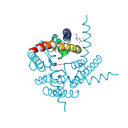
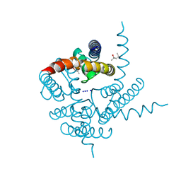
 | | Crystal Structure of TR3 LBD in complex with Molecule 3 | | 分子名称: | 1-(3,4,5-trihydroxyphenyl)octan-1-one, GLYCEROL, Nuclear receptor subfamily 4 group A member 1 | | 著者 | Li, F.W, Cai, Q.X, Li, A.Z, Tian, X.Y, Wang, W.J, Wang, Y, Hou, P.P, Wu, Q, Lin, T.W. | | 登録日 | 2014-09-22 | | 公開日 | 2015-09-09 | | 最終更新日 | 2024-03-20 | | 実験手法 | X-RAY DIFFRACTION (2.18 Å) | | 主引用文献 | Induction of Autophagic Death in Cancer Cells by Agonizing TR3 and Attenuating Akt2 Activity
Chem.Biol., 22, 2015
|
|
4WHQ
 | | Alkylperoxo reaction intermediate trapped in Protocatechuate 3,4-dioxygenase (pseudomonas putida) at pH 6.5 | | 分子名称: | (6S)-4-fluoro-6-hydroperoxy-6-hydroxycyclohexa-2,4-dien-1-one, 4-fluorobenzene-1,2-diol, BETA-MERCAPTOETHANOL, ... | | 著者 | Knoot, C.J, Purpero, V.M, Lipscomb, J.D. | | 登録日 | 2014-09-23 | | 公開日 | 2014-12-31 | | 最終更新日 | 2023-12-27 | | 実験手法 | X-RAY DIFFRACTION (1.78 Å) | | 主引用文献 | Crystal structures of alkylperoxo and anhydride intermediates in an intradiol ring-cleaving dioxygenase.
Proc.Natl.Acad.Sci.USA, 112, 2015
|
|
3JTL
 | |
4WFY
 | |
3JW6
 | |
4WIP
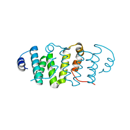
 | | DIX domain of human Dvl2 | | 分子名称: | PENTAETHYLENE GLYCOL, Segment polarity protein dishevelled homolog DVL-2 | | 著者 | Fiedler, M, Bienz, M, Madrzak, J, Chin, J.W. | | 登録日 | 2014-09-26 | | 公開日 | 2015-05-13 | | 最終更新日 | 2024-05-08 | | 実験手法 | X-RAY DIFFRACTION (2.691 Å) | | 主引用文献 | Ubiquitination of the Dishevelled DIX domain blocks its head-to-tail polymerization.
Nat Commun, 6, 2015
|
|
3SI0
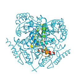
 | | Structure of glycosylated human glutaminyl cyclase | | 分子名称: | 2-acetamido-2-deoxy-beta-D-glucopyranose-(1-4)-2-acetamido-2-deoxy-beta-D-glucopyranose, CHLORIDE ION, Glutaminyl-peptide cyclotransferase, ... | | 著者 | Parthier, C, Carrillo, D, Stubbs, M.T. | | 登録日 | 2011-06-17 | | 公開日 | 2011-06-29 | | 最終更新日 | 2024-10-09 | | 実験手法 | X-RAY DIFFRACTION (2.1 Å) | | 主引用文献 | Structures of Glycosylated Mammalian Glutaminyl Cyclases Reveal Conformational Variability near the Active Center.
Biochemistry, 50, 2011
|
|
3JXG
 | | CA-like domain of mouse PTPRG | | 分子名称: | Receptor-type tyrosine-protein phosphatase gamma | | 著者 | Bouyain, S. | | 登録日 | 2009-09-19 | | 公開日 | 2009-12-22 | | 最終更新日 | 2024-10-30 | | 実験手法 | X-RAY DIFFRACTION (1.7 Å) | | 主引用文献 | The protein tyrosine phosphatases PTPRZ and PTPRG bind to distinct members of the contactin family of neural recognition molecules.
Proc.Natl.Acad.Sci.USA, 107, 2010
|
|
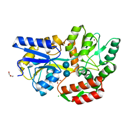
3JZ4
 | | Crystal structure of E. coli NADP dependent enzyme | | 分子名称: | NADP NICOTINAMIDE-ADENINE-DINUCLEOTIDE PHOSPHATE, Succinate-semialdehyde dehydrogenase [NADP+] | | 著者 | Langendorf, C.G, Key, T.L.G, Fenalti, G, Kan, W.T, Buckle, A.M, Caradoc-Davies, T, Tuck, K.L, Law, R.H.P, Whisstock, J.C. | | 登録日 | 2009-09-22 | | 公開日 | 2010-03-16 | | 最終更新日 | 2024-02-21 | | 実験手法 | X-RAY DIFFRACTION (2.3 Å) | | 主引用文献 | The X-ray crystal structure of Escherichia coli succinic semialdehyde dehydrogenase; structural insights into NADP+/enzyme interactions.
Plos One, 5, 2010
|
|
3K06
 | | Crystal Structure of CNG mimicking NaK mutant, NaK-NTPP, K+ complex | | 分子名称: | (4S)-2-METHYL-2,4-PENTANEDIOL, POTASSIUM ION, Potassium channel protein NaK | | 著者 | Jiang, Y, Derebe, M.G. | | 登録日 | 2009-09-24 | | 公開日 | 2011-01-12 | | 最終更新日 | 2024-02-21 | | 実験手法 | X-RAY DIFFRACTION (1.58 Å) | | 主引用文献 | Structural studies of ion permeation and Ca2+ blockage of a bacterial channel mimicking the cyclic nucleotide-gated channel pore.
Proc.Natl.Acad.Sci.USA, 108, 2011
|
|
3K0G
 | | Crystal Structure of CNG mimicking NaK mutant, NaK-ETPP, Na+ complex | | 分子名称: | (4S)-2-METHYL-2,4-PENTANEDIOL, Potassium channel protein NaK, SODIUM ION | | 著者 | Jiang, Y, Derebe, M.G. | | 登録日 | 2009-09-24 | | 公開日 | 2011-01-12 | | 最終更新日 | 2024-02-21 | | 実験手法 | X-RAY DIFFRACTION (1.95 Å) | | 主引用文献 | Structural studies of ion permeation and Ca2+ blockage of a bacterial channel mimicking the cyclic nucleotide-gated channel pore.
Proc.Natl.Acad.Sci.USA, 108, 2011
|
|
3K0K
 | | Crystal Structure of BRCA1 BRCT in complex with a minimal recognition tetrapeptide with a free carboxy C-terminus. | | 分子名称: | Breast cancer type 1 susceptibility protein, CHLORIDE ION, NICKEL (II) ION, ... | | 著者 | Campbell, S.J, Edwards, R.A, Glover, J.N. | | 登録日 | 2009-09-24 | | 公開日 | 2010-03-02 | | 最終更新日 | 2024-11-20 | | 実験手法 | X-RAY DIFFRACTION (2.7 Å) | | 主引用文献 | Comparison of the Structures and Peptide Binding Specificities of the BRCT Domains of MDC1 and BRCA1
Structure, 18, 2010
|
|
4WHO
 | |
3K86
 | |
4WJT
 | | Stationary Phase Survival Protein YuiC from B.subtilis complexed with NAG | | 分子名称: | (2S)-2-{[(2S)-2-{[(2R)-2-hydroxypropyl]oxy}propyl]oxy}propan-1-ol, 2-acetamido-2-deoxy-beta-D-glucopyranose, DIMETHYL SULFOXIDE, ... | | 著者 | Quay, D.H.X, Cole, A.R, Cryar, A, Thalassinos, K, Williams, M.A, Bhakta, S, Keep, N.H. | | 登録日 | 2014-10-01 | | 公開日 | 2015-07-08 | | 最終更新日 | 2024-01-10 | | 実験手法 | X-RAY DIFFRACTION (1.21 Å) | | 主引用文献 | Structure of the stationary phase survival protein YuiC from B.subtilis.
Bmc Struct.Biol., 15, 2015
|
|
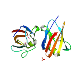
3JRR
 | | Crystal structure of the ligand binding suppressor domain of type 3 inositol 1,4,5-trisphosphate receptor | | 分子名称: | Inositol 1,4,5-trisphosphate receptor type 3 | | 著者 | Chan, J, Ishiyama, N, Ikura, M. | | 登録日 | 2009-09-08 | | 公開日 | 2010-09-15 | | 最終更新日 | 2023-09-06 | | 実験手法 | X-RAY DIFFRACTION (1.9 Å) | | 主引用文献 | A 1.9 angstrom crystal structure of the suppressor domain of type 3 inositol 1,4,5-trisphosphate receptor
To be Published
|
|


