1E48
 
 | |
1M7S
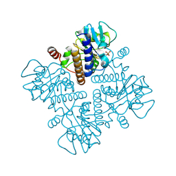
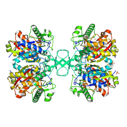
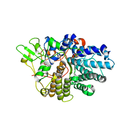
 | | Crystal Structure Analysis of Catalase CatF of Pseudomonas syringae | | 分子名称: | Catalase, PROTOPORPHYRIN IX CONTAINING FE | | 著者 | Carpena, X, Soriano, M, Klotz, M.G, Duckworth, H.W, Donald, L.J, Melik-Adamyan, W, Fita, I, Loewen, P.C. | | 登録日 | 2002-07-22 | | 公開日 | 2002-08-28 | | 最終更新日 | 2024-02-14 | | 実験手法 | X-RAY DIFFRACTION (1.8 Å) | | 主引用文献 | Structure of the Clade 1 catalase, CatF of Pseudomonas syringae, at 1.8 A resolution
Proteins, 50, 2003
|
|
2AXZ
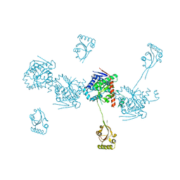
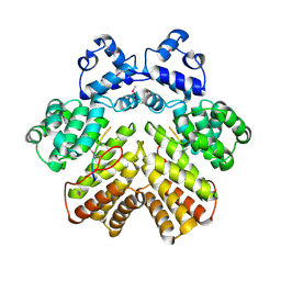 | | Crystal structure of PrgX/cCF10 complex | | 分子名称: | LVTLVFV peptide, PrgX, TPPKEVT(MSE) peptide | | 著者 | Shi, K, Brown, C.K, Gu, Z.Y, Kozlowicz, B.K, Dunny, G.M, Ohlendorf, D.H, Earhart, C.A. | | 登録日 | 2005-09-06 | | 公開日 | 2005-12-06 | | 最終更新日 | 2011-07-13 | | 実験手法 | X-RAY DIFFRACTION (3 Å) | | 主引用文献 | Structure of peptide sex pheromone receptor PrgX and PrgX/pheromone complexes and regulation of conjugation in Enterococcus faecalis.
Proc.Natl.Acad.Sci.Usa, 102, 2005
|
|
1M1O
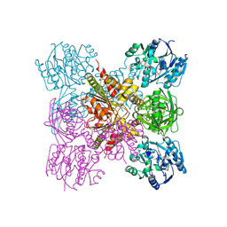
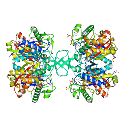 | | Crystal structure of biosynthetic thiolase, C89A mutant, complexed with acetoacetyl-CoA | | 分子名称: | ACETOACETYL-COENZYME A, Acetyl-CoA acetyltransferase, SULFATE ION | | 著者 | Kursula, P, Ojala, J, Lambeir, A.-M, Wierenga, R.K. | | 登録日 | 2002-06-20 | | 公開日 | 2002-11-29 | | 最終更新日 | 2024-02-14 | | 実験手法 | X-RAY DIFFRACTION (1.95 Å) | | 主引用文献 | The catalytic cycle of biosynthetic thiolase: A conformational journey of an acetyl group through four binding modes and two oxyanion holes
Biochemistry, 41, 2002
|
|
1MHL
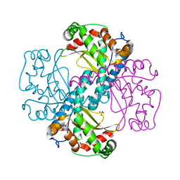
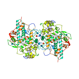 | | CRYSTAL STRUCTURE OF HUMAN MYELOPEROXIDASE ISOFORM C CRYSTALLIZED IN SPACE GROUP P2(1) AT PH 5.5 AND 20 DEG C | | 分子名称: | 2-acetamido-2-deoxy-beta-D-glucopyranose, CALCIUM ION, CHLORIDE ION, ... | | 著者 | Fenna, R.E, Zeng, J, Davey, C. | | 登録日 | 1995-06-09 | | 公開日 | 1996-01-06 | | 最終更新日 | 2020-07-29 | | 実験手法 | X-RAY DIFFRACTION (2.25 Å) | | 主引用文献 | Structure of the green heme in myeloperoxidase.
Arch.Biochem.Biophys., 316, 1995
|
|
1M4T
 | | Biosynthetic thiolase, Cys89 butyrylated | | 分子名称: | Acetyl-CoA acetyltransferase, GLYCEROL, SULFATE ION | | 著者 | Kursula, P, Ojala, J, Lambeir, A.-M, Wierenga, R.K. | | 登録日 | 2002-07-03 | | 公開日 | 2002-11-29 | | 最終更新日 | 2018-03-07 | | 実験手法 | X-RAY DIFFRACTION (1.77 Å) | | 主引用文献 | The catalytic cycle of biosynthetic thiolase: A conformational
journey of an acetyl group through four binding modes and two oxyanion holes
Biochemistry, 41, 2002
|
|
2AXV
 | | Structure of PrgX Y153C mutant | | 分子名称: | PrgX | | 著者 | Shi, K, Brown, C.K, Gu, Z.Y, Kozlowicz, B.K, Dunny, G.M, Ohlendorf, D.H, Earhart, C.A. | | 登録日 | 2005-09-06 | | 公開日 | 2005-12-06 | | 最終更新日 | 2024-02-14 | | 実験手法 | X-RAY DIFFRACTION (3 Å) | | 主引用文献 | Structure of peptide sex pheromone receptor PrgX and PrgX/pheromone complexes and regulation of conjugation in Enterococcus faecalis.
Proc.Natl.Acad.Sci.Usa, 102, 2005
|
|
1M3K
 | | biosynthetic thiolase, inactive C89A mutant | | 分子名称: | Acetyl-CoA acetyltransferase, GLYCEROL, SULFATE ION | | 著者 | Kursula, P, Ojala, J, Lambeir, A.-M, Wierenga, R.K. | | 登録日 | 2002-06-28 | | 公開日 | 2002-11-29 | | 最終更新日 | 2024-02-14 | | 実験手法 | X-RAY DIFFRACTION (1.7 Å) | | 主引用文献 | The catalytic cycle of biosynthetic thiolase: A conformational
journey of an acetyl group through four binding modes and two oxyanion holes
Biochemistry, 41, 2002
|
|
1M6S
 | |
1E58
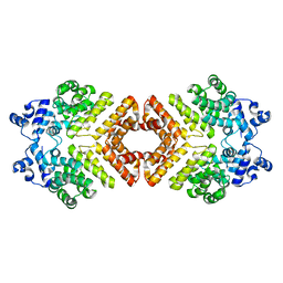
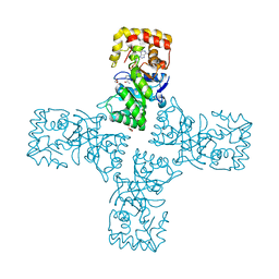
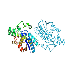 | | E.coli cofactor-dependent phosphoglycerate mutase | | 分子名称: | CHLORIDE ION, PHOSPHOGLYCERATE MUTASE, SULFATE ION | | 著者 | Bond, C.S, Hunter, W.N. | | 登録日 | 2000-07-19 | | 公開日 | 2001-03-20 | | 最終更新日 | 2019-05-22 | | 実験手法 | X-RAY DIFFRACTION (1.25 Å) | | 主引用文献 | High Resolution Structure of the Phosphohistidine-Activated Form of Escherichia Coli Cofactor-Dependent Phosphoglycerate Mutase.
J.Biol.Chem., 276, 2001
|
|
1M5Y
 | |
2FIV
 | | Crystal structure of feline immunodeficiency virus protease complexed with a substrate | | 分子名称: | ACE-ALN-VAL-STA-GLU-ALN-NH2, FELINE IMMUNODEFICIENCY VIRUS PROTEASE, SULFATE ION | | 著者 | Schalk-Hihi, C, Lubkowski, J, Zdanov, A, Wlodawer, A, Gustchina, A. | | 登録日 | 1997-07-21 | | 公開日 | 1997-11-12 | | 最終更新日 | 2023-08-09 | | 実験手法 | X-RAY DIFFRACTION (2 Å) | | 主引用文献 | Crystal structures of the inactive D30N mutant of feline immunodeficiency virus protease complexed with a substrate and an inhibitor.
Biochemistry, 36, 1997
|
|
4HIF
 | | Ultrahigh-resolution crystal structure of Z-DNA in complex with Zn2+ ions | | 分子名称: | CHLORIDE ION, DNA (5'-D(*CP*GP*CP*GP*CP*G)-3'), SPERMINE (FULLY PROTONATED FORM), ... | | 著者 | Drozdzal, P, Gilski, M, Kierzek, R, Lomozik, L, Jaskolski, M. | | 登録日 | 2012-10-11 | | 公開日 | 2013-06-05 | | 最終更新日 | 2023-09-20 | | 実験手法 | X-RAY DIFFRACTION (0.85 Å) | | 主引用文献 | Ultrahigh-resolution crystal structures of Z-DNA in complex with Mn(2+) and Zn(2+) ions.
Acta Crystallogr.,Sect.D, 69, 2013
|
|
3EHN
 | | BT1043 with N-acetyllactosamine | | 分子名称: | SusD homolog, beta-D-galactopyranose-(1-4)-2-acetamido-2-deoxy-alpha-D-glucopyranose | | 著者 | Smith, T.J, Koropatkin, N.M. | | 登録日 | 2008-09-13 | | 公開日 | 2009-05-12 | | 最終更新日 | 2024-02-21 | | 実験手法 | X-RAY DIFFRACTION (2.8 Å) | | 主引用文献 | Structure of a SusD homologue, BT1043, involved in mucin O-glycan utilization in a prominent human gut symbiont.
Biochemistry, 48, 2009
|
|
3E4B
 | | Crystal structure of AlgK from Pseudomonas fluorescens WCS374r | | 分子名称: | AlgK, CHLORIDE ION, GLYCEROL | | 著者 | Keiski, C.-L, Harwich, M, Jain, S, Neculai, A.M, Yip, P, Robinson, H, Whitney, J.C, Burrows, L.L, Ohman, D.E, Howell, P.L. | | 登録日 | 2008-08-11 | | 公開日 | 2009-08-25 | | 最終更新日 | 2011-07-13 | | 実験手法 | X-RAY DIFFRACTION (2.5 Å) | | 主引用文献 | AlgK is a TPR-containing protein and the periplasmic component of a novel exopolysaccharide secretin.
Structure, 18, 2010
|
|
6LRH
 | |
4X9Q
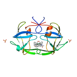
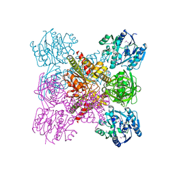
 | | MnSOD-3 Room Temperature Structure | | 分子名称: | MALONATE ION, MANGANESE (II) ION, SULFATE ION, ... | | 著者 | Hunter, G.J, Trinh, C.H, Hunter, T, Bonetta, R, Stewart, E.E. | | 登録日 | 2014-12-11 | | 公開日 | 2015-11-25 | | 最終更新日 | 2024-01-10 | | 実験手法 | X-RAY DIFFRACTION (1.77 Å) | | 主引用文献 | The structure of the Caenorhabditis elegans manganese superoxide dismutase MnSOD-3-azide complex.
Protein Sci., 24, 2015
|
|
6OW0
 | | Crystal structure of mithramycin 3-side chain keto-reductase MtmW in complex with NAD+ and PEG | | 分子名称: | DI(HYDROXYETHYL)ETHER, GLYCEROL, MtmW, ... | | 著者 | Hou, C, Yu, X, Rohr, J, Tsodikov, O.V. | | 登録日 | 2019-05-08 | | 公開日 | 2019-11-27 | | 最終更新日 | 2023-10-11 | | 実験手法 | X-RAY DIFFRACTION (2.67 Å) | | 主引用文献 | Discovery of a Cryptic Intermediate in Late Steps of Mithramycin Biosynthesis.
Angew.Chem.Int.Ed.Engl., 59, 2020
|
|
4XFO
 | |
6LRF
 | | Crystal structure of unliganded AgrE | | 分子名称: | Alr4995 protein | | 著者 | Lee, H, Rhee, S. | | 登録日 | 2020-01-16 | | 公開日 | 2020-04-01 | | 最終更新日 | 2020-05-13 | | 実験手法 | X-RAY DIFFRACTION (2.05466056 Å) | | 主引用文献 | Structural and mutational analyses of the bifunctional arginine dihydrolase and ornithine cyclodeaminase AgrE from the cyanobacteriumAnabaena.
J.Biol.Chem., 295, 2020
|
|
6O4L
 | | Structure of ALDH7A1 mutant E399D complexed with NAD | | 分子名称: | 1,2-ETHANEDIOL, Alpha-aminoadipic semialdehyde dehydrogenase, CHLORIDE ION, ... | | 著者 | Tanner, J.J, Korasick, D.A, Laciak, A.R. | | 登録日 | 2019-02-28 | | 公開日 | 2019-11-06 | | 最終更新日 | 2023-10-11 | | 実験手法 | X-RAY DIFFRACTION (1.85 Å) | | 主引用文献 | Structural analysis of pathogenic mutations targeting Glu427 of ALDH7A1, the hot spot residue of pyridoxine-dependent epilepsy.
J. Inherit. Metab. Dis., 43, 2020
|
|
4HIG
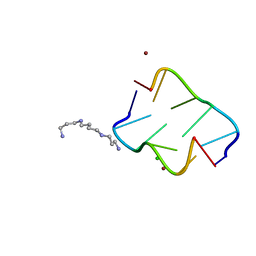
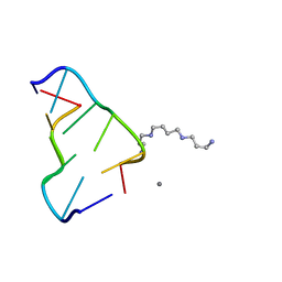 | | Ultrahigh-resolution crystal structure of Z-DNA in complex with Mn2+ ion. | | 分子名称: | DNA (5'-D(*CP*GP*CP*GP*CP*G)-3'), MANGANESE (II) ION, SPERMINE (FULLY PROTONATED FORM) | | 著者 | Drozdzal, P, Gilski, M, Kierzek, R, Lomozik, L, Jaskolski, M. | | 登録日 | 2012-10-11 | | 公開日 | 2013-06-05 | | 最終更新日 | 2023-09-20 | | 実験手法 | X-RAY DIFFRACTION (0.75 Å) | | 主引用文献 | Ultrahigh-resolution crystal structures of Z-DNA in complex with Mn(2+) and Zn(2+) ions.
Acta Crystallogr.,Sect.D, 69, 2013
|
|
3VTX
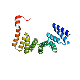 | | Crystal structure of MamA protein | | 分子名称: | GLYCEROL, MamA | | 著者 | Zeytuni, N, Baran, D, Davidov, G, Zarivach, R. | | 登録日 | 2012-06-08 | | 公開日 | 2012-10-31 | | 最終更新日 | 2024-03-20 | | 実験手法 | X-RAY DIFFRACTION (1.75 Å) | | 主引用文献 | Inter-phylum structural conservation of the magnetosome-associated TPR-containing protein, MamA
J.Struct.Biol., 180, 2012
|
|
2DU3
 | |
3W37
 | | Sugar beet alpha-glucosidase with acarbose | | 分子名称: | 2-acetamido-2-deoxy-beta-D-glucopyranose, 2-acetamido-2-deoxy-beta-D-glucopyranose-(1-4)-2-acetamido-2-deoxy-beta-D-glucopyranose, 4,6-dideoxy-4-{[(1S,4R,5S,6S)-4,5,6-trihydroxy-3-(hydroxymethyl)cyclohex-2-en-1-yl]amino}-alpha-D-glucopyranose-(1-4)-alpha-D-glucopyranose-(1-4)-alpha-D-glucopyranose, ... | | 著者 | Tagami, T, Yamashita, K, Okuyama, M, Mori, H, Yao, M, Kimura, A. | | 登録日 | 2012-12-13 | | 公開日 | 2013-05-29 | | 最終更新日 | 2020-07-29 | | 実験手法 | X-RAY DIFFRACTION (1.7 Å) | | 主引用文献 | Molecular basis for the recognition of long-chain substrates by plant & alpha-glucosidase
J.Biol.Chem., 288, 2013
|
|


