6XZL
 
 | |
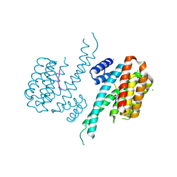
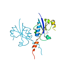
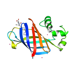
7L15
 | | Marsupial T cell receptor Spl_118 | | 分子名称: | 2-acetamido-2-deoxy-beta-D-glucopyranose, 2-acetamido-2-deoxy-beta-D-glucopyranose-(1-4)-2-acetamido-2-deoxy-beta-D-glucopyranose, T cell receptor gamma chain, ... | | 著者 | Wegrecki, M, Kannan Sivaraman, K, Praveena, T, Le Nours, J, Rossjohn, J. | | 登録日 | 2020-12-14 | | 公開日 | 2021-03-24 | | 最終更新日 | 2024-04-03 | | 実験手法 | X-RAY DIFFRACTION (2.25 Å) | | 主引用文献 | The molecular assembly of the marsupial gamma mu T cell receptor defines a third T cell lineage.
Science, 371, 2021
|
|
5V8G
 | | Pekin duck lysozyme isoform I (DEL-I) | | 分子名称: | CHLORIDE ION, THIOCYANATE ION, lysozyme isoform I | | 著者 | Langley, D.B, Christ, D. | | 登録日 | 2017-03-22 | | 公開日 | 2017-11-15 | | 最終更新日 | 2023-10-04 | | 実験手法 | X-RAY DIFFRACTION (1.2 Å) | | 主引用文献 | Structural basis of antigen recognition: crystal structure of duck egg lysozyme.
Acta Crystallogr D Struct Biol, 73, 2017
|
|
6Y17
 | |
6FBB
 | | Crystal structure of 14-3-3 sigma in complex with wild-type Shroom3 | | 分子名称: | 14-3-3 protein sigma, CHLORIDE ION, MAGNESIUM ION, ... | | 著者 | Leysen, S, Meijer, F.A, Milroy, L.G, Ottmann, C. | | 登録日 | 2017-12-18 | | 公開日 | 2018-03-14 | | 最終更新日 | 2018-05-09 | | 実験手法 | X-RAY DIFFRACTION (1.3 Å) | | 主引用文献 | Characterization of Coding/Noncoding Variants forSHROOM3in Patients with CKD.
J. Am. Soc. Nephrol., 29, 2018
|
|
7ZLW
 | | NME1 in complex with ADP | | 分子名称: | ADENOSINE-5'-DIPHOSPHATE, Nucleoside diphosphate kinase A | | 著者 | Garcia-Saez, I, Iuso, D, Khochbin, S, Petosa, C. | | 登録日 | 2022-04-15 | | 公開日 | 2023-08-09 | | 最終更新日 | 2023-09-20 | | 実験手法 | X-RAY DIFFRACTION (2.2 Å) | | 主引用文献 | Nucleoside diphosphate kinases 1 and 2 regulate a protective liver response to a high-fat diet.
Sci Adv, 9, 2023
|
|
7ZL8
 | | NME1 in complex with succinyl-CoA | | 分子名称: | Nucleoside diphosphate kinase A, SUCCINYL-COENZYME A | | 著者 | Garcia-Saez, I, Iuso, D, Khochbin, S, Petosa, C. | | 登録日 | 2022-04-14 | | 公開日 | 2023-08-09 | | 最終更新日 | 2023-09-20 | | 実験手法 | X-RAY DIFFRACTION (1.96 Å) | | 主引用文献 | Nucleoside diphosphate kinases 1 and 2 regulate a protective liver response to a high-fat diet.
Sci Adv, 9, 2023
|
|
6CCN
 | | Crystal structure of E.coli Phosphopantetheine Adenylyltransferase (PPAT/CoaD) in complex with (R)-2,4-dihydroxy-N-(2-(4-hydroxy-1H-benzo[d]imidazol-2-yl)ethyl)-3,3-dimethylbutanamide | | 分子名称: | (2R)-2,4-dihydroxy-N-[2-(7-hydroxy-1H-benzimidazol-2-yl)ethyl]-3,3-dimethylbutanamide, Phosphopantetheine adenylyltransferase, SULFATE ION | | 著者 | Mamo, M, Appleton, B.A. | | 登録日 | 2018-02-07 | | 公開日 | 2018-03-14 | | 最終更新日 | 2024-03-13 | | 実験手法 | X-RAY DIFFRACTION (1.87 Å) | | 主引用文献 | Fragment-Based Drug Discovery of Inhibitors of Phosphopantetheine Adenylyltransferase from Gram-Negative Bacteria.
J. Med. Chem., 61, 2018
|
|
7Q2K
 | | Crystal structure of the C-terminal catalytic domain of Plasmodium falciparum CTP:phosphocholine cytidylyltransferase with 2-pyrrolidinone | | 分子名称: | Cholinephosphate cytidylyltransferase, pyrrolidin-2-one | | 著者 | Duclovel, C, Gelin, M, Krimm, I, Cerdan, R, Guichou, J.-F. | | 登録日 | 2021-10-25 | | 公開日 | 2022-11-09 | | 最終更新日 | 2024-01-31 | | 実験手法 | X-RAY DIFFRACTION (1.94 Å) | | 主引用文献 | Crystallographic screening using ultra-low-molecular-weight ligands to guide drug design of PfCCT inhibitors.
To Be Published
|
|
6Y24
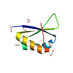 | | Crystal structure of fourth KH domain of FUBP1 | | 分子名称: | 1,2-ETHANEDIOL, Far upstream element-binding protein 1 | | 著者 | Ni, X, Joerger, A.C, Chaikuad, A, Arrowsmith, C.H, Edwards, A.M, Bountra, C, Knapp, S, Structural Genomics Consortium (SGC) | | 登録日 | 2020-02-14 | | 公開日 | 2020-03-25 | | 最終更新日 | 2024-01-24 | | 実験手法 | X-RAY DIFFRACTION (1.86 Å) | | 主引用文献 | Comparative structural analyses and nucleotide-binding characterization of the four KH domains of FUBP1.
Sci Rep, 10, 2020
|
|
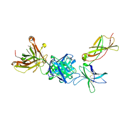
5GIJ
 | | Crystal structure of TDR-TDIF complex | | 分子名称: | 2-acetamido-2-deoxy-beta-D-glucopyranose, 2-acetamido-2-deoxy-beta-D-glucopyranose-(1-4)-2-acetamido-2-deoxy-beta-D-glucopyranose, Leucine-rich repeat receptor-like protein kinase TDR, ... | | 著者 | Morita, J, Kato, K, Ishitani, R, Nishimasu, H, Nureki, O. | | 登録日 | 2016-06-23 | | 公開日 | 2016-08-24 | | 最終更新日 | 2023-11-08 | | 実験手法 | X-RAY DIFFRACTION (3 Å) | | 主引用文献 | Crystal structure of the plant receptor-like kinase TDR in complex with the TDIF peptide
Nat Commun, 7, 2016
|
|
7KQ7
 | |
6FG1
 | |
8SA9
 | |
7Q3W
 | | Crystal structure of the C-terminal catalytic domain of Plasmodium falciparum CTP:phosphocholine cytidylyltransferase with (R)-2-Aminobutanamide hydrochloride | | 分子名称: | (R)-2-Aminobutanamide, Cholinephosphate cytidylyltransferase, Guanidinium | | 著者 | Duclovel, C, Gelin, M, Krimm, I, Cerdan, R, Guichou, J.-F. | | 登録日 | 2021-10-28 | | 公開日 | 2022-11-09 | | 最終更新日 | 2024-01-31 | | 実験手法 | X-RAY DIFFRACTION (1.75 Å) | | 主引用文献 | Crystallographic screening using ultra-low-molecular-weight ligands to guide drug design of PfCCT inhibitors.
To Be Published
|
|
5V91
 | | Crystal structure of fosfomycin resistance protein from Klebsiella pneumoniae | | 分子名称: | Fosfomycin resistance protein, ZINC ION | | 著者 | Klontz, E, Guenther, S, Silverstein, Z, Sundberg, E. | | 登録日 | 2017-03-22 | | 公開日 | 2017-08-30 | | 最終更新日 | 2023-10-04 | | 実験手法 | X-RAY DIFFRACTION (1.3 Å) | | 主引用文献 | Structure and Dynamics of FosA-Mediated Fosfomycin Resistance in Klebsiella pneumoniae and Escherichia coli.
Antimicrob. Agents Chemother., 61, 2017
|
|
6FBN
 | | Human Methionine Adenosyltransferase II mutant (Q113A) | | 分子名称: | (DIPHOSPHONO)AMINOPHOSPHONIC ACID, 1,2-ETHANEDIOL, DI(HYDROXYETHYL)ETHER, ... | | 著者 | Panmanee, J, Antonyuk, S.V, Hasnain, S.S. | | 登録日 | 2017-12-19 | | 公開日 | 2019-01-30 | | 最終更新日 | 2024-01-17 | | 実験手法 | X-RAY DIFFRACTION (2.7 Å) | | 主引用文献 | Control and regulation of S-Adenosylmethionine biosynthesis by the regulatory beta subunit and quinolone-based compounds.
Febs J., 286, 2019
|
|
6CCU
 | | Complex between a GID4 fragment and a short peptide | | 分子名称: | Glucose-induced degradation protein 4 homolog, Short peptide, UNKNOWN ATOM OR ION | | 著者 | Dong, C, Tempel, W, Bountra, C, Arrowsmith, C.H, Edwards, A.M, Min, J, Structural Genomics Consortium (SGC) | | 登録日 | 2018-02-07 | | 公開日 | 2018-03-07 | | 最終更新日 | 2023-10-04 | | 実験手法 | X-RAY DIFFRACTION (1.75 Å) | | 主引用文献 | Molecular basis of GID4-mediated recognition of degrons for the Pro/N-end rule pathway.
Nat. Chem. Biol., 14, 2018
|
|
7PX0
 | |
8SBY
 | | Crystal Structure of 2,3-dihydro-2,3-dihydroxybenzoate dehydrogenase from Klebsiella aerogenes (NAD and sulfate bound, hexagonal form) | | 分子名称: | 2,3-dihydroxybenzoate-2,3-dehydrogenase, CHLORIDE ION, NICOTINAMIDE-ADENINE-DINUCLEOTIDE, ... | | 著者 | Seattle Structural Genomics Center for Infectious Disease (SSGCID) | | 登録日 | 2023-04-04 | | 公開日 | 2023-04-12 | | 最終更新日 | 2024-05-22 | | 実験手法 | X-RAY DIFFRACTION (2.3 Å) | | 主引用文献 | Crystal Structure of 2,3-dihydro-2,3-dihydroxybenzoate dehydrogenase from Klebsiella aerogenes (NAD and sulfate bound, hexagonal form)
To be published
|
|
7KYL
 | | Powassan virus Envelope protein DIII in complex with neutralizing Fab POWV-80 | | 分子名称: | CHLORIDE ION, Envelope protein domain III, POWV-80 Fab heavy chain, ... | | 著者 | Errico, J.M, Nelson, C.A, Fremont, D.H, Center for Structural Genomics of Infectious Diseases (CSGID) | | 登録日 | 2020-12-08 | | 公開日 | 2021-04-07 | | 最終更新日 | 2023-10-18 | | 実験手法 | X-RAY DIFFRACTION (2 Å) | | 主引用文献 | Broadly neutralizing monoclonal antibodies protect against multiple tick-borne flaviviruses.
J.Exp.Med., 218, 2021
|
|
6PZN
 | |
8SA7
 | |
6JAQ
 | | Crystal structure of ABC transporter alpha-glycoside-binding mutant protein R356A in complex with glucose | | 分子名称: | 1,2-ETHANEDIOL, ABC transporter, periplasmic substrate-binding protein, ... | | 著者 | Kanaujia, S.P, Chandravanshi, M, Gogoi, P. | | 登録日 | 2019-01-24 | | 公開日 | 2019-10-30 | | 最終更新日 | 2023-11-22 | | 実験手法 | X-RAY DIFFRACTION (1.95 Å) | | 主引用文献 | Structural and thermodynamic correlation illuminates the selective transport mechanism of disaccharide alpha-glycosides through ABC transporter.
Febs J., 287, 2020
|
|
6FGT
 | | Crystal Structure of BAZ2B bromodomain in complex with 1-methylpyridinone compound 3 | | 分子名称: | Bromodomain adjacent to zinc finger domain protein 2B, ~{N}-[(1-azanylcyclohexyl)methyl]-1-methyl-6-oxidanylidene-pyridine-3-carboxamide | | 著者 | Dalle Vedove, A, Spiliotopoulos, D, Lolli, G, Caflisch, A. | | 登録日 | 2018-01-11 | | 公開日 | 2018-05-30 | | 最終更新日 | 2024-01-17 | | 実験手法 | X-RAY DIFFRACTION (2 Å) | | 主引用文献 | Structural Analysis of Small-Molecule Binding to the BAZ2A and BAZ2B Bromodomains.
ChemMedChem, 13, 2018
|
|


