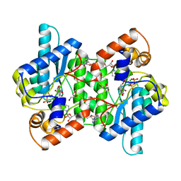
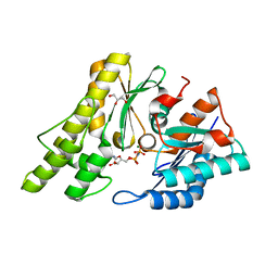
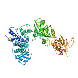
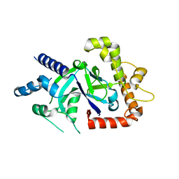
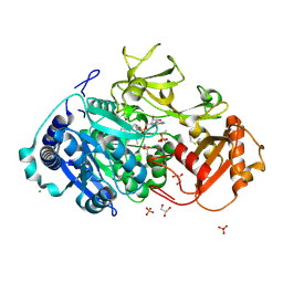
3U55
 
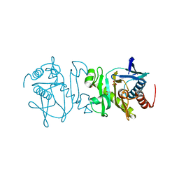 | | Crystal structure (Type-2) of SAICAR synthetase from Pyrococcus horikoshii OT3 | | 分子名称: | ACETATE ION, Phosphoribosylaminoimidazole-succinocarboxamide synthase, SULFATE ION | | 著者 | Manjunath, K, Kanaujia, S.P, Kanagaraj, S, Jeyakanthan, J, Sekar, K. | | 登録日 | 2011-10-11 | | 公開日 | 2012-10-17 | | 最終更新日 | 2023-11-01 | | 実験手法 | X-RAY DIFFRACTION (1.9 Å) | | 主引用文献 | Structure of SAICAR synthetase from Pyrococcus horikoshii OT3: insights into thermal stability
Int.J.Biol.Macromol., 53, 2013
|
|
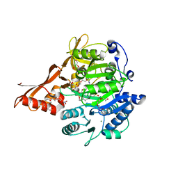
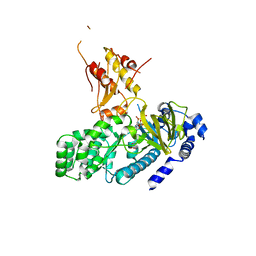
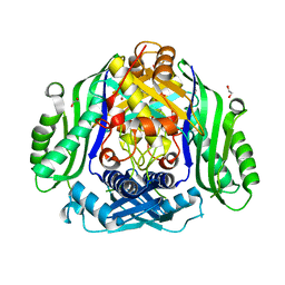
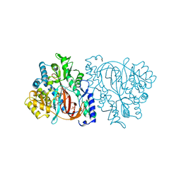
2R85
 | | Crystal structure of PurP from Pyrococcus furiosus complexed with AMP | | 分子名称: | (4S)-2-METHYL-2,4-PENTANEDIOL, ADENOSINE MONOPHOSPHATE, CHLORIDE ION, ... | | 著者 | Zhang, Y, White, R.H, Ealick, S.E. | | 登録日 | 2007-09-10 | | 公開日 | 2007-12-04 | | 最終更新日 | 2023-08-30 | | 実験手法 | X-RAY DIFFRACTION (1.7 Å) | | 主引用文献 | Crystal structure and function of 5-formaminoimidazole-4-carboxamide ribonucleotide synthetase from Methanocaldococcus jannaschii.
Biochemistry, 47, 2008
|
|
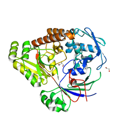
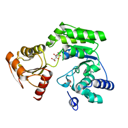
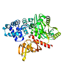
2C6I
 | | Crystal structure of the human CDK2 complexed with the triazolopyrimidine inhibitor | | 分子名称: | 4-{[5-(CYCLOHEXYLMETHOXY)[1,2,4]TRIAZOLO[1,5-A]PYRIMIDIN-7-YL]AMINO}BENZENESULFONAMIDE, CELL DIVISION PROTEIN KINASE 2 | | 著者 | Richardson, C.M, Dokurno, P, Murray, J.B, Surgenor, A.E. | | 登録日 | 2005-11-10 | | 公開日 | 2005-12-07 | | 最終更新日 | 2023-12-13 | | 実験手法 | X-RAY DIFFRACTION (1.8 Å) | | 主引用文献 | Triazolo[1,5-A]Pyrimidines as Novel Cdk2 Inhibitors: Protein Structure-Guided Design and Sar.
Bioorg.Med.Chem.Lett., 16, 2006
|
|
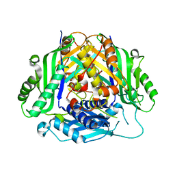
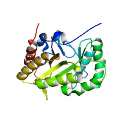
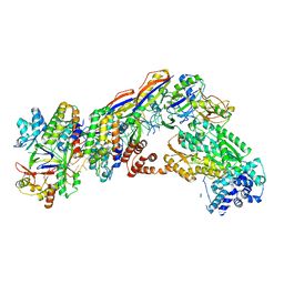
3W5O
 | | Crystal Structure of Human DNA ligase IV | | 分子名称: | ADENOSINE-5'-TRIPHOSPHATE, DNA ligase 4, SULFATE ION | | 著者 | Gu, X, Ochi, T, Blundell, T.L. | | 登録日 | 2013-02-02 | | 公開日 | 2013-04-03 | | 最終更新日 | 2023-11-08 | | 実験手法 | X-RAY DIFFRACTION (2.84 Å) | | 主引用文献 | Structure of the catalytic region of DNA ligase IV in complex with an artemis fragment sheds light on double-strand break repair
Structure, 21, 2013
|
|
2NSY
 | | CRYSTAL STRUCTURE OF NH3-DEPENDENT NAD+ SYNTHETASE FROM BACILLUS SUBTILIS IN COMPLEX WITH NAD-ADENYLATE | | 分子名称: | ADENOSINE MONOPHOSPHATE, GLYCEROL, MAGNESIUM ION, ... | | 著者 | Rizzi, M, Bolognesi, M, Coda, A. | | 登録日 | 1998-07-14 | | 公開日 | 1999-01-13 | | 最終更新日 | 2023-08-30 | | 実験手法 | X-RAY DIFFRACTION (2 Å) | | 主引用文献 | A novel deamido-NAD+-binding site revealed by the trapped NAD-adenylate intermediate in the NAD+ synthetase structure.
Structure, 6, 1998
|
|
3ETC
 | | 2.1 A structure of acyl-adenylate synthetase from Methanosarcina acetivorans containing a link between Lys256 and Cys298 | | 分子名称: | 4-(2-HYDROXYETHYL)-1-PIPERAZINE ETHANESULFONIC ACID, AMP-binding protein, FORMYL GROUP, ... | | 著者 | Shah, M.B, Gulick, A.M, Smith, K.S, Ingram-Smith, C. | | 登録日 | 2008-10-07 | | 公開日 | 2009-07-07 | | 最終更新日 | 2023-12-27 | | 実験手法 | X-RAY DIFFRACTION (2.1 Å) | | 主引用文献 | The 2.1 A crystal structure of an acyl-CoA synthetase from Methanosarcina acetivorans reveals an alternate acyl-binding pocket for small branched acyl substrates.
Proteins, 77, 2009
|
|
3HJ7
 | | Crystal structure of TILS C-terminal domain | | 分子名称: | CHLORIDE ION, tRNA(Ile)-lysidine synthase | | 著者 | Nakanishi, K, Bonnefond, L, Kimura, S, Suzuki, T, Ishitani, R, Nureki, O. | | 登録日 | 2009-05-21 | | 公開日 | 2009-10-20 | | 最終更新日 | 2023-11-01 | | 実験手法 | X-RAY DIFFRACTION (2.2 Å) | | 主引用文献 | Structural basis for translational fidelity ensured by transfer RNA lysidine synthetase.
Nature, 461, 2009
|
|
4DIT
 | |
2IVN
 | | Structure of UP1 protein | | 分子名称: | GLYCEROL, MAGNESIUM ION, O-SIALOGLYCOPROTEIN ENDOPEPTIDASE, ... | | 著者 | Hecker, A, Leulliot, N, Graille, M, Dorlet, P, Quevillon-Cheruel, S, Ulryck, N, Van Tilbeurgh, H, Forterre, P. | | 登録日 | 2006-06-14 | | 公開日 | 2007-07-31 | | 最終更新日 | 2024-05-08 | | 実験手法 | X-RAY DIFFRACTION (1.65 Å) | | 主引用文献 | An Archaeal Orthologue of the Universal Protein Kae1 is an Iron Metalloprotein which Exhibits Atypical DNA-Binding Properties and Apurinic-Endonuclease Activity in Vitro.
Nucleic Acids Res., 35, 2007
|
|
4L39
 | | Crystal structure of GH3.12 from Arabidopsis thaliana in complex with AMPCPP and salicylate | | 分子名称: | 2-HYDROXYBENZOIC ACID, 4-substituted benzoates-glutamate ligase GH3.12, DIPHOSPHOMETHYLPHOSPHONIC ACID ADENOSYL ESTER, ... | | 著者 | Zubieta, C, Jez, J.M, Brown, E, Marcellin, R, Kapp, U, Round, A, Westfall, C. | | 登録日 | 2013-06-05 | | 公開日 | 2013-10-02 | | 最終更新日 | 2023-09-20 | | 実験手法 | X-RAY DIFFRACTION (2.81 Å) | | 主引用文献 | Determination of the GH3.12 protein conformation through HPLC-integrated SAXS measurements combined with X-ray crystallography.
Acta Crystallogr.,Sect.D, 69, 2013
|
|
8UPI
 | | Structure of a periplasmic peptide binding protein from Mesorhizobium sp. AP09 bound to aminoserine | | 分子名称: | 1,2-ETHANEDIOL, AMINOSERINE, CALCIUM ION, ... | | 著者 | Frkic, R.L, Smith, O.B, Rahman, M, Kaczmarski, J.A, Jackson, C.J. | | 登録日 | 2023-10-22 | | 公開日 | 2023-11-08 | | 最終更新日 | 2024-06-05 | | 実験手法 | X-RAY DIFFRACTION (1.55 Å) | | 主引用文献 | Identification and Characterization of a Bacterial Periplasmic Solute Binding Protein That Binds l-Amino Acid Amides.
Biochemistry, 63, 2024
|
|
6SW6
 | |
3W1G
 | |
6SW5
 | | Crystal structure of the human S-adenosylmethionine synthetase 1 (ligand-free form) | | 分子名称: | 1,2-ETHANEDIOL, DI(HYDROXYETHYL)ETHER, S-adenosylmethionine synthase isoform type-1 | | 著者 | Panmanee, J, Antoyuk, S.V, Hasnain, S.S. | | 登録日 | 2019-09-19 | | 公開日 | 2020-06-17 | | 最終更新日 | 2024-01-24 | | 実験手法 | X-RAY DIFFRACTION (2.35 Å) | | 主引用文献 | Structural basis of the dominant inheritance of hypermethioninemia associated with the Arg264His mutation in the MAT1A gene.
Acta Crystallogr D Struct Biol, 76, 2020
|
|
6K8C
 | |
6UBD
 | | Crystal structure of a GH128 (subgroup VII) oligosaccharide-binding protein from Trichoderma gamsii (TgGH128_VII) | | 分子名称: | Glyco_hydro_cc domain-containing protein | | 著者 | Santos, C.R, Costa, P.A.C.R, Souza, B.P, Murakami, M.T. | | 登録日 | 2019-09-11 | | 公開日 | 2020-05-20 | | 最終更新日 | 2020-08-05 | | 実験手法 | X-RAY DIFFRACTION (1.25 Å) | | 主引用文献 | Structural insights into beta-1,3-glucan cleavage by a glycoside hydrolase family.
Nat.Chem.Biol., 16, 2020
|
|
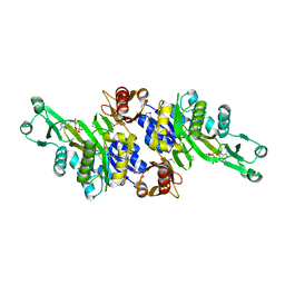
1I5O
 | | CRYSTAL STRUCTURE OF MUTANT R105A OF E. COLI ASPARTATE TRANSCARBAMOYLASE | | 分子名称: | ASPARTATE TRANSCARBAMOYLASE CATALYTIC CHAIN, ASPARTATE TRANSCARBAMOYLASE REGULATORY CHAIN, N-(PHOSPHONACETYL)-L-ASPARTIC ACID, ... | | 著者 | Macol, C.P, Tsuruta, H, Stec, B, Kantrowitz, E.R. | | 登録日 | 2001-02-28 | | 公開日 | 2001-05-02 | | 最終更新日 | 2023-08-09 | | 実験手法 | X-RAY DIFFRACTION (2.8 Å) | | 主引用文献 | Direct structural evidence for a concerted allosteric transition in Escherichia coli aspartate transcarbamoylase.
Nat.Struct.Biol., 8, 2001
|
|
5FO4
 | | Crystal structure of the P.falciparum cytosolic leucyl-tRNA synthetase editing domain (space group P1) | | 分子名称: | LEUCYL TRNA SYNTHASE | | 著者 | Palencia, A, Sonoiki, E, Guo, D, Ahyong, V, Dong, C, Li, X, Hernandez, V.S, Gut, J, Legac, J, Cooper, R, Alley, M.R.K, Freund, Y.R, DeRisi, J, Cusack, S, Rosenthal, P.J. | | 登録日 | 2015-11-18 | | 公開日 | 2016-06-22 | | 最終更新日 | 2024-05-01 | | 実験手法 | X-RAY DIFFRACTION (1.85 Å) | | 主引用文献 | Anti-Malarial Benzoxaboroles Target P. Falciparum Leucyl-tRNA Synthetase.
Antimicrob.Agents Chemother., 60, 2016
|
|
2HGS
 | | HUMAN GLUTATHIONE SYNTHETASE | | 分子名称: | ADENOSINE-5'-DIPHOSPHATE, GLUTATHIONE, MAGNESIUM ION, ... | | 著者 | Polekhina, G, Board, P, Rossjohn, J, Parker, M.W. | | 登録日 | 1999-01-04 | | 公開日 | 1999-06-22 | | 最終更新日 | 2023-12-27 | | 実験手法 | X-RAY DIFFRACTION (2.1 Å) | | 主引用文献 | Molecular basis of glutathione synthetase deficiency and a rare gene permutation event.
EMBO J., 18, 1999
|
|
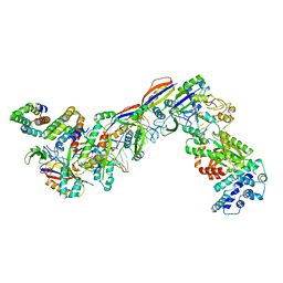
4EQ5
 | | DNA ligase from the archaeon Thermococcus sibiricus | | 分子名称: | ADENOSINE MONOPHOSPHATE, DNA ligase | | 著者 | Petrova, T, Bezsudnova, E.Y, Dorokhov, B.D, Slutskaya, E.S, Polyakov, K.M, Dorovatovskiy, P.V, Ravin, N.V, Skryabin, K.G, Kovalchuk, M.V, Popov, V.O. | | 登録日 | 2012-04-18 | | 公開日 | 2012-05-09 | | 最終更新日 | 2023-09-13 | | 実験手法 | X-RAY DIFFRACTION (2.85 Å) | | 主引用文献 | Expression, purification, crystallization and preliminary crystallographic analysis of a thermostable DNA ligase from the archaeon Thermococcus sibiricus.
Acta Crystallogr.,Sect.F, 68, 2012
|
|
5HM3
 | | 2.25 Angstrom Resolution Crystal Structure of Long-chain-fatty-acid-AMP Ligase FadD32 from Mycobacterium tuberculosis in complex with Inhibitor 5'-O-[(11-phenoxyundecanoyl)sulfamoyl]adenosine | | 分子名称: | 5'-O-[(11-phenoxyundecanoyl)sulfamoyl]adenosine, CHLORIDE ION, DI(HYDROXYETHYL)ETHER, ... | | 著者 | Minasov, G, Warwrzak, Z, Kuhn, M.L, Shuvalova, L, Flores, K.J, Wilson, D.J, Grimes, K.D, Aldrich, C.C, Anderson, W.A, Center for Structural Genomics of Infectious Diseases (CSGID) | | 登録日 | 2016-01-15 | | 公開日 | 2016-08-03 | | 最終更新日 | 2016-09-07 | | 実験手法 | X-RAY DIFFRACTION (2.25 Å) | | 主引用文献 | Structure of the Essential Mtb FadD32 Enzyme: A Promising Drug Target for Treating Tuberculosis.
Acs Infect Dis., 2, 2016
|
|
3AX6
 | | Crystal structure of N5-carboxyaminoimidazole ribonucleotide synthetase from Thermotoga maritima | | 分子名称: | ADENOSINE-5'-DIPHOSPHATE, Phosphoribosylaminoimidazole carboxylase, ATPase subunit | | 著者 | Miyazawa, R, Kanagawa, M, Baba, S, Nakagawa, N, Ebihara, A, Kuramitsu, S, Yokoyama, S, Kawai, G, Sampei, G, RIKEN Structural Genomics/Proteomics Initiative (RSGI) | | 登録日 | 2011-03-30 | | 公開日 | 2012-04-25 | | 実験手法 | X-RAY DIFFRACTION (2.2 Å) | | 主引用文献 | Crystal structures of N5-carboxyaminoimidazole ribonucleotide synthetase, PurK, from thermophilic bacteria
To be Published
|
|
7UZY
 | | Staphylococcus epidermidis RP62A CRISPR effector complex with non-self target RNA 2 | | 分子名称: | CRISPR system Cms endoribonuclease Csm3, CRISPR system Cms protein Csm2, CRISPR system Cms protein Csm4, ... | | 著者 | Smith, E.M, Ferrell, S.H, Tokars, V.L, Mondragon, A. | | 登録日 | 2022-05-09 | | 公開日 | 2022-07-06 | | 最終更新日 | 2022-08-17 | | 実験手法 | ELECTRON MICROSCOPY (4.05 Å) | | 主引用文献 | Structures of an active type III-A CRISPR effector complex.
Structure, 30, 2022
|
|
7V02
 | | Staphylococcus epidermidis RP62A CRISPR short effector complex | | 分子名称: | CRISPR system Cms endoribonuclease Csm3, CRISPR system Cms protein Csm2, CRISPR system Cms protein Csm4, ... | | 著者 | Smith, E.M, Ferrell, S.H, Tokars, V.L, Mondragon, A. | | 登録日 | 2022-05-09 | | 公開日 | 2022-07-06 | | 最終更新日 | 2022-08-17 | | 実験手法 | ELECTRON MICROSCOPY (4.97 Å) | | 主引用文献 | Structures of an active type III-A CRISPR effector complex.
Structure, 30, 2022
|
|
7UZZ
 | | Staphylococcus epidermidis RP62a CRISPR tall effector complex | | 分子名称: | CRISPR system Cms endoribonuclease Csm3, CRISPR system Cms protein Csm2, CRISPR system Cms protein Csm4, ... | | 著者 | Smith, E.M, Ferrell, S.H, Tokars, V.L, Mondragon, A. | | 登録日 | 2022-05-09 | | 公開日 | 2022-07-06 | | 最終更新日 | 2022-08-17 | | 実験手法 | ELECTRON MICROSCOPY (4.45 Å) | | 主引用文献 | Structures of an active type III-A CRISPR effector complex.
Structure, 30, 2022
|
|


