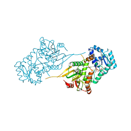
6V52
 
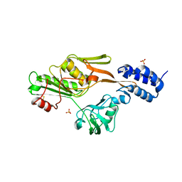
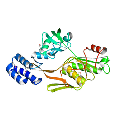
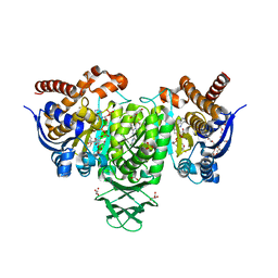
 | | IDO1 IN COMPLEX WITH COMPOUND 1 | | 分子名称: | 3-chloro-N-{4-[1-(propylcarbamoyl)cyclobutyl]phenyl}benzamide, Indoleamine 2,3-dioxygenase 1 | | 著者 | Lesburg, C.A, Koenig, K.V, Augustin, M.A. | | 登録日 | 2019-12-03 | | 公開日 | 2020-04-08 | | 最終更新日 | 2020-04-29 | | 実験手法 | X-RAY DIFFRACTION (1.78 Å) | | 主引用文献 | Strategic Incorporation of Polarity in Heme-Displacing Inhibitors of Indoleamine-2,3-dioxygenase-1 (IDO1).
Acs Med.Chem.Lett., 11, 2020
|
|
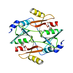
5BVU
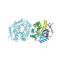
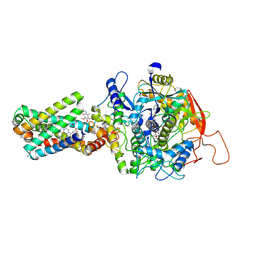
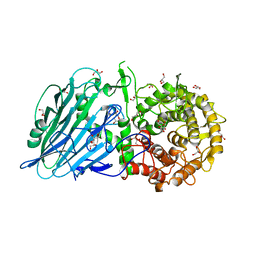 | | Crystal structure of Thermoanaerobacterium xylolyticum GH116 beta-glucosidase | | 分子名称: | 1,2-ETHANEDIOL, CALCIUM ION, GLYCEROL, ... | | 著者 | Charoenwattanasatien, R, Pengthaisong, S, Sansenya, S, Mutoh, R, Tanaka, H, Kurisu, G, Ketudat Cairns, J.R. | | 登録日 | 2015-06-05 | | 公開日 | 2016-05-18 | | 最終更新日 | 2024-03-20 | | 実験手法 | X-RAY DIFFRACTION (1.61 Å) | | 主引用文献 | Bacterial beta-Glucosidase Reveals the Structural and Functional Basis of Genetic Defects in Human Glucocerebrosidase 2 (GBA2).
Acs Chem.Biol., 11, 2016
|
|
6B4C
 | |
6V2M
 | |
6VA9
 | |
5C0P
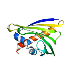
 | | The crystal structure of endo-arabinase from Bacteroides thetaiotaomicron VPI-5482 | | 分子名称: | 2-AMINO-2-HYDROXYMETHYL-PROPANE-1,3-DIOL, CHLORIDE ION, Endo-arabinase, ... | | 著者 | Tan, K, Cuff, M, Joachimiak, G, Endres, M, Joachimiak, A, Midwest Center for Structural Genomics (MCSG) | | 登録日 | 2015-06-12 | | 公開日 | 2015-07-01 | | 最終更新日 | 2019-12-25 | | 実験手法 | X-RAY DIFFRACTION (1.532 Å) | | 主引用文献 | The crystal structure of endo-arabinase from Bacteroides thetaiotaomicron VPI-5482
To Be Published
|
|
4Y0L
 | | Mycobacterial membrane protein MmpL11D2 | | 分子名称: | IODIDE ION, Putative membrane protein mmpL11, SULFATE ION | | 著者 | Torres, R, Chim, N, Goulding, C.W. | | 登録日 | 2015-02-06 | | 公開日 | 2015-08-12 | | 最終更新日 | 2024-02-28 | | 実験手法 | X-RAY DIFFRACTION (2.4 Å) | | 主引用文献 | The Structure and Interactions of Periplasmic Domains of Crucial MmpL Membrane Proteins from Mycobacterium tuberculosis.
Chem.Biol., 22, 2015
|
|
4C6C
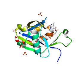
 | | Crystal structure of the dihydroorotase domain of human CAD in apo- form obtained recombinantly from HEK293 cells. | | 分子名称: | CAD PROTEIN, FORMIC ACID, ZINC ION | | 著者 | Ramon-Maiques, S, Lallous, N, Grande-Garcia, A. | | 登録日 | 2013-09-18 | | 公開日 | 2014-01-08 | | 最終更新日 | 2023-12-20 | | 実験手法 | X-RAY DIFFRACTION (1.451 Å) | | 主引用文献 | Structure, Functional Characterization and Evolution of the Dihydroorotase Domain of Human Cad.
Structure, 22, 2014
|
|
4Y0X
 | |
5C3J
 | | Crystal structure of Mitochondrial rhodoquinol-fumarate reductase from Ascaris suum with Ubiquinone-1 | | 分子名称: | Cytochrome b-large subunit, FE2/S2 (INORGANIC) CLUSTER, FE3-S4 CLUSTER, ... | | 著者 | Harada, S, Shiba, T, Sato, D, Yamamoto, A, Nagahama, M, Yone, A, Inaoka, D.K, Sakamoto, K, Inoue, M, Honma, T, Kita, K. | | 登録日 | 2015-06-17 | | 公開日 | 2016-06-22 | | 最終更新日 | 2023-11-08 | | 実験手法 | X-RAY DIFFRACTION (2.8 Å) | | 主引用文献 | Structural Insights into the Molecular Design of Flutolanil Derivatives Targeted for Fumarate Respiration of Parasite Mitochondria
To Be Published
|
|
1FCF
 | | PHOTOSYSTEM II D1 C-TERMINAL PROCESSING PROTEASE | | 分子名称: | PHOTOSYSTEM II D1 PROTEASE, SULFATE ION | | 著者 | Liao, D.I, Qian, J, Chisholm, D.A, Jordan, D.B, Diner, B.A. | | 登録日 | 2000-07-18 | | 公開日 | 2001-01-18 | | 最終更新日 | 2018-10-10 | | 実験手法 | X-RAY DIFFRACTION (2.1 Å) | | 主引用文献 | Crystal structures of the photosystem II D1 C-terminal processing protease.
Nat.Struct.Biol., 7, 2000
|
|
1FRO
 | |
6UTE
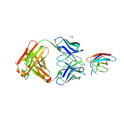 | | Crystal structure of Z032 Fab in complex with WNV EDIII | | 分子名称: | Envelope domain III, GLYCEROL, Z032 Fab heavy chain, ... | | 著者 | Esswein, S.R, Gristick, H.B, Keeffe, J.R, Bjorkman, P.J. | | 登録日 | 2019-10-29 | | 公開日 | 2020-04-15 | | 最終更新日 | 2023-10-11 | | 実験手法 | X-RAY DIFFRACTION (2.9 Å) | | 主引用文献 | Structural basis for Zika envelope domain III recognition by a germline version of a recurrent neutralizing antibody.
Proc.Natl.Acad.Sci.USA, 117, 2020
|
|
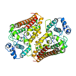
2DWU
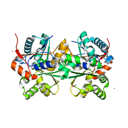 | | Crystal Structure of Glutamate Racemase Isoform RacE1 from Bacillus anthracis | | 分子名称: | D-GLUTAMIC ACID, GLYCEROL, Glutamate racemase, ... | | 著者 | Mehboob, S, Santarsiero, B.D, Johnson, M.E. | | 登録日 | 2006-08-17 | | 公開日 | 2007-06-19 | | 最終更新日 | 2023-10-25 | | 実験手法 | X-RAY DIFFRACTION (1.6 Å) | | 主引用文献 | Structural and Functional Analysis of Two Glutamate Racemase Isozymes from Bacillus anthracis and Implications for Inhibitor Design
J.Mol.Biol., 371, 2007
|
|
1FC6
 | | PHOTOSYSTEM II D1 C-TERMINAL PROCESSING PROTEASE | | 分子名称: | PHOTOSYSTEM II D1 PROTEASE | | 著者 | Liao, D.I, Qian, J, Chisholm, D.A, Jordan, D.B, Diner, B.A. | | 登録日 | 2000-07-18 | | 公開日 | 2001-01-18 | | 最終更新日 | 2021-11-03 | | 実験手法 | X-RAY DIFFRACTION (1.8 Å) | | 主引用文献 | Crystal structures of the photosystem II D1 C-terminal processing protease.
Nat.Struct.Biol., 7, 2000
|
|
6BAF
 | | Structure of the chromophore binding domain of Stigmatella aurantiaca phytochrome P1, wild-type | | 分子名称: | 3-[5-[(Z)-(4-ethenyl-3-methyl-5-oxidanylidene-pyrrol-2-ylidene)methyl]-2-[[5-[(Z)-(3-ethenyl-4-methyl-5-oxidanylidene-pyrrol-2-ylidene)methyl]-3-(3-hydroxy-3-oxopropyl)-4-methyl-1H-pyrrol-2-yl]methyl]-4-methyl-1H-pyrrol-3-yl]propanoic acid, Photoreceptor-histidine kinase BphP | | 著者 | Schmidt, M, Stojkovic, E. | | 登録日 | 2017-10-12 | | 公開日 | 2018-09-19 | | 最終更新日 | 2023-10-04 | | 実験手法 | X-RAY DIFFRACTION (1.85 Å) | | 主引用文献 | Structural basis for light control of cell development revealed by crystal structures of a myxobacterial phytochrome.
IUCrJ, 5, 2018
|
|
4C6O
 | | Crystal structure of the dihydroorotase domain of human CAD C1613S mutant in apo-form at pH 6.0 | | 分子名称: | CAD PROTEIN, FORMIC ACID, ZINC ION | | 著者 | Ramon-Maiques, S, Lallous, N, Grande-Garcia, A. | | 登録日 | 2013-09-18 | | 公開日 | 2014-02-05 | | 最終更新日 | 2023-12-20 | | 実験手法 | X-RAY DIFFRACTION (1.65 Å) | | 主引用文献 | Structure, Functional Characterization and Evolution of the Dihydroorotase Domain of Human Cad.
Structure, 22, 2014
|
|
6BAP
 | | Stigmatella aurantiaca bacterial phytochrome PAS-GAF-PHY, T289H mutant | | 分子名称: | 3-[5-[(Z)-(4-ethenyl-3-methyl-5-oxidanylidene-pyrrol-2-ylidene)methyl]-2-[[5-[(Z)-(3-ethenyl-4-methyl-5-oxidanylidene-pyrrol-2-ylidene)methyl]-3-(3-hydroxy-3-oxopropyl)-4-methyl-1H-pyrrol-2-yl]methyl]-4-methyl-1H-pyrrol-3-yl]propanoic acid, Photoreceptor-histidine kinase BphP | | 著者 | Schmidt, M, Stojkovic, E. | | 登録日 | 2017-10-14 | | 公開日 | 2018-09-19 | | 最終更新日 | 2023-10-04 | | 実験手法 | X-RAY DIFFRACTION (2.65 Å) | | 主引用文献 | Structural basis for light control of cell development revealed by crystal structures of a myxobacterial phytochrome.
IUCrJ, 5, 2018
|
|
5C9Y
 | |
6BAY
 | |
6VCQ
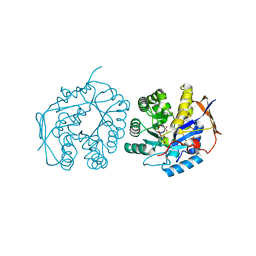
 | | Crystal structure of E.coli RppH in complex with GTP | | 分子名称: | GUANOSINE-5'-TRIPHOSPHATE, RNA pyrophosphohydrolase, SULFATE ION | | 著者 | Gao, A, Vasilyev, N, Kaushik, A, Duan, W, Serganov, A. | | 登録日 | 2019-12-21 | | 公開日 | 2020-02-05 | | 最終更新日 | 2023-10-11 | | 実験手法 | X-RAY DIFFRACTION (1.6 Å) | | 主引用文献 | Principles of RNA and nucleotide discrimination by the RNA processing enzyme RppH.
Nucleic Acids Res., 48, 2020
|
|
6VEI
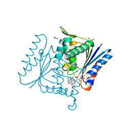
 | | Crystal Structure of Human Cytosolic Isocitrate Dehydrogenase (IDH1) R132H Mutant in Complex with NADPH and AG-881 (Vorasidenib) Inhibitor | | 分子名称: | 3,6,9,12,15,18,21,24,27,30,33,36-dodecaoxaoctatriacontane-1,38-diol, 6-(6-chloropyridin-2-yl)-N2,N4-bis[(2R)-1,1,1-trifluoropropan-2-yl]-1,3,5-triazine-2,4-diamine, ACETATE ION, ... | | 著者 | Padyana, A, Jin, L. | | 登録日 | 2020-01-02 | | 公開日 | 2020-02-05 | | 最終更新日 | 2023-10-11 | | 実験手法 | X-RAY DIFFRACTION (2.1 Å) | | 主引用文献 | Vorasidenib (AG-881): A First-in-Class, Brain-Penetrant Dual Inhibitor of Mutant IDH1 and 2 for Treatment of Glioma.
Acs Med.Chem.Lett., 11, 2020
|
|
8GS8
 | | cryo-EM structure of the human respiratory complex II | | 分子名称: | (1S)-2-{[(2-AMINOETHOXY)(HYDROXY)PHOSPHORYL]OXY}-1-[(PALMITOYLOXY)METHYL]ETHYL STEARATE, FE2/S2 (INORGANIC) CLUSTER, FE3-S4 CLUSTER, ... | | 著者 | Du, Z, Zhou, X, Lai, Y, Xu, J, Zhang, Y, Zhou, S, Liu, F, Gao, Y, Gong, H, Rao, Z. | | 登録日 | 2022-09-05 | | 公開日 | 2023-05-10 | | 最終更新日 | 2024-07-03 | | 実験手法 | ELECTRON MICROSCOPY (2.86 Å) | | 主引用文献 | Structure of the human respiratory complex II.
Proc.Natl.Acad.Sci.USA, 120, 2023
|
|
4C6N
 | | Crystal structure of the dihydroorotase domain of human CAD E1637T mutant bound to substrate at pH 6.0 | | 分子名称: | CAD PROTEIN, FORMIC ACID, N-CARBAMOYL-L-ASPARTATE, ... | | 著者 | Ramon-Maiques, S, Lallous, N, Grande-Garcia, A. | | 登録日 | 2013-09-18 | | 公開日 | 2014-02-05 | | 最終更新日 | 2023-12-20 | | 実験手法 | X-RAY DIFFRACTION (1.899 Å) | | 主引用文献 | Structure, Functional Characterization and Evolution of the Dihydroorotase Domain of Human Cad.
Structure, 22, 2014
|
|
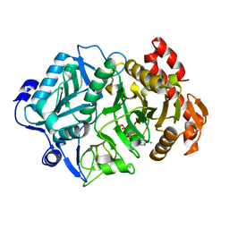
4C0X
 | | The crystal strucuture of PpAzoR in complex with anthraquinone-2- sulfonate | | 分子名称: | 9,10-dioxo-9,10-dihydroanthracene-2-sulfonic acid, FLAVIN MONONUCLEOTIDE, FMN-DEPENDENT NADH-AZOREDUCTASE 1, ... | | 著者 | Goncalves, A.M.D, de Sanctis, D, Bento, I. | | 登録日 | 2013-08-08 | | 公開日 | 2013-10-30 | | 最終更新日 | 2023-12-20 | | 実験手法 | X-RAY DIFFRACTION (1.499 Å) | | 主引用文献 | The Crystal Structure of Pseudomonas Putida Azor: The Active Site Revisited.
FEBS J., 280, 2013
|
|


