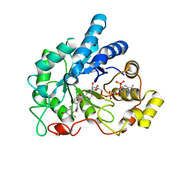
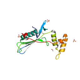
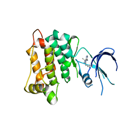
4LDG
 
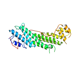
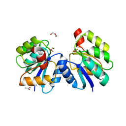
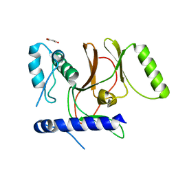 | | Crystal Structure of CpSET8 from Cryptosporidium, cgd4_370 | | 分子名称: | MALONATE ION, Protein with a SET domain within carboxy region | | 著者 | Wernimont, A.K, Tempel, W, Loppnau, P, Bountra, C, Arrowsmith, C.H, Edwards, A.M, Hui, R, El Bakkouri, M, Structural Genomics Consortium (SGC) | | 登録日 | 2013-06-24 | | 公開日 | 2013-07-24 | | 最終更新日 | 2024-02-28 | | 実験手法 | X-RAY DIFFRACTION (2.31 Å) | | 主引用文献 | Crystal Structure of CpSET8 from Cryptosporidium, cgd4_370
To be Published
|
|
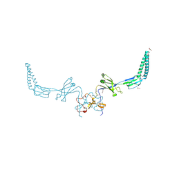
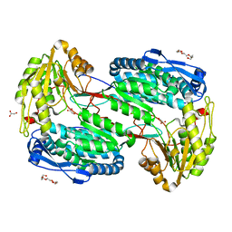
4LGM
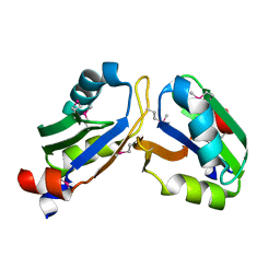
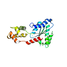 | | Crystal Structure of Sulfolobus solfataricus Vps4 | | 分子名称: | CHLORIDE ION, Vps4 AAA ATPase | | 著者 | Han, H, Hill, C.P, Whitby, F.G, Monroe, N. | | 登録日 | 2013-06-28 | | 公開日 | 2013-11-06 | | 最終更新日 | 2024-02-28 | | 実験手法 | X-RAY DIFFRACTION (2.711 Å) | | 主引用文献 | The Oligomeric State of the Active Vps4 AAA ATPase.
J.Mol.Biol., 426, 2014
|
|
4LGZ
 | |
4LGQ
 | |
4LIU
 | | Structure of YcfD, a Ribosomal oxygenase from Escherichia coli. | | 分子名称: | 50S ribosomal protein L16 arginine hydroxylase, PHOSPHATE ION, TRIS(HYDROXYETHYL)AMINOMETHANE | | 著者 | Brissett, N.C, Doherty, A.J, Fox, G.C. | | 登録日 | 2013-07-03 | | 公開日 | 2014-05-14 | | 最終更新日 | 2024-11-27 | | 実験手法 | X-RAY DIFFRACTION (2.7 Å) | | 主引用文献 | Ribosomal oxygenases are structurally conserved from prokaryotes to humans.
Nature, 509, 2014
|
|
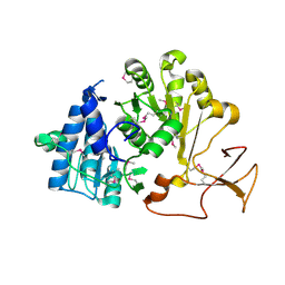
4LMU
 | | Crystal structure of Pim1 in complex with the inhibitor Quercetin (resulting from displacement of SKF86002) | | 分子名称: | 3,5,7,3',4'-PENTAHYDROXYFLAVONE, GLYCEROL, Serine/threonine-protein kinase pim-1 | | 著者 | Parker, L.J, Tanaka, A, Handa, N, Honda, K, Tomabechi, Y, Shirouzu, M, Yokoyama, S. | | 登録日 | 2013-07-11 | | 公開日 | 2014-02-12 | | 最終更新日 | 2023-11-08 | | 実験手法 | X-RAY DIFFRACTION (2.38 Å) | | 主引用文献 | Kinase crystal identification and ATP-competitive inhibitor screening using the fluorescent ligand SKF86002.
Acta Crystallogr.,Sect.D, 70, 2014
|
|
4LNZ
 | |
4L8J
 | |
4L9T
 | |
4LBA
 | |
4LE0
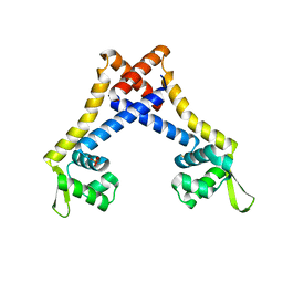
 | | Crystal structure of the receiver domain of DesR in complex with beryllofluoride and magnesium | | 分子名称: | ACETATE ION, BERYLLIUM TRIFLUORIDE ION, GLYCEROL, ... | | 著者 | Trajtenberg, F, Larrieux, N, Buschiazzo, A. | | 登録日 | 2013-06-25 | | 公開日 | 2014-07-16 | | 最終更新日 | 2024-02-28 | | 実験手法 | X-RAY DIFFRACTION (2.27 Å) | | 主引用文献 | Allosteric activation of bacterial response regulators: the role of the cognate histidine kinase beyond phosphorylation.
MBio, 5, 2014
|
|
4LIR
 | |
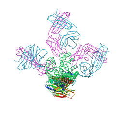
4V55
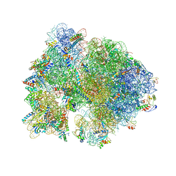 | | Crystal structure of the bacterial ribosome from Escherichia coli in complex with gentamicin and ribosome recycling factor (RRF). | | 分子名称: | (2R,3R,4R,5R)-2-((1S,2S,3R,4S,6R)-4,6-DIAMINO-3-((2R,3R,6S)-3-AMINO-6-(AMINOMETHYL)-TETRAHYDRO-2H-PYRAN-2-YLOXY)-2-HYDR OXYCYCLOHEXYLOXY)-5-METHYL-4-(METHYLAMINO)-TETRAHYDRO-2H-PYRAN-3,5-DIOL, 16S rRNA, 23S rRNA, ... | | 著者 | Borovinskaya, M.A, Pai, R.D, Zhang, W, Schuwirth, B.-S, Holton, J.M, Hirokawa, G, Kaji, H, Kaji, A, Cate, J.H.D. | | 登録日 | 2007-06-17 | | 公開日 | 2014-07-09 | | 最終更新日 | 2023-09-20 | | 実験手法 | X-RAY DIFFRACTION (4 Å) | | 主引用文献 | Structural basis for aminoglycoside inhibition of bacterial ribosome recycling.
Nat.Struct.Mol.Biol., 14, 2007
|
|
4LCF
 | | Crystal structure of the Pseudomonas aeruginosa LPXC/LPC-014 complex | | 分子名称: | NITRATE ION, Nalpha-{4-[4-(4-aminophenyl)buta-1,3-diyn-1-yl]benzoyl}-N-hydroxy-L-histidinamide, UDP-3-O-[3-hydroxymyristoyl] N-acetylglucosamine deacetylase, ... | | 著者 | Lee, C.-J, Zhou, P. | | 登録日 | 2013-06-21 | | 公開日 | 2013-08-21 | | 最終更新日 | 2024-02-28 | | 実験手法 | X-RAY DIFFRACTION (1.599 Å) | | 主引用文献 | Synthesis, Structure, and Antibiotic Activity of Aryl-Substituted LpxC Inhibitors.
J.Med.Chem., 56, 2013
|
|
4LBS
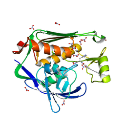
 | | Crystal structure of human AR complexed with NADP+ and {2-[(4-bromo-2,6-difluorobenzyl)carbamoyl]-5-chlorophenoxy}acetic acid | | 分子名称: | Aldose reductase, NADP NICOTINAMIDE-ADENINE-DINUCLEOTIDE PHOSPHATE, {2-[(4-bromo-2,6-difluorobenzyl)carbamoyl]-5-chlorophenoxy}acetic acid | | 著者 | Cousido-Siah, A, Mitschler, A, Ruiz, F.X, Fanfrlik, J, Kolar, M, Hobza, P, Podjarny, A. | | 登録日 | 2013-06-21 | | 公開日 | 2014-04-30 | | 最終更新日 | 2023-09-20 | | 実験手法 | X-RAY DIFFRACTION (0.76 Å) | | 主引用文献 | Modulation of aldose reductase inhibition by halogen bond tuning.
Acs Chem.Biol., 8, 2013
|
|
4LCU
 | | Structure of KcsA with E118A mutation | | 分子名称: | DIACYL GLYCEROL, Fab heavy chain, Fab light chain, ... | | 著者 | Nimigean, C.M, Posson, D.J, McCoy, J.M. | | 登録日 | 2013-06-23 | | 公開日 | 2013-10-30 | | 最終更新日 | 2024-11-20 | | 実験手法 | X-RAY DIFFRACTION (2.752 Å) | | 主引用文献 | Molecular interactions involved in proton-dependent gating in KcsA potassium channels.
J.Gen.Physiol., 142, 2013
|
|
4LFL
 | |
4LFS
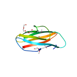
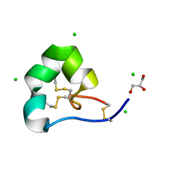 | | High resolution x-ray structure of racemic ShK toxin | | 分子名称: | CHLORIDE ION, GLYCEROL, Potassium channel toxin ShK | | 著者 | Dang, B, Kubota, T, Mandal, K, Bezanilla, F, Kent, S.B.H. | | 登録日 | 2013-06-27 | | 公開日 | 2013-08-14 | | 最終更新日 | 2024-11-20 | | 実験手法 | X-RAY DIFFRACTION (0.97 Å) | | 主引用文献 | Native Chemical Ligation at Asx-Cys, Glx-Cys: Chemical Synthesis and High-Resolution X-ray Structure of ShK Toxin by Racemic Protein Crystallography.
J.Am.Chem.Soc., 135, 2013
|
|
4LG3
 | |
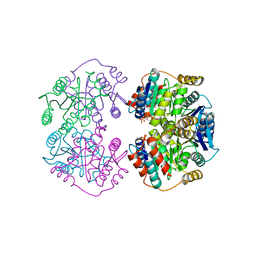
4LF1
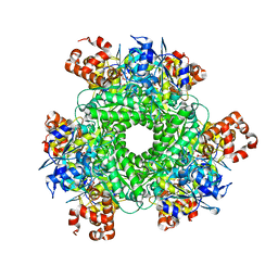 | | Hexameric Form II RuBisCO from Rhodopseudomonas palustris, activated and complexed with 2-CABP | | 分子名称: | 2-CARBOXYARABINITOL-1,5-DIPHOSPHATE, MAGNESIUM ION, Ribulose bisphosphate carboxylase | | 著者 | Chan, S, Satagopan, S, Sawaya, M.R, Eisenberg, D, Tabita, F.R, Perry, L.J. | | 登録日 | 2013-06-26 | | 公開日 | 2014-06-25 | | 最終更新日 | 2025-03-26 | | 実験手法 | X-RAY DIFFRACTION (2.38 Å) | | 主引用文献 | Structure-function studies with the unique hexameric form II ribulose-1,5-bisphosphate carboxylase/oxygenase (Rubisco) from Rhodopseudomonas palustris.
J.Biol.Chem., 289, 2014
|
|
4LFK
 | |
4LFQ
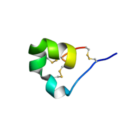 | | High resolution x-ray crystal structure of L-ShK toxin | | 分子名称: | Potassium channel toxin ShK | | 著者 | Dang, B, Kubota, T, Mandal, K, Bezanilla, F, Kent, S.B.H. | | 登録日 | 2013-06-27 | | 公開日 | 2013-08-14 | | 最終更新日 | 2024-11-20 | | 実験手法 | X-RAY DIFFRACTION (1.059 Å) | | 主引用文献 | Native Chemical Ligation at Asx-Cys, Glx-Cys: Chemical Synthesis and High-Resolution X-ray Structure of ShK Toxin by Racemic Protein Crystallography.
J.Am.Chem.Soc., 135, 2013
|
|
4LGC
 | |
4LGG
 | |
4LH0
 | |


