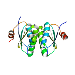
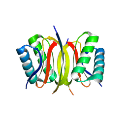
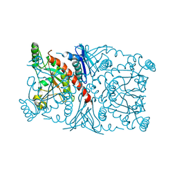
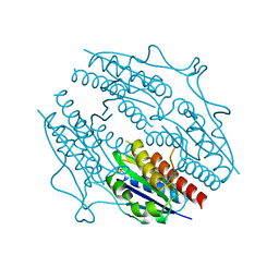
4QHF
 
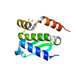 | | Crystal structure of Methanocaldococcus jannaschii monomeric selecase | | 分子名称: | GLYCEROL, NICKEL (II) ION, Uncharacterized protein MJ1213 | | 著者 | Lopez-pelegrin, M, Cerda-costa, N, Cintas-pedrola, A, Herranz-trillo, F, Bernado, P, Peinado, J.R, Arolas, J.L, Gomis-ruth, F.X. | | 登録日 | 2014-05-28 | | 公開日 | 2014-07-16 | | 最終更新日 | 2024-04-03 | | 実験手法 | X-RAY DIFFRACTION (2.1 Å) | | 主引用文献 | Multiple stable conformations account for reversible concentration-dependent oligomerization and autoinhibition of a metamorphic metallopeptidase
Angew.Chem.Int.Ed.Engl., 53, 2014
|
|
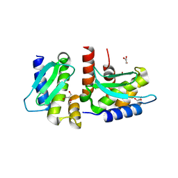
1E92
 | | Pteridine reductase 1 from Leishmania major complexed with NADP+ and dihydrobiopterin | | 分子名称: | 1,2-ETHANEDIOL, 7,8-DIHYDROBIOPTERIN, NADP NICOTINAMIDE-ADENINE-DINUCLEOTIDE PHOSPHATE, ... | | 著者 | Schuettelkopf, A.W, Hunter, W.N. | | 登録日 | 2000-10-04 | | 公開日 | 2001-10-05 | | 最終更新日 | 2023-12-13 | | 実験手法 | X-RAY DIFFRACTION (2.2 Å) | | 主引用文献 | Pteridine Reductase Mechanism Correlates Pterin Metabolism with Drug Resistance in Trypanosomatid Parasites
Nat.Struct.Biol., 8, 2001
|
|
1TTB
 | |
5EKE
 | | Structure of the polyisoprenyl-phosphate glycosyltransferase GtrB (F215A mutant) | | 分子名称: | MAGNESIUM ION, URIDINE-5'-DIPHOSPHATE, Uncharacterized glycosyltransferase sll0501 | | 著者 | Ardiccioni, C, Clarke, O.B, Tomasek, D, Banerjee, S, Rajashankar, K.R, Liu, Q, Shapiro, L, Mancia, F, New York Consortium on Membrane Protein Structure (NYCOMPS) | | 登録日 | 2015-11-03 | | 公開日 | 2016-01-06 | | 最終更新日 | 2024-03-06 | | 実験手法 | X-RAY DIFFRACTION (3.001 Å) | | 主引用文献 | Structure of the polyisoprenyl-phosphate glycosyltransferase GtrB and insights into the mechanism of catalysis.
Nat Commun, 7, 2016
|
|
4Q0S
 | | Crystal structure of Acinetobacter sp. DL28 L-ribose isomerase in complex with ribitol | | 分子名称: | COBALT (II) ION, COBALT HEXAMMINE(III), D-ribitol, ... | | 著者 | Yoshida, H, Yoshihara, A, Teraoka, M, Izumori, K, Kamitori, S. | | 登録日 | 2014-04-02 | | 公開日 | 2014-05-28 | | 最終更新日 | 2023-11-08 | | 実験手法 | X-RAY DIFFRACTION (1.93 Å) | | 主引用文献 | X-ray structure of a novel L-ribose isomerase acting on a non-natural sugar L-ribose as its ideal substrate.
Febs J., 281, 2014
|
|
6ITE
 | | Crystal structure of group A Streptococcal surface dehydrogenase (SDH) | | 分子名称: | Glyceraldehyde-3-phosphate dehydrogenase, NICOTINAMIDE-ADENINE-DINUCLEOTIDE, SULFATE ION | | 著者 | Yuan, C, Li, R, Huang, M.D. | | 登録日 | 2018-11-21 | | 公開日 | 2019-09-25 | | 最終更新日 | 2023-11-22 | | 実験手法 | X-RAY DIFFRACTION (1.739 Å) | | 主引用文献 | Structural determination of group A Streptococcal surface dehydrogenase and characterization of its interaction with urokinase-type plasminogen activator receptor.
Biochem.Biophys.Res.Commun., 510, 2019
|
|
4Q0P
 | | Crystal structure of Acinetobacter sp. DL28 L-ribose isomerase in complex with L-ribose | | 分子名称: | COBALT (II) ION, COBALT HEXAMMINE(III), L-Ribose isomerase, ... | | 著者 | Yoshida, H, Yoshihara, A, Teraoka, M, Izumori, K, Kamitori, S. | | 登録日 | 2014-04-02 | | 公開日 | 2014-05-28 | | 最終更新日 | 2024-04-03 | | 実験手法 | X-RAY DIFFRACTION (1.93 Å) | | 主引用文献 | X-ray structure of a novel L-ribose isomerase acting on a non-natural sugar L-ribose as its ideal substrate.
Febs J., 281, 2014
|
|
6SAT
 | |
7E5N
 | | crystal structure of cis assembled TROP-2 | | 分子名称: | Tumor-associated calcium signal transducer 2 | | 著者 | Sun, M, Zhang, H, Chai, Y, Qi, J, Gao, G.F, Tan, S. | | 登録日 | 2021-02-19 | | 公開日 | 2021-12-15 | | 最終更新日 | 2023-11-29 | | 実験手法 | X-RAY DIFFRACTION (3.2 Å) | | 主引用文献 | Structural insights into the cis and trans assembly of human trophoblast cell surface antigen 2.
Iscience, 24, 2021
|
|
5F1E
 | |
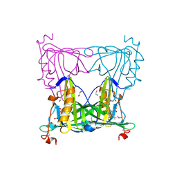
6S9R
 | | Crystal structure of SSDP from D. melanogaster | | 分子名称: | Sequence-specific single-stranded DNA-binding protein, isoform A | | 著者 | Renko, M, Bienz, M. | | 登録日 | 2019-07-15 | | 公開日 | 2019-10-09 | | 最終更新日 | 2024-05-15 | | 実験手法 | X-RAY DIFFRACTION (2.4 Å) | | 主引用文献 | Rotational symmetry of the structured Chip/LDB-SSDP core module of the Wnt enhanceosome.
Proc.Natl.Acad.Sci.USA, 116, 2019
|
|
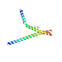
4QH8
 | | LC8 - Ana2 (237-246) Complex | | 分子名称: | Anastral spindle 2, Dynein light chain 1, cytoplasmic | | 著者 | Slevin, L.K, Romes, E.R, Dandulakis, M.G, Slep, K.C. | | 登録日 | 2014-05-27 | | 公開日 | 2014-06-11 | | 最終更新日 | 2023-09-20 | | 実験手法 | X-RAY DIFFRACTION (1.9 Å) | | 主引用文献 | The Mechanism of Dynein Light Chain LC8-mediated Oligomerization of the Ana2 Centriole Duplication Factor.
J.Biol.Chem., 289, 2014
|
|
4QHI
 | | Crystal structure of Methanocaldococcus jannaschii selecase mutant R36W | | 分子名称: | CHLORIDE ION, GLYCEROL, Uncharacterized protein MJ1213, ... | | 著者 | Lopez-pelegrin, M, Cerda-costa, N, Cintas-pedrola, A, Herranz-trillo, F, Bernado, P, Peinado, J.R, Arolas, J.L, Gomis-ruth, F.X. | | 登録日 | 2014-05-28 | | 公開日 | 2014-07-16 | | 最終更新日 | 2024-04-03 | | 実験手法 | X-RAY DIFFRACTION (2.3 Å) | | 主引用文献 | Multiple stable conformations account for reversible concentration-dependent oligomerization and autoinhibition of a metamorphic metallopeptidase
Angew.Chem.Int.Ed.Engl., 53, 2014
|
|
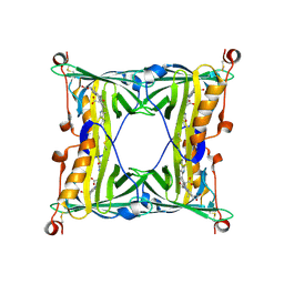
6S9S
 | | Dimerization domain of Xenopus laevis LDB1 in complex with darpin 10 | | 分子名称: | Darpin 10, LIM domain-binding protein 1 | | 著者 | Renko, M, Schaefer, J.V, Pluckthun, A, Bienz, M. | | 登録日 | 2019-07-15 | | 公開日 | 2019-10-09 | | 最終更新日 | 2019-10-23 | | 実験手法 | X-RAY DIFFRACTION (2.2 Å) | | 主引用文献 | Rotational symmetry of the structured Chip/LDB-SSDP core module of the Wnt enhanceosome.
Proc.Natl.Acad.Sci.USA, 116, 2019
|
|
6SCS
 | |
4Q0V
 | | Crystal structure of Acinetobacter sp. DL28 L-ribose isomerase mutant E204Q in complex with L-ribulose | | 分子名称: | COBALT (II) ION, COBALT HEXAMMINE(III), L-Ribose isomerase, ... | | 著者 | Yoshida, H, Yoshihara, A, Teraoka, M, Izumori, K, Kamitori, S. | | 登録日 | 2014-04-02 | | 公開日 | 2014-05-28 | | 最終更新日 | 2023-11-08 | | 実験手法 | X-RAY DIFFRACTION (1.98 Å) | | 主引用文献 | X-ray structure of a novel L-ribose isomerase acting on a non-natural sugar L-ribose as its ideal substrate.
Febs J., 281, 2014
|
|
1R53
 | | Crystal structure of the bifunctional chorismate synthase from Saccharomyces cerevisiae | | 分子名称: | Chorismate synthase | | 著者 | Quevillon-Cheruel, S, Leulliot, N, Meyer, P, Graille, M, Bremang, M, Blondeau, K, Sorel, I, Poupon, A, Janin, J, van Tilbeurgh, H. | | 登録日 | 2003-10-09 | | 公開日 | 2003-12-23 | | 最終更新日 | 2024-03-13 | | 実験手法 | X-RAY DIFFRACTION (2.2 Å) | | 主引用文献 | Crystal structure of the bifunctional chorismate synthase from Saccharomyces cerevisiae
J.Biol.Chem., 279, 2004
|
|
5EZ2
 | |
5F6Z
 | | Sandercyanin Fluorescent Protein purified from Sander vitreus | | 分子名称: | 2-acetamido-2-deoxy-beta-D-glucopyranose, BILIVERDINE IX ALPHA, Sandercyanin Fluorescent Protein | | 著者 | Ghosh, S, Yu, C.L, Ferraro, D, Sudha, S, Pal, S, Schaefer, W, Gibson, D.T, Subramanian, R. | | 登録日 | 2015-12-07 | | 公開日 | 2016-09-28 | | 最終更新日 | 2020-07-29 | | 実験手法 | X-RAY DIFFRACTION (2.248 Å) | | 主引用文献 | Blue protein with red fluorescence
Proc.Natl.Acad.Sci.USA, 113, 2016
|
|
4QHJ
 | | Crystal structure of Methanocaldococcus jannaschii selecase mutant I100F+H107F | | 分子名称: | ACETATE ION, GLYCEROL, Uncharacterized protein MJ1213, ... | | 著者 | Lopez-pelegrin, M, Cerda-costa, N, Cintas-pedrola, A, Herranz-trillo, F, Bernado, P, Peinado, J.R, Arolas, J.L, Gomis-ruth, F.X. | | 登録日 | 2014-05-28 | | 公開日 | 2014-07-16 | | 最終更新日 | 2024-04-03 | | 実験手法 | X-RAY DIFFRACTION (1.75 Å) | | 主引用文献 | Multiple stable conformations account for reversible concentration-dependent oligomerization and autoinhibition of a metamorphic metallopeptidase
Angew.Chem.Int.Ed.Engl., 53, 2014
|
|
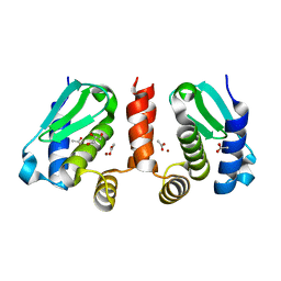
5F51
 | | Structure of B. abortus WrbA-related protein A (apo) | | 分子名称: | NAD(P)H dehydrogenase (quinone), SULFATE ION | | 著者 | Herrou, J, Czyz, D, Willett, J.W, Kim, H.S, Kim, Y, Crosson, S. | | 登録日 | 2015-12-03 | | 公開日 | 2016-03-09 | | 最終更新日 | 2023-09-27 | | 実験手法 | X-RAY DIFFRACTION (2.53 Å) | | 主引用文献 | WrpA Is an Atypical Flavodoxin Family Protein under Regulatory Control of the Brucella abortus General Stress Response System.
J.Bacteriol., 198, 2016
|
|
7BB3
 | | Structure of S. pombe YG-box oligomer | | 分子名称: | (4S)-2-METHYL-2,4-PENTANEDIOL, Survival motor neuron-like protein 1,Survival motor neuron-like protein 1 | | 著者 | Veepaschit, J, Grimm, C, Fischer, U. | | 登録日 | 2020-12-16 | | 公開日 | 2021-01-20 | | 最終更新日 | 2024-01-31 | | 実験手法 | X-RAY DIFFRACTION (2.158 Å) | | 主引用文献 | Identification and structural analysis of the Schizosaccharomyces pombe SMN complex.
Nucleic Acids Res., 49, 2021
|
|
7BPC
 | | Crystal structure of 2, 3-dihydroxybenzoic acid decarboxylase from Fusarium oxysporum in complex with 2,5-DHBA | | 分子名称: | 2,3-dihydroxybenzoate decarboxylase, 2,5-dihydroxybenzoic acid, ZINC ION | | 著者 | Song, M.K, Feng, J.H, Liu, W.D, Wu, Q.Q, Zhu, D.M. | | 登録日 | 2020-03-22 | | 公開日 | 2020-07-15 | | 最終更新日 | 2023-11-29 | | 実験手法 | X-RAY DIFFRACTION (2.45 Å) | | 主引用文献 | 2,3-Dihydroxybenzoic Acid Decarboxylase from Fusarium oxysporum: Crystal Structures and Substrate Recognition Mechanism.
Chembiochem, 21, 2020
|
|
5G0Y
 | |
1FM0
 | | MOLYBDOPTERIN SYNTHASE (MOAD/MOAE) | | 分子名称: | CHLORIDE ION, MOLYBDOPTERIN CONVERTING FACTOR, SUBUNIT 1, ... | | 著者 | Rudolph, M.J, Wuebbens, M.M, Rajagolpalan, K.V, Schindelin, H. | | 登録日 | 2000-08-15 | | 公開日 | 2001-01-17 | | 最終更新日 | 2024-02-07 | | 実験手法 | X-RAY DIFFRACTION (1.45 Å) | | 主引用文献 | Crystal structure of molybdopterin synthase and its evolutionary relationship to ubiquitin activation.
Nat.Struct.Biol., 8, 2001
|
|


