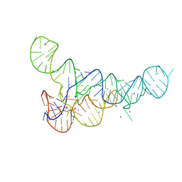
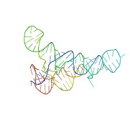
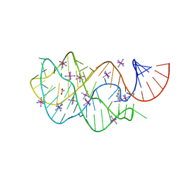
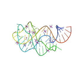
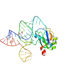
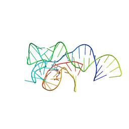
3E5E
 
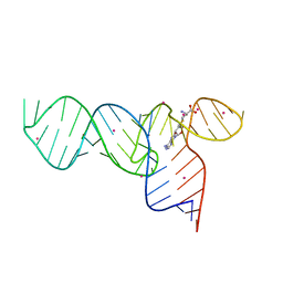 | | Crystal Structures of the SMK box (SAM-III) Riboswitch with SAH | | 分子名称: | S-ADENOSYL-L-HOMOCYSTEINE, SMK box (SAM-III) Riboswitch for RNA, STRONTIUM ION | | 著者 | Lu, C. | | 登録日 | 2008-08-13 | | 公開日 | 2008-10-07 | | 最終更新日 | 2023-08-30 | | 実験手法 | X-RAY DIFFRACTION (2.9 Å) | | 主引用文献 | Crystal structures of the SAM-III/S(MK) riboswitch reveal the SAM-dependent translation inhibition mechanism.
Nat.Struct.Mol.Biol., 15, 2008
|
|
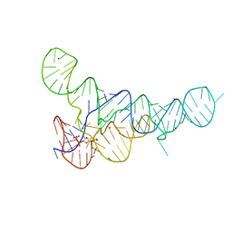
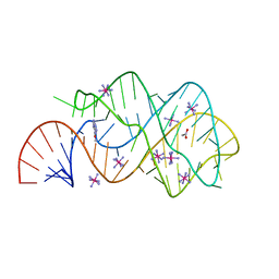
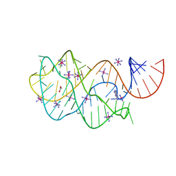
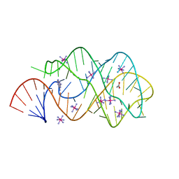
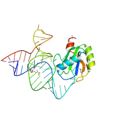
6DMC
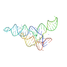 | | ppGpp Riboswitch bound to ppGpp, native structure | | 分子名称: | GUANOSINE-5',3'-TETRAPHOSPHATE, MAGNESIUM ION, POTASSIUM ION, ... | | 著者 | Peselis, A, Serganov, A. | | 登録日 | 2018-06-04 | | 公開日 | 2018-11-14 | | 最終更新日 | 2024-03-13 | | 実験手法 | X-RAY DIFFRACTION (2.2 Å) | | 主引用文献 | ykkC riboswitches employ an add-on helix to adjust specificity for polyanionic ligands.
Nat. Chem. Biol., 14, 2018
|
|
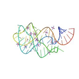
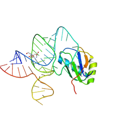
6DMD
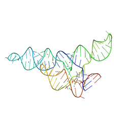 | | ppGpp Riboswitch bound to ppGpp, manganese chloride structure | | 分子名称: | GUANOSINE-5',3'-TETRAPHOSPHATE, MAGNESIUM ION, MANGANESE (II) ION, ... | | 著者 | Peselis, A, Serganov, A. | | 登録日 | 2018-06-05 | | 公開日 | 2018-11-14 | | 最終更新日 | 2024-05-22 | | 実験手法 | X-RAY DIFFRACTION (2.65 Å) | | 主引用文献 | ykkC riboswitches employ an add-on helix to adjust specificity for polyanionic ligands.
Nat. Chem. Biol., 14, 2018
|
|
6DNR
 | |
6DLQ
 | |
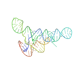
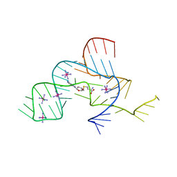
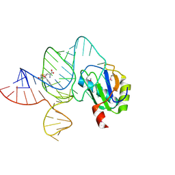
6DLT
 | | PRPP Riboswitch bound to PRPP, native structure | | 分子名称: | 1-O-pyrophosphono-5-O-phosphono-alpha-D-ribofuranose, MAGNESIUM ION, PRPP Riboswitch, ... | | 著者 | Peselis, A, Serganov, A. | | 登録日 | 2018-06-02 | | 公開日 | 2018-11-14 | | 最終更新日 | 2024-05-22 | | 実験手法 | X-RAY DIFFRACTION (2.9 Å) | | 主引用文献 | ykkC riboswitches employ an add-on helix to adjust specificity for polyanionic ligands.
Nat. Chem. Biol., 14, 2018
|
|
6DME
 | | ppGpp Riboswitch bound to ppGpp, thallium acetate structure | | 分子名称: | GUANOSINE-5',3'-TETRAPHOSPHATE, MAGNESIUM ION, THALLIUM (I) ION, ... | | 著者 | Peselis, A, Serganov, A. | | 登録日 | 2018-06-05 | | 公開日 | 2018-11-14 | | 最終更新日 | 2024-05-22 | | 実験手法 | X-RAY DIFFRACTION (2.702 Å) | | 主引用文献 | ykkC riboswitches employ an add-on helix to adjust specificity for polyanionic ligands.
Nat. Chem. Biol., 14, 2018
|
|
6DLR
 | |
6DLS
 | |
3GES
 | |
3GAO
 | |
3NPQ
 | |
3GER
 | |
3G4M
 | |
3NPN
 | |
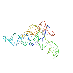
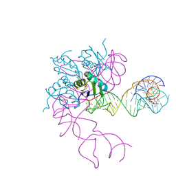
3IRW
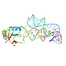 | | Structure of a c-di-GMP riboswitch from V. cholerae | | 分子名称: | 9,9'-[(2R,3R,3aS,5S,7aR,9R,10R,10aS,12S,14aR)-3,5,10,12-tetrahydroxy-5,12-dioxidooctahydro-2H,7H-difuro[3,2-d:3',2'-j][1,3,7,9,2,8]tetraoxadiphosphacyclododecine-2,9-diyl]bis(2-amino-1,9-dihydro-6H-purin-6-one), IRIDIUM HEXAMMINE ION, MAGNESIUM ION, ... | | 著者 | Smith, K.D. | | 登録日 | 2009-08-24 | | 公開日 | 2009-11-10 | | 最終更新日 | 2024-02-21 | | 実験手法 | X-RAY DIFFRACTION (2.7 Å) | | 主引用文献 | Structural basis of ligand binding by a c-di-GMP riboswitch.
Nat.Struct.Mol.Biol., 16, 2009
|
|
2G9C
 | | Modified pyrimidines Specifically bind the purine riboswitch | | 分子名称: | ACETATE ION, COBALT HEXAMMINE(III), PYRIMIDINE-2,4,6-TRIAMINE, ... | | 著者 | Gilbert, S.D, Mediatore, S.J, Batey, R.T. | | 登録日 | 2006-03-06 | | 公開日 | 2006-11-21 | | 最終更新日 | 2024-02-14 | | 実験手法 | X-RAY DIFFRACTION (1.7 Å) | | 主引用文献 | Modified pyrimidines specifically bind the purine riboswitch.
J.Am.Chem.Soc., 128, 2006
|
|
3EGZ
 | | Crystal structure of an in vitro evolved tetracycline aptamer and artificial riboswitch | | 分子名称: | 7-CHLOROTETRACYCLINE, MAGNESIUM ION, Tetracycline aptamer and artificial riboswitch, ... | | 著者 | Xiao, H, Edwards, T.E, Ferre-D'Amare, A.R. | | 登録日 | 2008-09-11 | | 公開日 | 2008-10-28 | | 最終更新日 | 2021-10-20 | | 実験手法 | X-RAY DIFFRACTION (2.2 Å) | | 主引用文献 | Structural basis for specific, high-affinity tetracycline binding by an in vitro evolved aptamer and artificial riboswitch
Chem.Biol., 15, 2008
|
|
2B57
 | | Guanine Riboswitch C74U mutant bound to 2,6-diaminopurine | | 分子名称: | 65-MER, 9H-PURINE-2,6-DIAMINE, ACETATE ION, ... | | 著者 | Gilbert, S.D, Stoddard, C.D, Wise, S.J, Batey, R.T. | | 登録日 | 2005-09-27 | | 公開日 | 2006-05-23 | | 最終更新日 | 2024-02-14 | | 実験手法 | X-RAY DIFFRACTION (2.15 Å) | | 主引用文献 | Thermodynamic and kinetic characterization of ligand binding to the purine riboswitch aptamer domain.
J.Mol.Biol., 359, 2006
|
|
2CKY
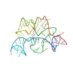 | | Structure of the Arabidopsis thaliana thiamine pyrophosphate riboswitch with its regulatory ligand | | 分子名称: | MAGNESIUM ION, NUCLEIC ACID, OSMIUM ION, ... | | 著者 | Thore, S, Leibundgut, M, Ban, N. | | 登録日 | 2006-04-24 | | 公開日 | 2006-05-08 | | 最終更新日 | 2024-05-08 | | 実験手法 | X-RAY DIFFRACTION (2.9 Å) | | 主引用文献 | Structure of the Eukaryotic Thiamine Pyrophosphate Riboswitch with its Regulatory Ligand.
Science, 312, 2006
|
|
6LAU
 | | the wildtype SAM-VI riboswitch bound to SAH | | 分子名称: | CESIUM ION, GUANOSINE-5'-TRIPHOSPHATE, RNA (54-MER), ... | | 著者 | Ren, A, Sun, A. | | 登録日 | 2019-11-13 | | 公開日 | 2020-01-01 | | 最終更新日 | 2023-11-22 | | 実験手法 | X-RAY DIFFRACTION (3.109 Å) | | 主引用文献 | SAM-VI riboswitch structure and signature for ligand discrimination.
Nat Commun, 10, 2019
|
|
6LAX
 | | the mutant SAM-VI riboswitch (U6C) bound to SAM | | 分子名称: | RNA (55-MER), S-ADENOSYLMETHIONINE, U1 small nuclear ribonucleoprotein A | | 著者 | Sun, A, Ren, A. | | 登録日 | 2019-11-13 | | 公開日 | 2020-01-01 | | 最終更新日 | 2023-11-22 | | 実験手法 | X-RAY DIFFRACTION (2.7 Å) | | 主引用文献 | SAM-VI riboswitch structure and signature for ligand discrimination.
Nat Commun, 10, 2019
|
|
6LAZ
 | | the wildtype SAM-VI riboswitch bound to a N-mustard SAM analog M1 | | 分子名称: | (2~{S})-4-[[(2~{R},3~{S},4~{R},5~{R})-5-(6-aminopurin-9-yl)-3,4-bis(oxidanyl)oxolan-2-yl]methyl-(2-hydroxyethyl)amino]-2-azaniumyl-butanoate, MAGNESIUM ION, RNA (55-MER), ... | | 著者 | Ren, A, Sun, A. | | 登録日 | 2019-11-13 | | 公開日 | 2020-01-01 | | 最終更新日 | 2023-11-22 | | 実験手法 | X-RAY DIFFRACTION (2.76 Å) | | 主引用文献 | SAM-VI riboswitch structure and signature for ligand discrimination.
Nat Commun, 10, 2019
|
|
6LAS
 | | the wildtype SAM-VI riboswitch bound to SAM | | 分子名称: | RNA (55-MER), S-ADENOSYLMETHIONINE, U1 small nuclear ribonucleoprotein A | | 著者 | Ren, A, Sun, A. | | 登録日 | 2019-11-13 | | 公開日 | 2020-01-01 | | 実験手法 | X-RAY DIFFRACTION (2.708 Å) | | 主引用文献 | SAM-VI riboswitch structure and signature for ligand discrimination.
Nat Commun, 10, 2019
|
|
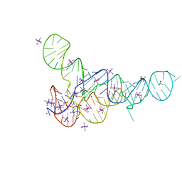
6OD9
 | | Co-crystal structure of the Fusobacterium ulcerans ZTP riboswitch using an X-ray free-electron laser | | 分子名称: | AMINOIMIDAZOLE 4-CARBOXAMIDE RIBONUCLEOTIDE, CESIUM ION, MAGNESIUM ION, ... | | 著者 | Jones, C.P, Tran, B, Ferre-D'Amare, A.R. | | 登録日 | 2019-03-26 | | 公開日 | 2019-07-17 | | 最終更新日 | 2023-10-11 | | 実験手法 | X-RAY DIFFRACTION (4.102 Å) | | 主引用文献 | Co-crystal structure of the Fusobacterium ulcerans ZTP riboswitch using an X-ray free-electron laser.
Acta Crystallogr.,Sect.F, 75, 2019
|
|


