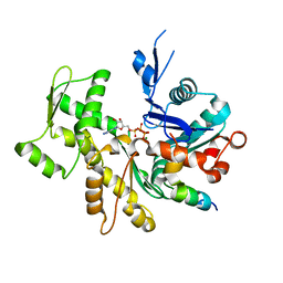
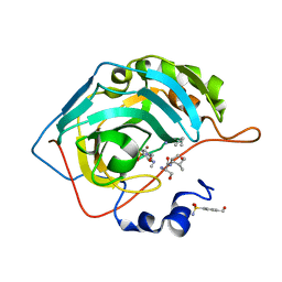
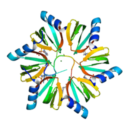
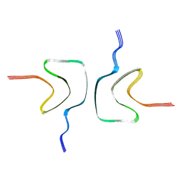
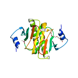
3TYI
 
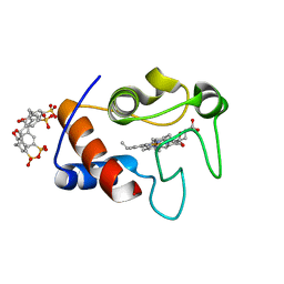 | | Crystal Structure of Cytochrome c - p-Sulfonatocalix[4]arene Complexes | | 分子名称: | 25,26,27,28-tetrahydroxypentacyclo[19.3.1.1~3,7~.1~9,13~.1~15,19~]octacosa-1(25),3(28),4,6,9(27),10,12,15(26),16,18,21,23-dodecaene-5,11,17,23-tetrasulfonic acid, Cytochrome c iso-1, PROTOPORPHYRIN IX CONTAINING FE | | 著者 | Mc Govern, R.E, Fernandes, H, Khan, A.R, Crowley, P.B. | | 登録日 | 2011-09-26 | | 公開日 | 2012-05-02 | | 最終更新日 | 2023-09-13 | | 実験手法 | X-RAY DIFFRACTION (1.399 Å) | | 主引用文献 | Protein camouflage in cytochrome c-calixarene complexes.
Nat Chem, 4, 2012
|
|
7PZN
 | |
7RJF
 | | MOPD-1 mutant-L47W | | 分子名称: | MALONATE ION, ZINC ION, [L47W]MOPD-1 | | 著者 | Huawu, Y, Conan, K.W, Gordon, J.K, Brett, M.C, Yen-Hua, H, David, J.C. | | 登録日 | 2021-07-20 | | 公開日 | 2021-10-27 | | 最終更新日 | 2023-10-18 | | 実験手法 | X-RAY DIFFRACTION (1.8 Å) | | 主引用文献 | Rational Design of Potent Peptide Inhibitors of the PD-1:PD-L1 Interaction for Cancer Immunotherapy.
J.Am.Chem.Soc., 143, 2021
|
|
7PCU
 | | Crystal structure of YTHDF1 YTH domain in complex with ebselen | | 分子名称: | N-phenyl-2-selanylbenzamide, YTH domain-containing family protein 1 | | 著者 | Dalle Vedove, A, Cazzanelli, G, Quattrone, A, Provenzani, A, Lolli, G. | | 登録日 | 2021-08-04 | | 公開日 | 2022-12-07 | | 最終更新日 | 2024-02-07 | | 実験手法 | X-RAY DIFFRACTION (2.65 Å) | | 主引用文献 | Small-Molecule Ebselen Binds to YTHDF Proteins Interfering with the Recognition of N 6 -Methyladenosine-Modified RNAs.
Acs Pharmacol Transl Sci, 5, 2022
|
|
3U8X
 | | Crystal Structure of a chimera containing the N-terminal domain (residues 8-29) of drosophila Ciboulot and the C-terminal domain (residues 18-44) of bovine Thymosin-beta4, bound to G-actin-ATP | | 分子名称: | ADENOSINE-5'-TRIPHOSPHATE, Actin, alpha skeletal muscle, ... | | 著者 | Renault, L, Husson, C, Carlier, M.F, Didry, D. | | 登録日 | 2011-10-17 | | 公開日 | 2012-01-25 | | 最終更新日 | 2023-09-13 | | 実験手法 | X-RAY DIFFRACTION (2 Å) | | 主引用文献 | How a single residue in individual beta-thymosin/WH2 domains controls their functions in actin assembly
Embo J., 31, 2012
|
|
7OX6
 | | Solution structure of human interleukin-9 | | 分子名称: | Interleukin-9 | | 著者 | Savvides, S.N, Tripsianes, K, De Vos, T, Papageorgiou, A, Evangelidis, T. | | 登録日 | 2021-06-22 | | 公開日 | 2022-12-28 | | 最終更新日 | 2023-01-11 | | 実験手法 | SOLUTION NMR | | 主引用文献 | Structural basis for the mechanism and antagonism of receptor signaling mediated by Interleukin-9 (IL-9)
Biorxiv, 2022
|
|
7OYN
 | | Carbonic anhydrase II in complex with Hit3 (MH57) | | 分子名称: | Carbonic anhydrase 2, Hit3 (MH57), ZINC ION | | 著者 | Kugler, M, Brynda, J, Rezacova, P. | | 登録日 | 2021-06-24 | | 公開日 | 2023-01-18 | | 最終更新日 | 2024-02-07 | | 実験手法 | X-RAY DIFFRACTION (0.98 Å) | | 主引用文献 | Identification of specific carbonic anhydrase inhibitors via in situ click chemistry, phage-display and synthetic peptide libraries: comparison of the methods and structural study.
Rsc Med Chem, 14, 2023
|
|
7OYP
 | | Carbonic anhydrase II in complex with Hit3-t1 (MH172) | | 分子名称: | (2S)-3-oxidanyl-2-[2-[(4-sulfamoylphenyl)methoxyamino]ethanoylamino]propanamide, 4-methylbenzenesulfonamide, Carbonic anhydrase 2, ... | | 著者 | Kugler, M, Brynda, J, Rezacova, P. | | 登録日 | 2021-06-24 | | 公開日 | 2023-01-18 | | 最終更新日 | 2024-02-07 | | 実験手法 | X-RAY DIFFRACTION (1.05 Å) | | 主引用文献 | Identification of specific carbonic anhydrase inhibitors via in situ click chemistry, phage-display and synthetic peptide libraries: comparison of the methods and structural study.
Rsc Med Chem, 14, 2023
|
|
7OYR
 | | Carbonic anhydrase II in complex with Hit3-t4 (MH181) | | 分子名称: | Carbonic anhydrase 2, Hit3-t4 (MH181), ZINC ION | | 著者 | Kugler, M, Brynda, J, Rezacova, P. | | 登録日 | 2021-06-24 | | 公開日 | 2023-01-18 | | 最終更新日 | 2024-02-07 | | 実験手法 | X-RAY DIFFRACTION (1.15 Å) | | 主引用文献 | Identification of specific carbonic anhydrase inhibitors via in situ click chemistry, phage-display and synthetic peptide libraries: comparison of the methods and structural study.
Rsc Med Chem, 14, 2023
|
|
7OYQ
 | | Carbonic anhydrase II in complex with Hit3-t2 (MH174) | | 分子名称: | Carbonic anhydrase 2, Hit3-t2 (MH174), ZINC ION | | 著者 | Kugler, M, Brynda, J, Rezacova, P. | | 登録日 | 2021-06-24 | | 公開日 | 2023-01-18 | | 最終更新日 | 2024-02-07 | | 実験手法 | X-RAY DIFFRACTION (1.15 Å) | | 主引用文献 | Identification of specific carbonic anhydrase inhibitors via in situ click chemistry, phage-display and synthetic peptide libraries: comparison of the methods and structural study.
Rsc Med Chem, 14, 2023
|
|
7OYM
 | | Carbonic anhydrase II in complex with Hit2 (MH65) | | 分子名称: | Carbonic anhydrase 2, Hit2 (MH65), ZINC ION | | 著者 | Kugler, M, Brynda, J, Rezacova, P. | | 登録日 | 2021-06-24 | | 公開日 | 2023-01-18 | | 最終更新日 | 2024-02-07 | | 実験手法 | X-RAY DIFFRACTION (0.98 Å) | | 主引用文献 | Identification of specific carbonic anhydrase inhibitors via in situ click chemistry, phage-display and synthetic peptide libraries: comparison of the methods and structural study.
Rsc Med Chem, 14, 2023
|
|
7OYO
 | | Carbonic anhydrase II in complex with Hit4 (MH70) | | 分子名称: | Carbonic anhydrase 2, DIMETHYL SULFOXIDE, ZINC ION, ... | | 著者 | Kugler, M, Brynda, J, Rezacova, P. | | 登録日 | 2021-06-24 | | 公開日 | 2023-01-18 | | 最終更新日 | 2024-02-07 | | 実験手法 | X-RAY DIFFRACTION (1.03 Å) | | 主引用文献 | Identification of specific carbonic anhydrase inhibitors via in situ click chemistry, phage-display and synthetic peptide libraries: comparison of the methods and structural study.
Rsc Med Chem, 14, 2023
|
|
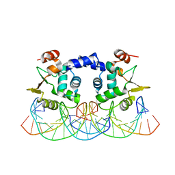
3RER
 | | Crystal structure of E. coli Hfq in complex with AU6A RNA and ADP | | 分子名称: | 5'-R(*AP*UP*UP*UP*UP*UP*UP*A)-3', ADENOSINE-5'-DIPHOSPHATE, MAGNESIUM ION, ... | | 著者 | Wang, W.W, Wu, J.H, Shi, Y.Y. | | 登録日 | 2011-04-05 | | 公開日 | 2011-10-19 | | 最終更新日 | 2023-11-01 | | 実験手法 | X-RAY DIFFRACTION (1.7 Å) | | 主引用文献 | Cooperation of Escherichia coli Hfq hexamers in DsrA binding.
Genes Dev., 25, 2011
|
|
3RI4
 | |
3R32
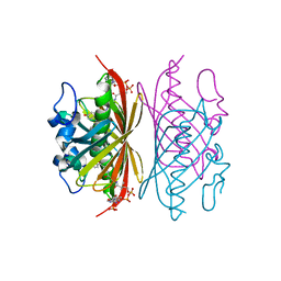 | | Crystal structure of Arthrobacter sp. strain SU 4-hydroxybenzoyl CoA thioesterase mutant E73A complexed with 4-hydroxyphenacyl CoA | | 分子名称: | 4-HYDROXYPHENACYL COENZYME A, 4-hydroxybenzoyl-CoA thioesterase | | 著者 | Holden, H.M, Thoden, J.B, Song, F, Zhuang, Z, Trujillo, M, Dunaway-Mariano, D. | | 登録日 | 2011-03-15 | | 公開日 | 2012-03-28 | | 最終更新日 | 2023-09-13 | | 実験手法 | X-RAY DIFFRACTION (1.8 Å) | | 主引用文献 | The Catalytic Mechanism of the Hotdog-fold Enzyme Superfamily 4-Hydroxybenzoyl-CoA Thioesterase from Arthrobacter sp. Strain SU.
Biochemistry, 51, 2012
|
|
3R3A
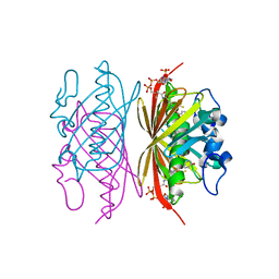 | | Crystal structure of Arthrobacter sp. strain SU 4-hydroxybenzoyl CoA thioesterase mutant Q58A complexed with 4-hydroxybenzoic acid and CoA | | 分子名称: | 4-hydroxybenzoyl-CoA thioesterase, COENZYME A, P-HYDROXYBENZOIC ACID | | 著者 | Holden, H.M, Thoden, J.B, Song, F, Zhuang, Z, Trujillo, M, Dunaway-Mariano, D. | | 登録日 | 2011-03-15 | | 公開日 | 2012-03-28 | | 最終更新日 | 2023-09-13 | | 実験手法 | X-RAY DIFFRACTION (1.8 Å) | | 主引用文献 | The Catalytic Mechanism of the Hotdog-fold Enzyme Superfamily 4-Hydroxybenzoyl-CoA Thioesterase from Arthrobacter sp. Strain SU.
Biochemistry, 51, 2012
|
|
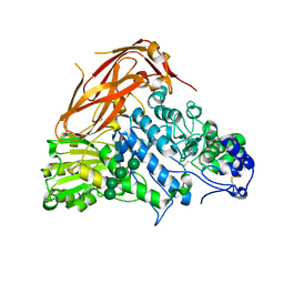
7YK2
 | | Cryo-EM structure of Apo-alpha-syn fibril | | 分子名称: | Alpha-synuclein | | 著者 | Xu, Q.H, Xia, W.C, Tao, Y.Q, Liu, C. | | 登録日 | 2022-07-21 | | 公開日 | 2023-03-22 | | 最終更新日 | 2024-05-08 | | 実験手法 | ELECTRON MICROSCOPY (2.8 Å) | | 主引用文献 | Conformational Dynamics of an alpha-Synuclein Fibril upon Receptor Binding Revealed by Insensitive Nuclei Enhanced by Polarization Transfer-Based Solid-State Nuclear Magnetic Resonance and Cryo-Electron Microscopy.
J.Am.Chem.Soc., 145, 2023
|
|
7XTJ
 | | Crystal structure of E88A mutant of GH3 beta-xylosidase from Aspergillus niger (AnBX) | | 分子名称: | 2-acetamido-2-deoxy-beta-D-glucopyranose, 2-acetamido-2-deoxy-beta-D-glucopyranose-(1-4)-2-acetamido-2-deoxy-beta-D-glucopyranose, GLYCEROL, ... | | 著者 | Kaenying, W, Kongsaeree, P.T, Tagami, T. | | 登録日 | 2022-05-17 | | 公開日 | 2023-03-15 | | 最終更新日 | 2023-11-29 | | 実験手法 | X-RAY DIFFRACTION (2.5 Å) | | 主引用文献 | Crystal structure and identification of amino acid residues for catalysis and binding of GH3 AnBX beta-xylosidase from Aspergillus niger.
Appl.Microbiol.Biotechnol., 107, 2023
|
|
7YK8
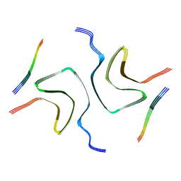 | | Cryo-EM structure of dLAG3-alpha-syn fibril | | 分子名称: | Alpha-synuclein | | 著者 | Xu, Q.H, Xia, W.C, Tao, Y.Q, Liu, C. | | 登録日 | 2022-07-22 | | 公開日 | 2023-03-22 | | 最終更新日 | 2024-05-08 | | 実験手法 | ELECTRON MICROSCOPY (2.8 Å) | | 主引用文献 | Conformational Dynamics of an alpha-Synuclein Fibril upon Receptor Binding Revealed by Insensitive Nuclei Enhanced by Polarization Transfer-Based Solid-State Nuclear Magnetic Resonance and Cryo-Electron Microscopy.
J.Am.Chem.Soc., 145, 2023
|
|
3R68
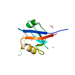 | | Molecular Analysis of the PDZ3 domain of PDZK1 | | 分子名称: | 1,2-ETHANEDIOL, ACETATE ION, CALCIUM ION, ... | | 著者 | Kocher, O, Birrane, G, Krieger, M. | | 登録日 | 2011-03-21 | | 公開日 | 2011-05-18 | | 最終更新日 | 2024-02-21 | | 実験手法 | X-RAY DIFFRACTION (1.3 Å) | | 主引用文献 | Identification of the PDZ3 Domain of the Adaptor Protein PDZK1 as a Second, Physiologically Functional Binding Site for the C Terminus of the High Density Lipoprotein Receptor Scavenger Receptor Class B Type I.
J.Biol.Chem., 286, 2011
|
|
7Q2U
 | | The crystal structure of the HINT1 Q62A mutant. | | 分子名称: | CACODYLATE ION, HEXAETHYLENE GLYCOL, Histidine triad nucleotide-binding protein 1, ... | | 著者 | Dolot, R.M, Strom, A.M, Wagner, C.R. | | 登録日 | 2021-10-26 | | 公開日 | 2021-11-03 | | 最終更新日 | 2024-02-07 | | 実験手法 | X-RAY DIFFRACTION (2.27 Å) | | 主引用文献 | Dynamic Long-Range Interactions Influence Substrate Binding and Catalysis by Human Histidine Triad Nucleotide-Binding Proteins (HINTs), Key Regulators of Multiple Cellular Processes and Activators of Antiviral ProTides.
Biochemistry, 61, 2022
|
|
7R8R
 | | Physachenolide C with Bromodomain (BRD3-BD1) | | 分子名称: | Bromodomain-containing protein 3, Physachenolide C | | 著者 | Fromme, R, Sivinski, J, Zerio, C, Gunatilaka, A.A.L, Chapman, E. | | 登録日 | 2021-06-27 | | 公開日 | 2022-09-21 | | 最終更新日 | 2023-10-25 | | 実験手法 | X-RAY DIFFRACTION (1.8 Å) | | 主引用文献 | Physachenolide C is a Potent, Selective BET Inhibitor.
J.Med.Chem., 66, 2023
|
|
3RFX
 | |
7R1Q
 | |
7R1P
 | |


