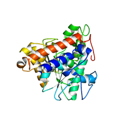
5GI7
 
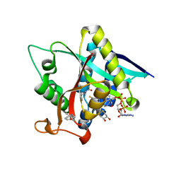 | | Crystal Structure of Drosophila melanogaster Dopamine N-Acetyltransferase in Ternary Complex with CoA and Acetyl-phenylethylamine | | 分子名称: | (4S,5S)-1,2-DITHIANE-4,5-DIOL, COENZYME A, Dopamine N-acetyltransferase, ... | | 著者 | Yang, Y.C, Lin, S.J, Cheng, K.C, Cheng, H.C, Lyu, P.C. | | 登録日 | 2016-06-22 | | 公開日 | 2017-07-05 | | 最終更新日 | 2024-03-20 | | 実験手法 | X-RAY DIFFRACTION (1.201 Å) | | 主引用文献 | An essential role of acetyl coenzyme A in the catalytic cycle of insect arylalkylamine N-acetyltransferase.
Commun Biol, 3, 2020
|
|
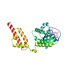
5GI9
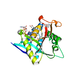 | | Crystal Structure of Drosophila melanogaster Dopamine N-Acetyltransferase in Ternary Complex with CoA and Acetyl-Tryptamine | | 分子名称: | (4S,5S)-1,2-DITHIANE-4,5-DIOL, COENZYME A, Dopamine N-acetyltransferase, ... | | 著者 | Yang, Y.C, Lin, S.J, Cheng, K.C, Cheng, H.C, Lyu, P.C. | | 登録日 | 2016-06-22 | | 公開日 | 2017-07-05 | | 最終更新日 | 2024-03-20 | | 実験手法 | X-RAY DIFFRACTION (1.4 Å) | | 主引用文献 | An essential role of acetyl coenzyme A in the catalytic cycle of insect arylalkylamine N-acetyltransferase.
Commun Biol, 3, 2020
|
|
5GIF
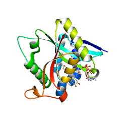 | | Crystal Structure of Drosophila melanogaster E47D Dopamine N-Acetyltransferase in Binary Complex with Acetyl-CoA | | 分子名称: | (4S,5S)-1,2-DITHIANE-4,5-DIOL, ACETYL COENZYME *A, Dopamine N-acetyltransferase | | 著者 | Yang, Y.C, Wu, C.Y, Cheng, H.C, Lyu, P.C. | | 登録日 | 2016-06-23 | | 公開日 | 2017-07-05 | | 最終更新日 | 2024-03-20 | | 実験手法 | X-RAY DIFFRACTION (1.3 Å) | | 主引用文献 | Crystal Structure of Drosophila melanogaster E47D Dopamine N-Acetyltransferase in Binary Complex with Acetyl-CoA
To Be Published
|
|
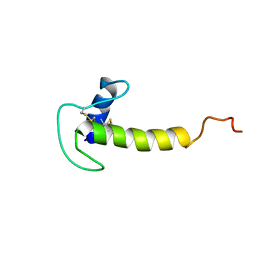
5GI5
 | |
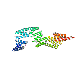
2LGW
 | |
5YXM
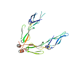
 | | Crystal structure of Chlamydomonas Outer Arm Dynein Light Chain 1 | | 分子名称: | Dynein light chain 1, axonemal, PHOSPHATE ION | | 著者 | Toda, A, Tanaka, H, Nishikawa, Y, Yagi, T, Kurisu, G. | | 登録日 | 2017-12-06 | | 公開日 | 2018-03-14 | | 最終更新日 | 2023-11-22 | | 実験手法 | X-RAY DIFFRACTION (1.545 Å) | | 主引用文献 | Structural atlas of dynein motors at atomic resolution.
Biophys Rev, 10, 2018
|
|
3AGY
 | |
3APQ
 | |
3AGZ
 | |
3AGX
 | |
7X89
 | | Tid1 | | 分子名称: | DnaJ homolog subfamily A member 3, mitochondrial | | 著者 | Jang, J, Lee, S.H, Kang, D.H, Sim, D.W, Jo, K.S, Ryu, H, Kim, E.H, Ryu, K.S, Lee, J.H, Kim, J.H, Won, H.S. | | 登録日 | 2022-03-11 | | 公開日 | 2023-03-22 | | 最終更新日 | 2024-05-15 | | 実験手法 | SOLUTION NMR | | 主引用文献 | Structural studies on the J-domain and GF-motif of the mitochondrial Hsp40, Tid1
To Be Published
|
|
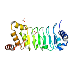
6CGH
 | | Solution structure of the four-helix bundle region of human J-protein Zuotin, a component of ribosome-associated complex (RAC) | | 分子名称: | DnaJ homolog subfamily C member 2 | | 著者 | Shrestha, O.K, Lee, W, Tonelli, M, Cornilescu, G, Markley, J.L, Ciesielski, S.J, Craig, E.A. | | 登録日 | 2018-02-20 | | 公開日 | 2019-06-12 | | 最終更新日 | 2024-05-15 | | 実験手法 | SOLUTION NMR | | 主引用文献 | Structure and evolution of the 4-helix bundle domain of Zuotin, a J-domain protein co-chaperone of Hsp70.
Plos One, 14, 2019
|
|
3APO
 | |
3APS
 | |
2QLD
 | | human Hsp40 Hdj1 | | 分子名称: | DnaJ homolog subfamily B member 1 | | 著者 | Hu, J, Wu, Y, Li, J, Fu, Z, Sha, B. | | 登録日 | 2007-07-12 | | 公開日 | 2008-07-15 | | 最終更新日 | 2024-04-03 | | 実験手法 | X-RAY DIFFRACTION (2.7 Å) | | 主引用文献 | The crystal structure of the putative peptide-binding fragment from the human Hsp40 protein Hdj1.
Bmc Struct.Biol., 8, 2008
|
|
7V6D
 | | Structure of lipase B from Lasiodiplodia theobromae | | 分子名称: | 2-acetamido-2-deoxy-beta-D-glucopyranose-(1-4)-2-acetamido-2-deoxy-beta-D-glucopyranose, CALCIUM ION, Lipase B | | 著者 | Xue, B, Zhang, H.F, Nguyen, G.K.T, Yew, W.S. | | 登録日 | 2021-08-20 | | 公開日 | 2021-10-13 | | 最終更新日 | 2023-11-29 | | 実験手法 | X-RAY DIFFRACTION (2.5 Å) | | 主引用文献 | A Novel Lipase from Lasiodiplodia theobromae Efficiently Hydrolyses C8-C10 Methyl Esters for the Preparation of Medium-Chain Triglycerides' Precursors.
Int J Mol Sci, 22, 2021
|
|
6JMG
 | | Crystal structure of xRbj | | 分子名称: | DnaJ homolog subfamily C member 27-A, GUANOSINE-5'-TRIPHOSPHATE, MAGNESIUM ION | | 著者 | Gao, Z.R, Qi, J.X. | | 登録日 | 2019-03-09 | | 公開日 | 2020-03-11 | | 最終更新日 | 2024-03-27 | | 実験手法 | X-RAY DIFFRACTION (2.701 Å) | | 主引用文献 | Crystal structure of Rbj
To Be Published
|
|
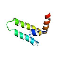
2KJQ
 | | Solution Structure Of Protein NMB1076 From Neisseria meningitidis. Northeast Structural Genomics Consortium Target MR101B. | | 分子名称: | DnaA-related protein | | 著者 | Wu, Y, Maglaqui, M, Eletsky, A, Ciccosanti, C, Sathyamoorthy, B, Jiang, M, Garcia, E, Nair, R, Rost, B, Swapna, G, Acton, T, Xiao, R, Everett, J, Montelione, G.T, Szyperski, T, Northeast Structural Genomics Consortium (NESG) | | 登録日 | 2009-06-08 | | 公開日 | 2010-04-28 | | 最終更新日 | 2024-05-08 | | 実験手法 | SOLUTION NMR | | 主引用文献 | Solution Structure Of Protein NMB1076 From Neisseria meningitidis. Northeast Structural Genomics Consortium Target MR101B.
To be Published
|
|
3IWR
 | | Crystal structure of class I chitinase from Oryza sativa L. japonica | | 分子名称: | (4S)-2-METHYL-2,4-PENTANEDIOL, 2-(N-MORPHOLINO)-ETHANESULFONIC ACID, Chitinase | | 著者 | Kezuka, Y, Watanabe, T, Nonaka, T. | | 登録日 | 2009-09-03 | | 公開日 | 2010-04-21 | | 最終更新日 | 2023-09-06 | | 実験手法 | X-RAY DIFFRACTION (2.57 Å) | | 主引用文献 | Structure of full-length class I chitinase from rice revealed by X-ray crystallography and small-angle X-ray scattering.
Proteins, 78, 2010
|
|
6PTG
 | |
6K80
 | | Crystal Structure of Drosophila melanogaster Dopamine N-Acetyltransferase in Complex with CoA and Tryptophol | | 分子名称: | 2-(1H-indol-3-yl)ethanol, ACETYL COENZYME *A, Dopamine N-acetyltransferase | | 著者 | Wu, C.Y, Hu, I.C, Yang, Y.C, Cheng, H.C, Lyu, P.C. | | 登録日 | 2019-06-11 | | 公開日 | 2020-06-17 | | 最終更新日 | 2023-11-22 | | 実験手法 | X-RAY DIFFRACTION (1.28 Å) | | 主引用文献 | An essential role of acetyl coenzyme A in the catalytic cycle of insect arylalkylamine N-acetyltransferase.
Commun Biol, 3, 2020
|
|
8IZT
 | |
8IZU
 | |
5YHN
 | | Solution structure of the LEKTI Domain 4 | | 分子名称: | cDNA FLJ60407, highly similar to Serine protease inhibitor Kazal-type 5 | | 著者 | Mok, Y.K, Ramesh, K. | | 登録日 | 2017-09-29 | | 公開日 | 2018-08-08 | | 最終更新日 | 2023-06-14 | | 実験手法 | SOLUTION NMR | | 主引用文献 | Homologous Lympho-Epithelial Kazal-type Inhibitor Domains Delay Blood Coagulation by Inhibiting Factor X and XI with Differential Specificity.
Structure, 26, 2018
|
|
3IEG
 | | Crystal Structure of P58(IPK) TPR Domain at 2.5 A | | 分子名称: | DnaJ homolog subfamily C member 3 | | 著者 | Tao, J, Sha, B. | | 登録日 | 2009-07-22 | | 公開日 | 2010-03-31 | | 最終更新日 | 2019-07-24 | | 実験手法 | X-RAY DIFFRACTION (2.51 Å) | | 主引用文献 | Crystal Structure of P58(IPK) TPR Fragment Reveals the Mechanism for its Molecular Chaperone Activity in UPR.
J.Mol.Biol., 64, 2010
|
|


