3LKK
 
 | |
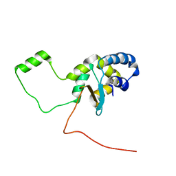
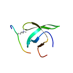
1QDP
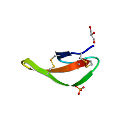
 | | SOLUTION STRUCTURE OF ROBUSTOXIN, THE LETHAL NEUROTOXIN FROM THE FUNNEL WEB SPIDER ATRAX ROBUSTUS, NMR, 20 STRUCTURES | | 分子名称: | ROBUSTOXIN | | 著者 | Pallaghy, P.K, Alewood, D, Alewood, P.F, Norton, R.S. | | 登録日 | 1997-10-09 | | 公開日 | 1998-01-14 | | 最終更新日 | 2022-03-02 | | 実験手法 | SOLUTION NMR | | 主引用文献 | Solution structure of robustoxin, the lethal neurotoxin from the funnel-web spider Atrax robustus.
FEBS Lett., 419, 1997
|
|
6ZGO
 | |
3LMM
 | | Crystal Structure of the DIP2311 protein from Corynebacterium diphtheriae, Northeast Structural Genomics Consortium Target CdR35 | | 分子名称: | CHLORIDE ION, COBALT (II) ION, Uncharacterized protein | | 著者 | Forouhar, F, Lew, S, Seetharaman, J, Mao, M, Xiao, R, Ciccosanti, C, Buchwald, W.A, Maglaqui, M, Everett, J.K, Nair, R, Acton, T.B, Rost, B, Montelione, G.T, Hunt, J.F, Tong, L, Northeast Structural Genomics Consortium (NESG) | | 登録日 | 2010-01-31 | | 公開日 | 2010-02-16 | | 最終更新日 | 2019-07-17 | | 実験手法 | X-RAY DIFFRACTION (3 Å) | | 主引用文献 | Northeast Structural Genomics Consortium Target CdR35
To be Published
|
|
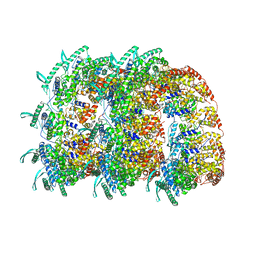
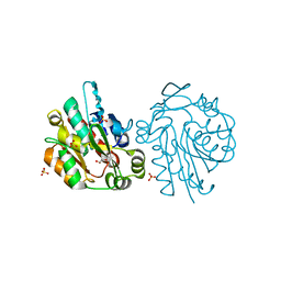
1QKI
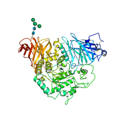
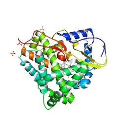
 | | X-RAY STRUCTURE OF HUMAN GLUCOSE 6-PHOSPHATE DEHYDROGENASE (VARIANT CANTON R459L) COMPLEXED WITH STRUCTURAL NADP+ | | 分子名称: | GLUCOSE-6-PHOSPHATE 1-DEHYDROGENASE, GLYCEROL, GLYCOLIC ACID, ... | | 著者 | Au, S.W.N, Gover, S, Lam, V.M.S, Adams, M.J. | | 登録日 | 1999-07-20 | | 公開日 | 2000-03-16 | | 最終更新日 | 2023-12-13 | | 実験手法 | X-RAY DIFFRACTION (3 Å) | | 主引用文献 | Human Glucose-6-Phosphate Dehydrogenase: The Crystal Structure Reveals a Structural Nadp+ Molecule and Provides Insights Into Enzyme Deficiency
Structure, 8, 2000
|
|
3LPO
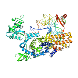
 | | Crystal structure of the N-terminal domain of sucrase-isomaltase | | 分子名称: | 2-acetamido-2-deoxy-beta-D-glucopyranose, 2-acetamido-2-deoxy-beta-D-glucopyranose-(1-4)-2-acetamido-2-deoxy-beta-D-glucopyranose, Sucrase-isomaltase, ... | | 著者 | Sim, L, Rose, D.R. | | 登録日 | 2010-02-05 | | 公開日 | 2010-03-31 | | 最終更新日 | 2020-07-29 | | 実験手法 | X-RAY DIFFRACTION (3.2 Å) | | 主引用文献 | Structural basis for substrate selectivity in human maltase-glucoamylase and sucrase-isomaltase N-terminal domains.
J.Biol.Chem., 285, 2010
|
|
1QJH
 | |
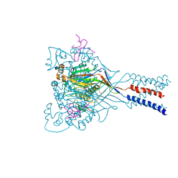
5LFB
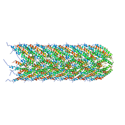 | | Structure of the bacterial sex F pilus (12.5 Angstrom rise) | | 分子名称: | Pilin, [(2~{S})-3-[[(2~{R})-2,3-bis(oxidanyl)propoxy]-oxidanyl-phosphoryl]oxy-2-hexadec-9-enoyloxy-propyl] hexadecanoate | | 著者 | Costa, T.R.D, Ilangovan, I, Ukleja, M, Redzej, A, Santini, J.M, Smith, T.K, Egelman, E.H, Waksman, G. | | 登録日 | 2016-06-30 | | 公開日 | 2016-11-02 | | 最終更新日 | 2024-05-15 | | 実験手法 | ELECTRON MICROSCOPY (5 Å) | | 主引用文献 | Structure of the Bacterial Sex F Pilus Reveals an Assembly of a Stoichiometric Protein-Phospholipid Complex.
Cell, 166, 2016
|
|
4BHF
 | | Three dimensional structure of human gamma-butyrobetaine hydroxylase in complex with 4-(Trimethylammonio)pentanoate | | 分子名称: | 4-(Trimethylammonio)pentanoic acid, GAMMA-BUTYROBETAINE DIOXYGENASE, HEXANE-1,6-DIAMINE, ... | | 著者 | Tars, K, Leitans, J, Kazaks, A. | | 登録日 | 2013-04-02 | | 公開日 | 2014-03-12 | | 最終更新日 | 2023-12-20 | | 実験手法 | X-RAY DIFFRACTION (2.05 Å) | | 主引用文献 | Targeting Carnitine Biosynthesis: Discovery of New Inhibitors Against Gamma-Butyrobetaine Hydroxylase.
J.Med.Chem., 57, 2014
|
|
4BKK
 | | The Respiratory Syncytial Virus nucleoprotein-RNA complex forms a left-handed helical nucleocapsid. | | 分子名称: | NUCLEOPROTEIN, RNA (161-MER) | | 著者 | Bakker, S.E, Duquerroy, S, Galloux, M, Loney, C, Conner, E, Eleouet, J.F, Rey, F.A, Bhella, D. | | 登録日 | 2013-04-26 | | 公開日 | 2013-10-30 | | 最終更新日 | 2024-05-08 | | 実験手法 | ELECTRON MICROSCOPY | | 主引用文献 | The Respiratory Syncytial Virus Nucleoprotein-RNA Complex Forms a Left-Handed Helical Nucleocapsid.
J.Gen.Virol., 94, 2013
|
|
5LGF
 | |
4FZ1
 | |
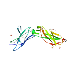
4FZ8
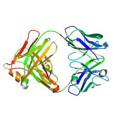
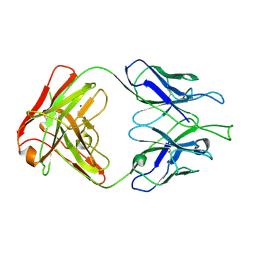 | | Crystal structure of C11 Fab, an ADCC mediating anti-HIV-1 antibody. | | 分子名称: | 2-acetamido-2-deoxy-beta-D-glucopyranose, FAB heavy chain of human ANTI-HIV-1 ENV ANTIBODY C11, FAB light chain of human ANTI-HIV-1 ENV ANTIBODY C11, ... | | 著者 | Wu, X, Tolbert, W.D, Pazgier, M. | | 登録日 | 2012-07-06 | | 公開日 | 2013-07-10 | | 最終更新日 | 2023-09-13 | | 実験手法 | X-RAY DIFFRACTION (2.66 Å) | | 主引用文献 | Recognition Patterns of the C1/C2 Epitopes Involved in Fc-Mediated Response in HIV-1 Natural Infection and the RV114 Vaccine Trial.
Mbio, 11, 2020
|
|
5LIV
 | | Crystal structure of myxobacterial CYP260A1 | | 分子名称: | 2-(N-MORPHOLINO)-ETHANESULFONIC ACID, Cytochrome P450 CYP260A1,Cytochrome P450 CYP260A1, DIMETHYL SULFOXIDE, ... | | 著者 | Carius, Y, Khatri, Y, Bernhardt, R, Lancaster, C.R.D. | | 登録日 | 2016-07-15 | | 公開日 | 2016-11-23 | | 最終更新日 | 2024-01-10 | | 実験手法 | X-RAY DIFFRACTION (2.67 Å) | | 主引用文献 | Structural characterization of CYP260A1 from Sorangium cellulosum to investigate the 1 alpha-hydroxylation of a mineralocorticoid.
FEBS Lett., 590, 2016
|
|
4AS1
 | | Ternary complex of E. coli leucyl-tRNA synthetase, tRNA(leu) and the benzoxaborole AN2679 in the editing conformation | | 分子名称: | LEUCINE--TRNA LIGASE, MAGNESIUM ION, TRNA-LEU5 (UAA ISOACEPTOR) | | 著者 | Palencia, A, Crepin, T, Vu, M.T, Lincecum Jr, T.L, Martinis, S.A, Cusack, S. | | 登録日 | 2012-04-27 | | 公開日 | 2012-06-13 | | 最終更新日 | 2024-01-31 | | 実験手法 | X-RAY DIFFRACTION (2.02 Å) | | 主引用文献 | Structural Dynamics of the Aminoacylation and Proofreading Functional Cycle of Bacterial Leucyl-tRNA Synthetase
Nat.Struct.Mol.Biol., 19, 2012
|
|
6ZLG
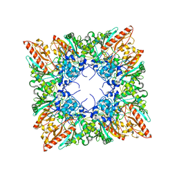
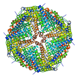 | | Folding of an iron binding peptide in response to sedimentation is resolved using ferritin as a nano-reactor | | 分子名称: | Ferritin | | 著者 | Davidov, G, Abelya, G, Zalk, R, Izbicki, B, Shaibi, S, Spektor, L, Meyron Holtz, E.G, Zarivach, R, Frank, G.A. | | 登録日 | 2020-06-30 | | 公開日 | 2021-07-07 | | 最終更新日 | 2024-07-10 | | 実験手法 | ELECTRON MICROSCOPY (3 Å) | | 主引用文献 | Folding of an Intrinsically Disordered Iron-Binding Peptide in Response to Sedimentation Revealed by Cryo-EM.
J.Am.Chem.Soc., 142, 2020
|
|
3LVX
 | |
4BD3
 | |
1QJ4
 | |
3MG0
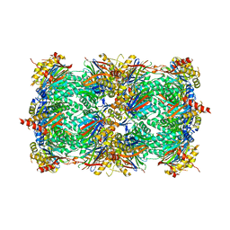 | | Structure of yeast 20S proteasome with bortezomib | | 分子名称: | N-[(1R)-1-(DIHYDROXYBORYL)-3-METHYLBUTYL]-N-(PYRAZIN-2-YLCARBONYL)-L-PHENYLALANINAMIDE, Proteasome component C1, Proteasome component C11, ... | | 著者 | Sintchak, M.D. | | 登録日 | 2010-04-05 | | 公開日 | 2011-05-18 | | 最終更新日 | 2023-09-06 | | 実験手法 | X-RAY DIFFRACTION (2.68 Å) | | 主引用文献 | Characterization of a new series of non-covalent proteasome inhibitors with exquisite potency and selectivity for the 20S beta5-subunit.
Biochem.J., 430, 2010
|
|
3LQM
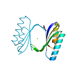
 | | Structure of the IL-10R2 Common Chain | | 分子名称: | GLYCEROL, Interleukin-10 receptor subunit beta, SULFATE ION | | 著者 | Yoon, S.I, Walter, M.R. | | 登録日 | 2010-02-09 | | 公開日 | 2010-05-26 | | 最終更新日 | 2021-10-13 | | 実験手法 | X-RAY DIFFRACTION (2.14 Å) | | 主引用文献 | Structure and mechanism of receptor sharing by the IL-10R2 common chain.
Structure, 18, 2010
|
|
3LS5
 | | Anti-tetrahydrocannabinol Fab Fragment, Free Form | | 分子名称: | Heavy chain of antibody Fab fragment, Light chain of antibody Fab fragment | | 著者 | Niemi, M.H, Rouvinen, J. | | 登録日 | 2010-02-12 | | 公開日 | 2010-06-02 | | 最終更新日 | 2014-01-22 | | 実験手法 | X-RAY DIFFRACTION (1.9 Å) | | 主引用文献 | A structural insight into the molecular recognition of a (-)-Delta9-tetrahydrocannabinol and the development of a sensitive, one-step, homogeneous immunocomplex-based assay for its detection
J.Mol.Biol., 400, 2010
|
|
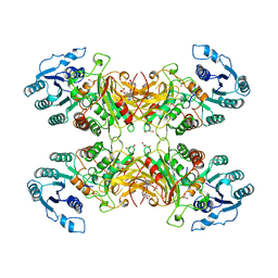
1QK1
 | | CRYSTAL STRUCTURE OF HUMAN UBIQUITOUS MITOCHONDRIAL CREATINE KINASE | | 分子名称: | CREATINE KINASE, UBIQUITOUS MITOCHONDRIAL, PHOSPHATE ION | | 著者 | Eder, M, Schlattner, U, Fritz-Wolf, K, Wallimann, T, Kabsch, W. | | 登録日 | 1999-07-08 | | 公開日 | 2000-04-11 | | 最終更新日 | 2023-12-13 | | 実験手法 | X-RAY DIFFRACTION (2.7 Å) | | 主引用文献 | Crystal Structure of Human Ubiquitous Mitochondrial Creatine Kinase
Proteins: Struct.,Funct., Genet., 39, 2000
|
|
1QMV
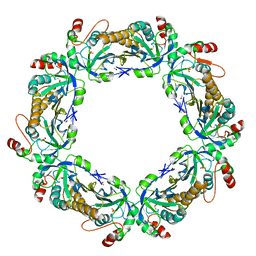 | | thioredoxin peroxidase B from red blood cells | | 分子名称: | PEROXIREDOXIN-2 | | 著者 | Isupov, M.N, Littlechild, J.A, Lebedev, A.A, Errington, N, Vagin, A.A, Schroder, E. | | 登録日 | 1999-10-07 | | 公開日 | 2000-07-28 | | 最終更新日 | 2023-12-13 | | 実験手法 | X-RAY DIFFRACTION (1.7 Å) | | 主引用文献 | Crystal Structure of Decameric 2-Cys Peroxiredoxin from Human Erythrocytes at 1.7 A Resolution.
Structure, 8, 2000
|
|
3LSB
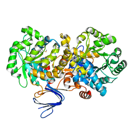 | | Crystal structure of the mutant E241Q of atrazine chlorohydrolase TrzN from Arthrobacter aurescens TC1 complexed with zinc and ametrin | | 分子名称: | N-ethyl-N'-(1-methylethyl)-6-(methylsulfanyl)-1,3,5-triazine-2,4-diamine, Triazine hydrolase, ZINC ION | | 著者 | Fedorov, A.A, Fedorov, E.V, Seffernick, J, Wackett, L.P, Almo, S.C. | | 登録日 | 2010-02-12 | | 公開日 | 2010-07-21 | | 最終更新日 | 2023-09-06 | | 実験手法 | X-RAY DIFFRACTION (1.932 Å) | | 主引用文献 | Crystal structure of the mutant E241Q of atrazine chlorohydrolase TrzN
from Arthrobacter aurescens TC1 complexed with zinc and Ametryn
To be Published
|
|


