Loading
PDBj
メニューPDBj@FacebookPDBj@TwitterPDBj@YouTubewwPDB FoundationwwPDB
RCSB PDBPDBeBMRBAdv. SearchSearch help

2QNF
DownloadVisualize
BU of 2qnf by Molmil
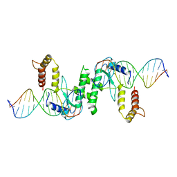
Crystal structure of T4 Endonuclease VII H43N mutant in complex with heteroduplex DNA containing base mismatches
分子名称: DNA (5'-D(*DCP*DAP*DCP*DAP*DTP*DCP*DGP*DAP*DTP*DGP*DGP*DAP*DGP*DCP*DCP*DG)-3'), DNA (5'-D(*DCP*DAP*DCP*DAP*DTP*DCP*DGP*DAP*DTP*DGP*DGP*DAP*DGP*DCP*DGP*DC)-3'), DNA (5'-D(*DCP*DGP*DGP*DCP*DTP*DCP*DCP*DAP*DTP*DCP*DGP*DAP*DTP*DGP*DTP*DG)-3'), ...
著者Biertumpfel, C, Yang, W, Suck, D.
登録日2007-07-18
公開日2008-01-29
最終更新日2023-08-30
実験手法X-RAY DIFFRACTION (3 Å)
主引用文献Crystal structure of T4 endonuclease VII resolving a Holliday junction.
Nature, 449, 2007
2P0H
DownloadVisualize
BU of 2p0h by Molmil
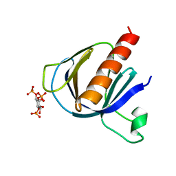
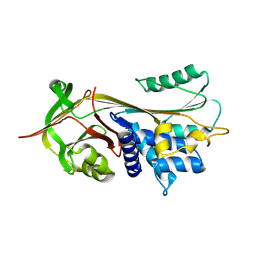
ArhGAP9 PH domain in complex with Ins(1,3,4)P3
分子名称: (1S,3S,4S)-1,3,4-TRIPHOSPHO-MYO-INOSITOL, Rho GTPase-activating protein 9
著者Ceccarelli, D.F.J, Blasutig, I, Goudreault, M, Ruston, J, Pawson, T, Sicheri, F.
登録日2007-02-28
公開日2007-03-27
最終更新日2023-08-30
実験手法X-RAY DIFFRACTION (1.9 Å)
主引用文献Non-canonical Interaction of Phosphoinositides with Pleckstrin Homology Domains of Tiam1 and ArhGAP9.
J.Biol.Chem., 282, 2007
2EG7
DownloadVisualize
BU of 2eg7 by Molmil
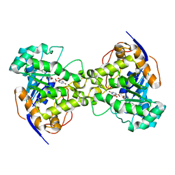
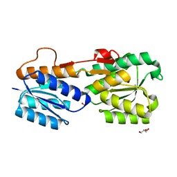
The crystal structure of E. coli dihydroorotase complexed with HDDP
分子名称: 2-OXO-1,2,3,6-TETRAHYDROPYRIMIDINE-4,6-DICARBOXYLIC ACID, Dihydroorotase, ZINC ION
著者Lee, M, Maher, M.J, Guss, J.M.
登録日2007-02-28
公開日2007-07-03
最終更新日2023-11-15
実験手法X-RAY DIFFRACTION (2 Å)
主引用文献Structures of Ligand-free and Inhibitor Complexes of Dihydroorotase from Escherichia coli: Implications for Loop Movement in Inhibitor Design
J.Mol.Biol., 370, 2007
2FV2
DownloadVisualize
BU of 2fv2 by Molmil
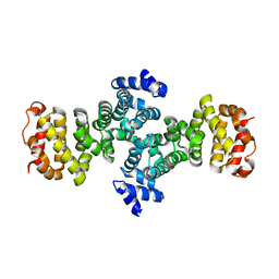
Crystal Structure Analysis of human Rcd-1 conserved region
分子名称: MANGANESE (II) ION, RCD1 required for cell differentiation1 homolog
著者Garces, R.G, Gillon, W, Pai, E.F.
登録日2006-01-28
公開日2007-01-09
最終更新日2024-02-14
実験手法X-RAY DIFFRACTION (2.2 Å)
主引用文献Atomic model of human Rcd-1 reveals an armadillo-like-repeat protein with in vitro nucleic acid binding properties.
Protein Sci., 16, 2007
2FN3
DownloadVisualize
BU of 2fn3 by Molmil
High resolution structure of s26a mutant of benzoylformate decarboxylase from pseudomonas putida complexed with thiamine thiazolone diphosphate
分子名称: 2-{3-[(4-AMINO-2-METHYLPYRIMIDIN-5-YL)METHYL]-4-METHYL-2-OXO-2,3-DIHYDRO-1,3-THIAZOL-5-YL}ETHYL TRIHYDROGEN DIPHOSPHATE, Benzoylformate decarboxylase, CALCIUM ION, ...
著者Bera, A.K, Hasson, M.S.
登録日2006-01-10
公開日2006-12-19
最終更新日2024-02-14
実験手法X-RAY DIFFRACTION (1 Å)
主引用文献High resolution structure of benzoylformate decarboxylase active site mutant s26a from pseudomonas putida complexed with thiamine thiazolone diphosphate
To be Published
2QEF
DownloadVisualize
BU of 2qef by Molmil
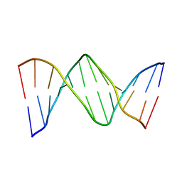
X-ray structure of 7-deaza-dG and Z3dU modified duplex CGCGAATXCZCG
分子名称: DNA (5'-D(*DCP*DGP*DCP*DGP*DAP*DAP*DTP*(ZDU)P*DCP*(7GU)P*DCP*DG)-3')
著者Wang, F, Li, F, Ganguly, M, Marky, L.A, Gold, B, Egli, M, Stone, M.P.
登録日2007-06-25
公開日2008-05-06
最終更新日2023-08-30
実験手法X-RAY DIFFRACTION (1.6 Å)
主引用文献A bridging water anchors the tethered 5-(3-aminopropyl)-2'-deoxyuridine amine in the DNA major groove proximate to the N+2 C.G base pair: implications for formation of interstrand 5'-GNC-3' cross-links by nitrogen mustards.
Biochemistry, 47, 2008
2G33
DownloadVisualize
BU of 2g33 by Molmil
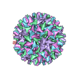
Human Hepatitis B Virus T=4 capsid, strain adyw
分子名称: Core antigen
著者Bourne, C.R, Zlotnick, A.
登録日2006-02-17
公開日2006-11-14
最終更新日2023-08-30
実験手法X-RAY DIFFRACTION (3.96 Å)
主引用文献Global Structural Changes in Hepatitis B Virus Capsids Induced by the Assembly Effector HAP1.
J.Virol., 80, 2006
2G54
DownloadVisualize
BU of 2g54 by Molmil
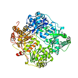
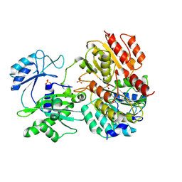
Crystal structure of Zn-bound human insulin-degrading enzyme in complex with insulin B chain
分子名称: 1,4-DIETHYLENE DIOXIDE, Insulin-degrading enzyme, ZINC ION, ...
著者Shen, Y, Tang, W.-J.
登録日2006-02-22
公開日2006-10-24
最終更新日2024-02-14
実験手法X-RAY DIFFRACTION (2.25 Å)
主引用文献Structures of human insulin-degrading enzyme reveal a new substrate recognition mechanism.
Nature, 443, 2006
2QK8
DownloadVisualize
BU of 2qk8 by Molmil
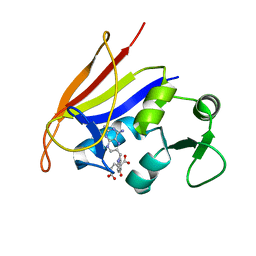
Crystal structure of the anthrax drug target, Bacillus anthracis dihydrofolate reductase
分子名称: Dihydrofolate reductase, METHOTREXATE
著者Bennett, B.C, Xu, H, Simmerman, R.F, Lee, R.E, Dealwis, C.G.
登録日2007-07-10
公開日2007-08-28
最終更新日2023-08-30
実験手法X-RAY DIFFRACTION (2.4 Å)
主引用文献Crystal structure of the anthrax drug target, Bacillus anthracis dihydrofolate reductase.
J.Med.Chem., 50, 2007
2R00
DownloadVisualize
BU of 2r00 by Molmil
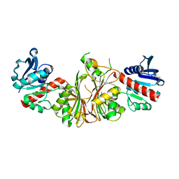
crystal structure of aspartate semialdehyde dehydrogenase II complexed with ASA from vibrio cholerae
分子名称: 2,2'-oxydiacetic acid, Aspartate-semialdehyde dehydrogenase
著者Viola, R.E, Liu, X, Ohren, J.F, Faehnle, C.R.
登録日2007-08-17
公開日2008-04-08
最終更新日2023-08-30
実験手法X-RAY DIFFRACTION (2.03 Å)
主引用文献The structure of a redundant enzyme: a second isoform of aspartate beta-semialdehyde dehydrogenase in Vibrio cholerae.
Acta Crystallogr.,Sect.D, 64, 2008
2QLE
DownloadVisualize
BU of 2qle by Molmil
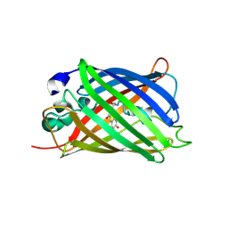
GFP/S205V mutant
分子名称: Green fluorescent protein, IMIDAZOLE
著者Shu, X, Remington, S.J.
登録日2007-07-12
公開日2008-02-12
最終更新日2023-11-15
実験手法X-RAY DIFFRACTION (1.59 Å)
主引用文献An alternative excited-state proton transfer pathway in green fluorescent protein variant S205V.
Protein Sci., 16, 2007
2D1L
DownloadVisualize
BU of 2d1l by Molmil
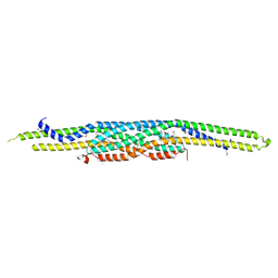
Structure of F-actin binding domain IMD of MIM (Missing In Metastasis)
分子名称: Metastasis suppressor protein 1
著者Lee, S.H, Kerff, F, Chereau, D, Ferron, F, Dominguez, R.
登録日2005-08-27
公開日2006-09-12
最終更新日2017-10-11
実験手法X-RAY DIFFRACTION (1.85 Å)
主引用文献Structural basis for the actin-binding function of missing-in-metastasis
Structure, 15, 2007
2QUG
DownloadVisualize
BU of 2qug by Molmil
Crystal structure of alpha-1-antitrypsin, crystal form A
分子名称: Alpha-1-antitrypsin
著者Hansen, G, Morton, C.J, Pearce, M.C, Feil, S.C, Adams, J.J, Parker, M.W, Bottomley, S.P.
登録日2007-08-05
公開日2008-08-12
最終更新日2011-07-13
実験手法X-RAY DIFFRACTION (2 Å)
主引用文献Preventing serpin aggregation: The molecular mechanism of citrate action upon antitrypsin unfolding.
Protein Sci., 17, 2008
2RTB
DownloadVisualize
BU of 2rtb by Molmil
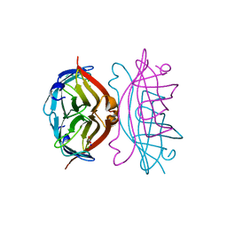
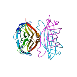
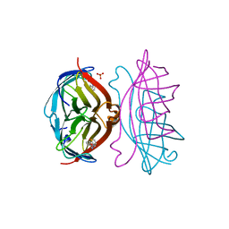
APOSTREPTAVIDIN, PH 3.32, SPACE GROUP I222
分子名称: ACETATE ION, CHLORIDE ION, SODIUM ION, ...
著者Katz, B.A.
登録日1997-09-11
公開日1998-10-14
最終更新日2024-02-21
実験手法X-RAY DIFFRACTION (1.5 Å)
主引用文献Binding of biotin to streptavidin stabilizes intersubunit salt bridges between Asp61 and His87 at low pH.
J.Mol.Biol., 274, 1997
2RTH
DownloadVisualize
BU of 2rth by Molmil
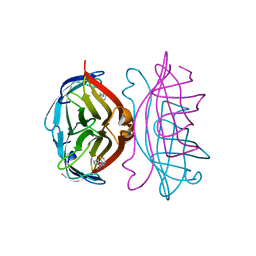
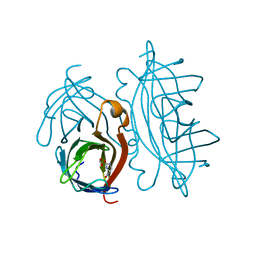
STREPTAVIDIN-GLYCOLURIL COMPLEX, PH 2.50, SPACE GROUP I222
分子名称: ACETATE ION, GLYCOLURIL, STREPTAVIDIN
著者Katz, B.A.
登録日1997-09-11
公開日1998-10-14
最終更新日2024-02-21
実験手法X-RAY DIFFRACTION (1.56 Å)
主引用文献Binding of biotin to streptavidin stabilizes intersubunit salt bridges between Asp61 and His87 at low pH.
J.Mol.Biol., 274, 1997
2RTP
DownloadVisualize
BU of 2rtp by Molmil
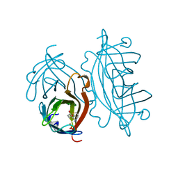
STREPTAVIDIN-2-IMINOBIOTIN COMPLEX, PH 3.25, SPACE GROUP I222
分子名称: 2-IMINOBIOTIN, STREPTAVIDIN
著者Katz, B.A.
登録日1997-09-11
公開日1998-10-14
最終更新日2024-02-21
実験手法X-RAY DIFFRACTION (1.5 Å)
主引用文献Binding of biotin to streptavidin stabilizes intersubunit salt bridges between Asp61 and His87 at low pH.
J.Mol.Biol., 274, 1997
2RTE
DownloadVisualize
BU of 2rte by Molmil
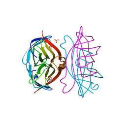
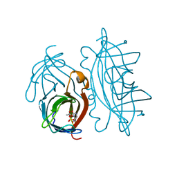
STREPTAVIDIN-BIOTIN COMPLEX, PH 1.90, SPACE GROUP I222
分子名称: BIOTIN, STREPTAVIDIN, SULFATE ION
著者Katz, B.A.
登録日1997-09-11
公開日1998-10-14
最終更新日2024-02-21
実験手法X-RAY DIFFRACTION (1.5 Å)
主引用文献Binding of biotin to streptavidin stabilizes intersubunit salt bridges between Asp61 and His87 at low pH.
J.Mol.Biol., 274, 1997
2RTM
DownloadVisualize
BU of 2rtm by Molmil
STREPTAVIDIN-2-IMINOBIOTIN-SULFATE COMPLEX, PH 3.50, SPACE GROUP I4122
分子名称: 2-IMINOBIOTIN, STREPTAVIDIN, SULFATE ION
著者Katz, B.A.
登録日1997-09-11
公開日1998-10-14
最終更新日2024-02-21
実験手法X-RAY DIFFRACTION (1.3 Å)
主引用文献Binding of biotin to streptavidin stabilizes intersubunit salt bridges between Asp61 and His87 at low pH.
J.Mol.Biol., 274, 1997
2FS6
DownloadVisualize
BU of 2fs6 by Molmil
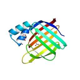
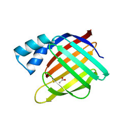
Crystal Structure of Apo-Cellular Retinoic Acid Binding Protein Type II At 1.35 Angstroms Resolution
分子名称: ACETATE ION, CHLORIDE ION, Cellular retinoic acid-binding protein 2, ...
著者Vaezeslami, S, Geiger, J.H.
登録日2006-01-21
公開日2006-09-19
最終更新日2024-02-14
実験手法X-RAY DIFFRACTION (1.35 Å)
主引用文献The structure of Apo-wild-type cellular retinoic acid binding protein II at 1.4 A and its relationship to ligand binding and nuclear translocation.
J.Mol.Biol., 363, 2006
2RTF
DownloadVisualize
BU of 2rtf by Molmil
STREPTAVIDIN-BIOTIN COMPLEX, PH 2.00, SPACE GROUP I222
分子名称: BIOTIN, STREPTAVIDIN, SULFATE ION
著者Katz, B.A.
登録日1997-09-11
公開日1998-10-14
最終更新日2024-02-21
実験手法X-RAY DIFFRACTION (1.47 Å)
主引用文献Binding of biotin to streptavidin stabilizes intersubunit salt bridges between Asp61 and His87 at low pH.
J.Mol.Biol., 274, 1997
2RTJ
DownloadVisualize
BU of 2rtj by Molmil
STREPTAVIDIN-GLYCOLURIL COMPLEX, PH 2.50, SPACE GROUP I4122
分子名称: FORMIC ACID, GLYCOLURIL, STREPTAVIDIN
著者Katz, B.A.
登録日1997-09-11
公開日1998-10-14
最終更新日2024-02-21
実験手法X-RAY DIFFRACTION (1.4 Å)
主引用文献Binding of biotin to streptavidin stabilizes intersubunit salt bridges between Asp61 and His87 at low pH.
J.Mol.Biol., 274, 1997
2RTK
DownloadVisualize
BU of 2rtk by Molmil
STREPTAVIDIN-GLYCOLURIL COMPLEX, PH 2.58, SPACE GROUP I4122 PREPARED FROM AN APOSTREPTAVIDIN CRYSTAL
分子名称: ACETATE ION, GLYCOLURIL, STREPTAVIDIN, ...
著者Katz, B.A.
登録日1997-09-11
公開日1998-10-14
最終更新日2024-02-21
実験手法X-RAY DIFFRACTION (1.82 Å)
主引用文献Binding of biotin to streptavidin stabilizes intersubunit salt bridges between Asp61 and His87 at low pH.
J.Mol.Biol., 274, 1997
2FVY
DownloadVisualize
BU of 2fvy by Molmil
High Resolution Glucose Bound Crystal Structure of GGBP
分子名称: ACETATE ION, CALCIUM ION, CARBON DIOXIDE, ...
著者Borrok, M.J, Kiessling, L.L, Forest, K.T.
登録日2006-01-31
公開日2007-02-06
最終更新日2024-04-03
実験手法X-RAY DIFFRACTION (0.92 Å)
主引用文献Conformational changes of glucose/galactose-binding protein illuminated by open, unliganded, and ultra-high-resolution ligand-bound structures.
Protein Sci., 16, 2007
2FS7
DownloadVisualize
BU of 2fs7 by Molmil
Crystal Structure of Apo-Cellular Retinoic Acid Binding Protein Type II At 1.55 Angstroms Resolution
分子名称: ACETATE ION, CHLORIDE ION, Cellular retinoic acid-binding protein 2
著者Vaezeslami, S, Geiger, J.H.
登録日2006-01-21
公開日2006-09-19
最終更新日2024-02-14
実験手法X-RAY DIFFRACTION (1.55 Å)
主引用文献The structure of Apo-wild-type cellular retinoic acid binding protein II at 1.4 A and its relationship to ligand binding and nuclear translocation.
J.Mol.Biol., 363, 2006
2FPI
DownloadVisualize
BU of 2fpi by Molmil
Crystal structure of pig GTP-specific succinyl-CoA synthetase from polyethylene glycol
分子名称: SULFATE ION, Succinyl-CoA ligase [GDP-forming] alpha-chain, mitochondrial, ...
著者Fraser, M.E.
登録日2006-01-16
公開日2006-02-21
最終更新日2017-10-18
実験手法X-RAY DIFFRACTION (2.7 Å)
主引用文献Interactions of GTP with the ATP-grasp Domain of GTP-specific Succinyl-CoA Synthetase
J.Biol.Chem., 281, 2006

223790

件を2024-08-14に公開中

PDB statisticsPDBj update infoContact PDBjnumon