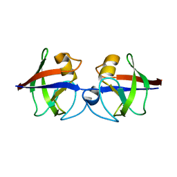
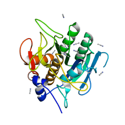
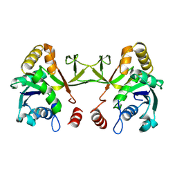
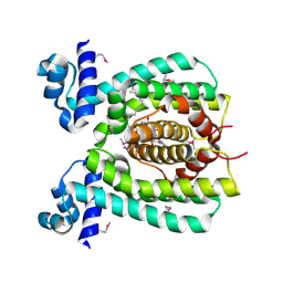
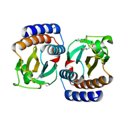
Loading
PDBj
メニューPDBj@FacebookPDBj@TwitterPDBj@YouTubewwPDB FoundationwwPDB
RCSB PDBPDBeBMRBAdv. SearchSearch help

1USL
DownloadVisualize
BU of 1usl by Molmil
Structure Of Mycobacterium tuberculosis Ribose-5-Phosphate Isomerase, RpiB, Rv2465c, Complexed With Phosphate.
分子名称: PHOSPHATE ION, RIBOSE 5-PHOSPHATE ISOMERASE B
著者Roos, A.K, Andersson, C.E, Unge, T, Jones, T.A, Mowbray, S.L.
登録日2003-11-25
公開日2004-01-02
最終更新日2023-12-13
実験手法X-RAY DIFFRACTION (1.88 Å)
主引用文献Mycobacterium Tuberculosis Ribose-5-Phosphate Isomerase Has a Known Fold, But a Novel Active Site
J.Mol.Biol., 335, 2004
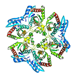
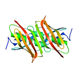
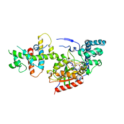
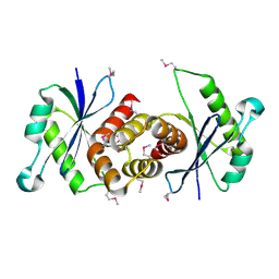
4AVR
DownloadVisualize
BU of 4avr by Molmil
Crystal structure of the hypothetical protein Pa4485 from Pseudomonas aeruginosa
分子名称: PA4485
著者McMahon, S.A, Moynie, L, Liu, H, Duthie, F, Alphey, M.S, Naismith, J.H.
登録日2012-05-29
公開日2013-01-09
最終更新日2024-05-08
実験手法X-RAY DIFFRACTION (1.08 Å)
主引用文献The Aeropath Project Targeting Pseudomonas Aeruginosa: Crystallographic Studies for Assessment of Potential Targets in Early-Stage Drug Discovery.
Acta Crystallogr.,Sect.F, 69, 2013
1SKJ
DownloadVisualize
BU of 1skj by Molmil
COCRYSTAL STRUCTURE OF UREA-SUBSTITUTED PHOSPHOPEPTIDE COMPLEX
分子名称: 4-[3-CARBOXYMETHYL-3-(4-PHOSPHONOOXY-BENZYL)-UREIDO]-4-[(3-CYCLOHEXYL-PROPYL)-METHYL-CARBAMOYL]BUTYRIC ACID, PP60 V-SRC TYROSINE KINASE TRANSFORMING PROTEIN
著者Holland, D.R, Rubin, J.R.
登録日1997-09-18
公開日1998-02-25
最終更新日2024-05-22
実験手法X-RAY DIFFRACTION (2 Å)
主引用文献Design, synthesis, and cocrystal structure of a nonpeptide Src SH2 domain ligand.
J.Med.Chem., 40, 1997
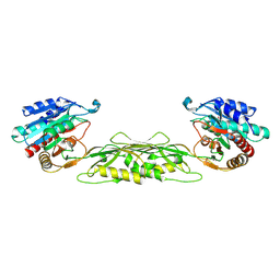
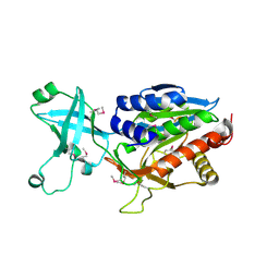
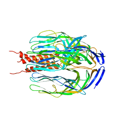
4AVF
DownloadVisualize
BU of 4avf by Molmil
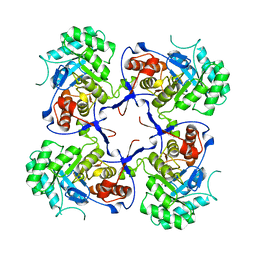
Crystal structure of Pseudomonas aeruginosa inosine 5'-monophosphate dehydrogenase
分子名称: INOSINE-5'-MONOPHOSPHATE DEHYDROGENASE
著者McMahon, S.A, Moynie, L, Liu, H, Duthie, F, Naismith, J.H.
登録日2012-05-25
公開日2013-01-09
最終更新日2023-12-20
実験手法X-RAY DIFFRACTION (2.23 Å)
主引用文献The Aeropath Project Targeting Pseudomonas Aeruginosa: Crystallographic Studies for Assessment of Potential Targets in Early-Stage Drug Discovery
Acta Crystallogr.,Sect.F, 69, 2013
4BIQ
DownloadVisualize
BU of 4biq by Molmil
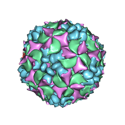
Homology model of coxsackievirus A7 (CAV7) empty capsid proteins.
分子名称: VP1, VP2, VP3
著者Seitsonen, J.J.T, Shakeel, S, Susi, P, Pandurangan, A.P, Sinkovits, R.S, Hyvonen, H, Laurinmaki, P, Yla-Pelto, J, Topf, M, Hyypia, T, Butcher, S.J.
登録日2013-04-12
公開日2013-10-02
最終更新日2024-05-08
実験手法ELECTRON MICROSCOPY (6.09 Å)
主引用文献Combined Approaches to Flexible Fitting and Assessment in Virus Capsids Undergoing Conformational Change.
J.Struct.Biol., 185, 2014
1SB7
DownloadVisualize
BU of 1sb7 by Molmil
Crystal structure of the E.coli pseudouridine synthase TruD
分子名称: GLYCEROL, PHOSPHATE ION, tRNA pseudouridine synthase D
著者Hoang, C, Ferre-D'Amare, A.R.
登録日2004-02-10
公開日2004-06-29
最終更新日2011-07-13
実験手法X-RAY DIFFRACTION (2.2 Å)
主引用文献Crystal structure of the highly divergent pseudouridine synthase TruD reveals a circular permutation of a conserved fold.
Rna, 10, 2004
1SCB
DownloadVisualize
BU of 1scb by Molmil
ENZYME CRYSTAL STRUCTURE IN A NEAT ORGANIC SOLVENT
分子名称: ACETONITRILE, CALCIUM ION, SUBTILISIN CARLSBERG
著者Fitzpatrick, P.A, Steinmetz, A.C.U, Ringe, D, Klibanov, A.M.
登録日1993-07-13
公開日1994-01-31
最終更新日2024-02-14
実験手法X-RAY DIFFRACTION (2.3 Å)
主引用文献Enzyme crystal structure in a neat organic solvent.
Proc.Natl.Acad.Sci.USA, 90, 1993
1VGU
DownloadVisualize
BU of 1vgu by Molmil
Crystal structure of 4-diphosphocytidyl-2C-methyl-D-erythritol synthase
分子名称: 2-C-methyl-D-erythritol 4-phosphate cytidylyltransferase
著者Structural GenomiX
登録日2003-11-03
公開日2003-11-25
最終更新日2023-12-27
実験手法X-RAY DIFFRACTION (2.8 Å)
主引用文献Structural analysis of a set of proteins resulting from a bacterial genomics project
Proteins, 60, 2005
1VH0
DownloadVisualize
BU of 1vh0 by Molmil
Crystal structure of a hypothetical protein
分子名称: Hypothetical UPF0247 protein SAV0024/SA0023
著者Structural GenomiX
登録日2003-11-03
公開日2003-11-25
最終更新日2023-12-27
実験手法X-RAY DIFFRACTION (2.31 Å)
主引用文献Structural analysis of a set of proteins resulting from a bacterial genomics project
Proteins, 60, 2005
1VHL
DownloadVisualize
BU of 1vhl by Molmil
Crystal structure of dephospho-CoA kinase with adenosine-5'-diphosphate
分子名称: ACETATE ION, ADENOSINE-5'-DIPHOSPHATE, Dephospho-CoA kinase
著者Structural GenomiX
登録日2003-12-01
公開日2003-12-30
最終更新日2023-12-27
実験手法X-RAY DIFFRACTION (1.65 Å)
主引用文献Structural analysis of a set of proteins resulting from a bacterial genomics project
Proteins, 60, 2005
1VI1
DownloadVisualize
BU of 1vi1 by Molmil
Crystal structure of a fatty acid/phospholipid synthesis protein
分子名称: Fatty acid/phospholipid synthesis protein plsX
著者Structural GenomiX
登録日2003-12-01
公開日2003-12-30
最終更新日2023-12-27
実験手法X-RAY DIFFRACTION (2.95 Å)
主引用文献Structural analysis of a set of proteins resulting from a bacterial genomics project
Proteins, 60, 2005
1VGY
DownloadVisualize
BU of 1vgy by Molmil
Crystal structure of succinyl diaminopimelate desuccinylase
分子名称: succinyl-diaminopimelate desuccinylase
著者Structural GenomiX
登録日2003-11-03
公開日2003-11-25
最終更新日2023-12-27
実験手法X-RAY DIFFRACTION (1.9 Å)
主引用文献Structural analysis of a set of proteins resulting from a bacterial genomics project
Proteins, 60, 2005
1VHJ
DownloadVisualize
BU of 1vhj by Molmil
Crystal structure of purine nucleoside phosphorylase
分子名称: purine nucleoside phosphorylase
著者Structural GenomiX
登録日2003-12-01
公開日2003-12-30
最終更新日2023-12-27
実験手法X-RAY DIFFRACTION (2.23 Å)
主引用文献Structural analysis of a set of proteins resulting from a bacterial genomics project
Proteins, 60, 2005
1VGV
DownloadVisualize
BU of 1vgv by Molmil
Crystal structure of UDP-N-acetylglucosamine_2 epimerase
分子名称: UDP-N-acetylglucosamine 2-epimerase, URIDINE-DIPHOSPHATE-N-ACETYLGLUCOSAMINE
著者Structural GenomiX
登録日2003-11-03
公開日2003-11-25
最終更新日2023-12-27
実験手法X-RAY DIFFRACTION (2.31 Å)
主引用文献Structural analysis of a set of proteins resulting from a bacterial genomics project
Proteins, 60, 2005
1VIX
DownloadVisualize
BU of 1vix by Molmil
Crystal structure of a putative peptidase T
分子名称: Peptidase T, SULFATE ION, ZINC ION
著者Structural GenomiX
登録日2003-12-01
公開日2003-12-30
最終更新日2023-12-27
実験手法X-RAY DIFFRACTION (2.5 Å)
主引用文献Structural analysis of a set of proteins resulting from a bacterial genomics project
Proteins, 60, 2005
1VGT
DownloadVisualize
BU of 1vgt by Molmil
Crystal structure of 4-diphosphocytidyl-2C-methyl-D-erythritol synthase
分子名称: 2-C-methyl-D-erythritol 4-phosphate cytidylyltransferase
著者Structural GenomiX
登録日2003-11-03
公開日2003-11-25
最終更新日2023-12-27
実験手法X-RAY DIFFRACTION (1.8 Å)
主引用文献Structural analysis of a set of proteins resulting from a bacterial genomics project
Proteins, 60, 2005
1VH5
DownloadVisualize
BU of 1vh5 by Molmil
Crystal structure of a putative thioesterase
分子名称: Hypothetical protein ydiI
著者Burley, S.K, New York SGX Research Center for Structural Genomics (NYSGXRC)
登録日2003-12-01
公開日2003-12-30
最終更新日2023-12-27
実験手法X-RAY DIFFRACTION (1.34 Å)
主引用文献Structural analysis of a set of proteins resulting from a bacterial genomics project
Proteins, 60, 2005
1VHE
DownloadVisualize
BU of 1vhe by Molmil
Crystal structure of a aminopeptidase/glucanase homolog
分子名称: ZINC ION, aminopeptidase/glucanase homolog
著者Structural GenomiX
登録日2003-12-01
公開日2003-12-30
最終更新日2023-12-27
実験手法X-RAY DIFFRACTION (1.9 Å)
主引用文献Structural analysis of a set of proteins resulting from a bacterial genomics project
Proteins, 60, 2005
1VHS
DownloadVisualize
BU of 1vhs by Molmil
Crystal structure of a putative phosphinothricin N-acetyltransferase
分子名称: similar to phosphinothricin acetyltransferase
著者Structural GenomiX
登録日2003-12-01
公開日2003-12-30
最終更新日2023-12-27
実験手法X-RAY DIFFRACTION (1.8 Å)
主引用文献Structural analysis of a set of proteins resulting from a bacterial genomics project
Proteins, 60, 2005
1VI0
DownloadVisualize
BU of 1vi0 by Molmil
Crystal structure of a transcriptional regulator
分子名称: DODECYL-COA, transcriptional regulator
著者Structural GenomiX
登録日2003-12-01
公開日2003-12-30
最終更新日2023-12-27
実験手法X-RAY DIFFRACTION (1.65 Å)
主引用文献Structural analysis of a set of proteins resulting from a bacterial genomics project
Proteins, 60, 2005
1SHZ
DownloadVisualize
BU of 1shz by Molmil
Crystal Structure of the p115RhoGEF rgRGS Domain in A Complex with Galpha(13):Galpha(i1) Chimera
分子名称: GUANOSINE-5'-DIPHOSPHATE, Guanine Nucleotide-Binding Protein Galpha(13):Galpha(i1) Chimera, MAGNESIUM ION, ...
著者Chen, Z, Singer, W.D, Sternweis, P.C, Sprang, S.R.
登録日2004-02-26
公開日2005-01-18
最終更新日2023-08-23
実験手法X-RAY DIFFRACTION (2.85 Å)
主引用文献Structure of the p115RhoGEF rgRGS domain-Galpha13/i1 chimera complex suggests convergent evolution of a GTPase activator.
Nat.Struct.Mol.Biol., 12, 2005
1SLQ
DownloadVisualize
BU of 1slq by Molmil
Crystal structure of the trimeric state of the rhesus rotavirus VP4 membrane interaction domain, VP5CT
分子名称: VP4
著者Dormitzer, P.R, Nason, E.B, Prasad, B.V.V, Harrison, S.C.
登録日2004-03-06
公開日2004-08-31
最終更新日2024-02-14
実験手法X-RAY DIFFRACTION (3.2 Å)
主引用文献Structural rearrangements in the membrane penetration protein of a non-enveloped virus.
Nature, 430, 2004
1VH1
DownloadVisualize
BU of 1vh1 by Molmil
Crystal structure of CMP-KDO synthetase
分子名称: 3-deoxy-manno-octulosonate cytidylyltransferase
著者Structural GenomiX
登録日2003-12-01
公開日2003-12-30
最終更新日2023-12-27
実験手法X-RAY DIFFRACTION (2.6 Å)
主引用文献Structural analysis of a set of proteins resulting from a bacterial genomics project
Proteins, 60, 2005
1VHM
DownloadVisualize
BU of 1vhm by Molmil
Crystal structure of an hypothetical protein
分子名称: 2-(N-MORPHOLINO)-ETHANESULFONIC ACID, Protein yebR
著者Structural GenomiX
登録日2003-12-01
公開日2003-12-30
最終更新日2023-12-27
実験手法X-RAY DIFFRACTION (2.1 Å)
主引用文献Structural analysis of a set of proteins resulting from a bacterial genomics project
Proteins, 60, 2005
1VHX
DownloadVisualize
BU of 1vhx by Molmil
Crystal structure of Putative Holliday junction resolvase
分子名称: Putative Holliday junction resolvase
著者Structural GenomiX
登録日2003-12-01
公開日2003-12-30
最終更新日2023-12-27
実験手法X-RAY DIFFRACTION (1.96 Å)
主引用文献Structural analysis of a set of proteins resulting from a bacterial genomics project
Proteins, 60, 2005

224004

件を2024-08-21に公開中

PDB statisticsPDBj update infoContact PDBjnumon