2WKG
 
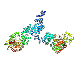
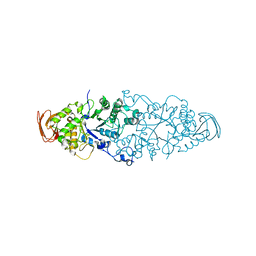 | | Nostoc punctiforme Debranching Enzyme (NPDE)(Native form) | | 分子名称: | ALPHA AMYLASE, CATALYTIC REGION | | 著者 | Dumbrepatil, A.B, Choi, J.H, Song, H.N, Park, K.H, Woo, E.J. | | 登録日 | 2009-06-11 | | 公開日 | 2009-09-29 | | 最終更新日 | 2023-12-13 | | 実験手法 | X-RAY DIFFRACTION (3 Å) | | 主引用文献 | Structural Features of the Nostoc Punctiforme Debranching Enzyme Reveal the Basis of its Mechanism and Substrate Specificity.
Proteins, 78, 2010
|
|
4UT5
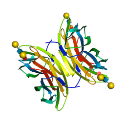 | | Crystal structure of the LecB lectin from Pseudomonas aeruginosa strain PA7 in complex with lewis a tetrasaccharide | | 分子名称: | CALCIUM ION, LECB LECTIN, beta-D-galactopyranose, ... | | 著者 | Boukerb, A.M, Decor, A, Tabaroni, R, Varrot, A, Debentzmann, S, Vidal, S, Imberty, A, Cournoyer, B. | | 登録日 | 2014-07-18 | | 公開日 | 2015-07-22 | | 最終更新日 | 2024-01-10 | | 実験手法 | X-RAY DIFFRACTION (1.75 Å) | | 主引用文献 | Genomic Rearrangements and Functional Diversification of Leca and Lecb Lectin-Coding Regions Impacting the Efficacy of Glycomimetics Directed Against Pseudomonas Aeruginosa.
Front.Microbiol., 7, 2016
|
|
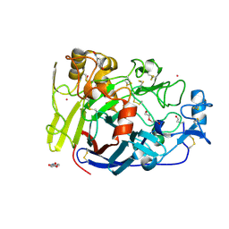
4UTS
 | | Room temperature crystal structure of the fast switching M159T mutant of fluorescent protein Dronpa | | 分子名称: | FLUORESCENT PROTEIN DRONPA | | 著者 | Kaucikas, M, Fitzpatrick, A, Bryan, E, Struve, A, Henning, R, Kosheleva, I, Srajer, V, van Thor, J.J. | | 登録日 | 2014-07-22 | | 公開日 | 2015-06-03 | | 最終更新日 | 2024-01-10 | | 実験手法 | X-RAY DIFFRACTION (2.03 Å) | | 主引用文献 | Room Temperature Crystal Structure of the Fast Switching M159T Mutant of the Fluorescent Protein Dronpa.
Proteins, 83, 2015
|
|
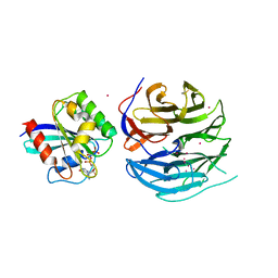
4UWT
 | | Hypocrea jecorina Cel7A E212Q mutant in complex with p-nitrophenyl cellobioside | | 分子名称: | 2-acetamido-2-deoxy-beta-D-glucopyranose, CELLOBIOHYDROLASE CEL7A, COBALT (II) ION, ... | | 著者 | Nutt, A, Momeni, M.H, Johansson, G, Stahlberg, J. | | 登録日 | 2014-08-14 | | 公開日 | 2015-08-26 | | 最終更新日 | 2024-01-31 | | 実験手法 | X-RAY DIFFRACTION (1.2 Å) | | 主引用文献 | Enzyme kinetics by GH7 cellobiohydrolases on chromogenic substrates is dictated by non-productive binding: insights from crystal structures and MD simulation.
Febs J., 2022
|
|
4V0N
 | |
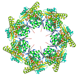
8G7M
 | | ATP-bound mtHsp60 V72I focus | | 分子名称: | 60 kDa heat shock protein, mitochondrial, ADENOSINE-5'-TRIPHOSPHATE, ... | | 著者 | Braxton, J.R, Shao, H, Tse, E, Gestwicki, J.E, Southworth, D.R. | | 登録日 | 2023-02-16 | | 公開日 | 2023-07-12 | | 最終更新日 | 2024-06-19 | | 実験手法 | ELECTRON MICROSCOPY (3.2 Å) | | 主引用文献 | Asymmetric apical domain states of mitochondrial Hsp60 coordinate substrate engagement and chaperonin assembly.
Biorxiv, 2023
|
|
7NTF
 | |
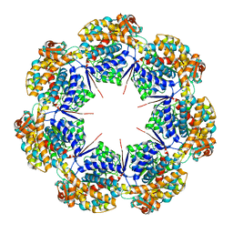
8G7L
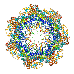
 | | ATP-bound mtHsp60 V72I | | 分子名称: | 60 kDa heat shock protein, mitochondrial, ADENOSINE-5'-TRIPHOSPHATE, ... | | 著者 | Braxton, J.R, Shao, H, Tse, E, Gestwicki, J.E, Southworth, D.R. | | 登録日 | 2023-02-16 | | 公開日 | 2023-07-12 | | 最終更新日 | 2024-06-19 | | 実験手法 | ELECTRON MICROSCOPY (2.5 Å) | | 主引用文献 | Asymmetric apical domain states of mitochondrial Hsp60 coordinate substrate engagement and chaperonin assembly.
Biorxiv, 2023
|
|
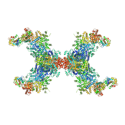
8G7O
 | | ATP- and mtHsp10-bound mtHsp60 V72I focus | | 分子名称: | 10 kDa heat shock protein, mitochondrial, 60 kDa heat shock protein, ... | | 著者 | Braxton, J.R, Shao, H, Tse, E, Gestwicki, J.E, Southworth, D.R. | | 登録日 | 2023-02-16 | | 公開日 | 2023-07-12 | | 最終更新日 | 2024-06-19 | | 実験手法 | ELECTRON MICROSCOPY (3.4 Å) | | 主引用文献 | Asymmetric apical domain states of mitochondrial Hsp60 coordinate substrate engagement and chaperonin assembly.
Biorxiv, 2023
|
|
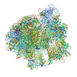
4V6X
 | | Structure of the human 80S ribosome | | 分子名称: | 18S ribosomal RNA, 28S ribosomal RNA, 40S ribosomal protein S10, ... | | 著者 | Anger, A.M, Armache, J.-P, Berninghausen, O, Habeck, M, Subklewe, M, Wilson, D.N, Beckmann, R. | | 登録日 | 2013-02-27 | | 公開日 | 2014-07-09 | | 最終更新日 | 2023-02-01 | | 実験手法 | ELECTRON MICROSCOPY (5 Å) | | 主引用文献 | Structures of the human and Drosophila 80S ribosome.
Nature, 497, 2013
|
|
1X24
 | | Prl-1 (ptp4a) | | 分子名称: | protein tyrosine phosphatase 4a1 | | 著者 | Zhang, Z.Y, Sun, J.P, Liu, S, Wang, W.Q, Yang, H. | | 登録日 | 2005-04-20 | | 公開日 | 2005-10-11 | | 最終更新日 | 2011-07-13 | | 実験手法 | X-RAY DIFFRACTION (3.2 Å) | | 主引用文献 | Structure and Biochemical Properties of PRL-1, a Phosphatase Implicated in Cell Growth, Differentiation, and Tumor Invasion(,)
Biochemistry, 44, 2005
|
|
8G7N
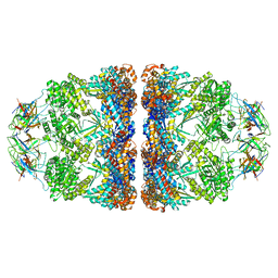 | | ATP- and mtHsp10-bound mtHsp60 V72I | | 分子名称: | 10 kDa heat shock protein, mitochondrial, 60 kDa heat shock protein, ... | | 著者 | Braxton, J.R, Shao, H, Tse, E, Gestwicki, J.E, Southworth, D.R. | | 登録日 | 2023-02-16 | | 公開日 | 2023-07-12 | | 最終更新日 | 2024-06-19 | | 実験手法 | ELECTRON MICROSCOPY (2.7 Å) | | 主引用文献 | Asymmetric apical domain states of mitochondrial Hsp60 coordinate substrate engagement and chaperonin assembly.
Biorxiv, 2023
|
|
8G7J
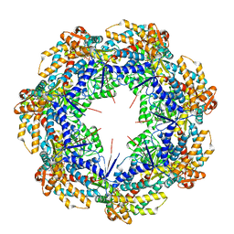
 | | mtHsp60 V72I apo | | 分子名称: | 60 kDa heat shock protein, mitochondrial | | 著者 | Braxton, J.R, Shao, H, Tse, E, Gestwicki, J.E, Southworth, D.R. | | 登録日 | 2023-02-16 | | 公開日 | 2023-07-12 | | 最終更新日 | 2024-06-19 | | 実験手法 | ELECTRON MICROSCOPY (3.4 Å) | | 主引用文献 | Asymmetric apical domain states of mitochondrial Hsp60 coordinate substrate engagement and chaperonin assembly.
Biorxiv, 2023
|
|
8G7K
 | | mtHsp60 V72I apo focus | | 分子名称: | 60 kDa heat shock protein, mitochondrial | | 著者 | Braxton, J.R, Shao, H, Tse, E, Gestwicki, J.E, Southworth, D.R. | | 登録日 | 2023-02-16 | | 公開日 | 2023-07-12 | | 最終更新日 | 2024-06-19 | | 実験手法 | ELECTRON MICROSCOPY (3.8 Å) | | 主引用文献 | Asymmetric apical domain states of mitochondrial Hsp60 coordinate substrate engagement and chaperonin assembly.
Biorxiv, 2023
|
|
8JFK
 | |
7NWT
 | | Initiated 70S ribosome in complex with 2A protein from encephalomyocarditis virus (EMCV) | | 分子名称: | 16S ribosomal RNA, 23S ribosomal RNA, 30S ribosomal protein S10, ... | | 著者 | Hill, C.H, Napthine, S, Pekarek, L, Kibe, A, Firth, A.E, Graham, S.C, Caliskan, N, Brierley, I. | | 登録日 | 2021-03-17 | | 公開日 | 2021-12-15 | | 最終更新日 | 2024-04-24 | | 実験手法 | ELECTRON MICROSCOPY (2.66 Å) | | 主引用文献 | Structural and molecular basis for Cardiovirus 2A protein as a viral gene expression switch
Nat Commun, 12, 2021
|
|
8ATJ
 | | Crystal Structure of Shank2-SAM domain | | 分子名称: | CHLORIDE ION, FORMIC ACID, Isoform 4 of SH3 and multiple ankyrin repeat domains protein 2, ... | | 著者 | Bento, I, Gracia Alai, M, Kreienkamp, J.-H. | | 登録日 | 2022-08-23 | | 公開日 | 2022-11-30 | | 最終更新日 | 2024-01-31 | | 実験手法 | X-RAY DIFFRACTION (2.117 Å) | | 主引用文献 | Structural deficits in key domains of Shank2 lead to alterations in postsynaptic nanoclusters and to a neurodevelopmental disorder in humans.
Mol Psychiatry, 2022
|
|
8B10
 | | Crystal Structure of Shank2-SAM mutant domain - L1800W | | 分子名称: | 2-[BIS-(2-HYDROXY-ETHYL)-AMINO]-2-HYDROXYMETHYL-PROPANE-1,3-DIOL, CHLORIDE ION, DI(HYDROXYETHYL)ETHER, ... | | 著者 | Bento, I, Gracia Alai, M, Kreienkamp, J.-H. | | 登録日 | 2022-09-08 | | 公開日 | 2022-11-30 | | 最終更新日 | 2024-01-31 | | 実験手法 | X-RAY DIFFRACTION (1.95 Å) | | 主引用文献 | Structural deficits in key domains of Shank2 lead to alterations in postsynaptic nanoclusters and to a neurodevelopmental disorder in humans.
Mol Psychiatry, 2022
|
|
2M02
 | | 3D structure of cap-gly domain of mammalian dynactin determined by magic angle spinning NMR spectroscopy | | 分子名称: | Dynactin subunit 1 | | 著者 | Yan, S, Hou, G, Schwieters, C.D, Ahmed, S, Williams, J.C, Polenova, T. | | 登録日 | 2012-10-15 | | 公開日 | 2013-05-08 | | 最終更新日 | 2024-05-15 | | 実験手法 | SOLID-STATE NMR | | 主引用文献 | Three-Dimensional Structure of CAP-Gly Domain of Mammalian Dynactin Determined by Magic Angle Spinning NMR Spectroscopy: Conformational Plasticity and Interactions with End-Binding Protein EB1.
J.Mol.Biol., 425, 2013
|
|
2WC7
 | | Crystal structure of Nostoc Punctiforme Debranching Enzyme(NPDE)(Acarbose soaked) | | 分子名称: | ALPHA AMYLASE, CATALYTIC REGION | | 著者 | Dumbrepatil, A.-B, Song, H.-N, Choi, J.-H, Park, K.-H, Woo, E.-J. | | 登録日 | 2009-03-10 | | 公開日 | 2009-09-29 | | 最終更新日 | 2023-12-13 | | 実験手法 | X-RAY DIFFRACTION (2.37 Å) | | 主引用文献 | Structural Features of the Nostoc Punctiforme Debranching Enzyme Reveal the Basis of its Mechanism and Substrate Specificity.
Proteins, 78, 2010
|
|
2M5J
 | | Solution structure of the periplasmic signaling domain of HasR, a TonB-dependent outer membrane heme transporter | | 分子名称: | HasR protein | | 著者 | Malki, I, Cardoso de Amorim, G, Prochnicka-Chalufour, A, Simenel, C, Delepierre, M, Izadi-Pruneyre, N. | | 登録日 | 2013-02-26 | | 公開日 | 2014-03-05 | | 最終更新日 | 2024-05-15 | | 実験手法 | SOLUTION NMR | | 主引用文献 | Interaction of a Partially Disordered Antisigma Factor with Its Partner, the Signaling Domain of the TonB-Dependent Transporter HasR
Plos One, 9, 2014
|
|
6PNP
 | | Crystal structure of the splice insert-free neurexin-1 LNS2 domain in complex with neurexophilin-1 | | 分子名称: | Neurexin-1, Neurexophilin-1 | | 著者 | Wilson, S.C, White, K.I, Zhou, Q, Brunger, A.T. | | 登録日 | 2019-07-02 | | 公開日 | 2019-10-09 | | 最終更新日 | 2023-10-11 | | 実験手法 | X-RAY DIFFRACTION (1.94 Å) | | 主引用文献 | Structures of neurexophilin-neurexin complexes reveal a regulatory mechanism of alternative splicing.
Embo J., 38, 2019
|
|
8J9F
 | | Structure of STG-hydrolyzing beta-glucosidase 1 (PSTG1) | | 分子名称: | Beta-glucosidase, GLYCEROL | | 著者 | Yanai, T, Imaizumi, R, Takahashi, Y, Katsumura, E, Yamamoto, M, Nakayama, T, Yamashita, S, Takeshita, K, Sakai, N, Matsuura, H. | | 登録日 | 2023-05-03 | | 公開日 | 2024-04-10 | | 最終更新日 | 2024-07-17 | | 実験手法 | X-RAY DIFFRACTION (2.85 Å) | | 主引用文献 | Structural insights into a bacterial beta-glucosidase capable of degrading sesaminol triglucoside to produce sesaminol: toward the understanding of the aglycone recognition mechanism by the C-terminal lid domain.
J.Biochem., 174, 2023
|
|
2WCS
 | | Crystal Structure of Debranching enzyme from Nostoc punctiforme (NPDE) | | 分子名称: | ALPHA AMYLASE, CATALYTIC REGION | | 著者 | Dumbrepatil, A.B, Choi, J.H, Nam, S.H, Park, K.H, Woo, E.J. | | 登録日 | 2009-03-16 | | 公開日 | 2009-09-29 | | 最終更新日 | 2023-12-13 | | 実験手法 | X-RAY DIFFRACTION (2.8 Å) | | 主引用文献 | Structural Features of the Nostoc Punctiforme Debranching Enzyme Reveal the Basis of its Mechanism and Substrate Specificity.
Proteins, 78, 2010
|
|
2W94
 | | Native structure of the Discoidin I from Dictyostelium discoideum at 1.8 angstrom resolution | | 分子名称: | (4R)-2-METHYLPENTANE-2,4-DIOL, (4S)-2-METHYL-2,4-PENTANEDIOL, CALCIUM ION, ... | | 著者 | Saboia Aragao, K, Imberty, A, Varrot, A. | | 登録日 | 2009-01-21 | | 公開日 | 2010-03-31 | | 最終更新日 | 2023-12-13 | | 実験手法 | X-RAY DIFFRACTION (1.8 Å) | | 主引用文献 | Discoidin I from Dictyostelium Discoideum and Interactions with Oligosaccharides: Specificity, Affinity, Crystal Structures and Comparison with Discoidin II.
J.Mol.Biol., 400, 2010
|
|


