1KS8
 
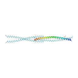
 | | The structure of Endoglucanase from termite, Nasutitermes takasagoensis, at pH 2.5. | | 分子名称: | Endo-b-1,4-glucanase, SULFATE ION | | 著者 | Khademi, S, Guarino, L.A, Watanabe, H, Tokuda, G, Meyer, E.F. | | 登録日 | 2002-01-11 | | 公開日 | 2003-01-21 | | 最終更新日 | 2024-10-30 | | 実験手法 | X-RAY DIFFRACTION (1.4 Å) | | 主引用文献 | Structure of an endoglucanase from termite, Nasutitermes takasagoensis.
Acta Crystallogr.,Sect.D, 58, 2002
|
|
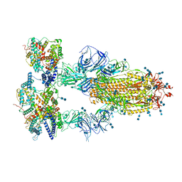
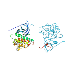
7V85
 | | Cryo-EM structure of SARS-CoV-2 S-Kappa variant (B.1.617.1) in complex with Angiotensin-converting enzyme 2 (ACE2) ectodomain, two ACE2-bound form | | 分子名称: | 2-acetamido-2-deoxy-beta-D-glucopyranose, 2-acetamido-2-deoxy-beta-D-glucopyranose-(1-4)-2-acetamido-2-deoxy-beta-D-glucopyranose, Angiotensin-converting enzyme 2,Green fluorescent protein, ... | | 著者 | Yang, T.J, Yu, P.Y, Chang, Y.C, Hsu, S.T.D. | | 登録日 | 2021-08-22 | | 公開日 | 2021-10-06 | | 最終更新日 | 2024-10-16 | | 実験手法 | ELECTRON MICROSCOPY (3.3 Å) | | 主引用文献 | Cryo-EM structure of SARS-CoV-2 S-Kappa variant (B.1.617.1) in complex with Angiotensin-converting enzyme 2 (ACE2) ectodomain, two ACE2-bound form
To Be Published
|
|
2PLJ
 | |
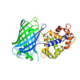
2P28
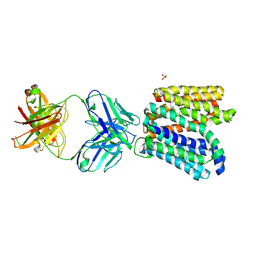
 | | Structure of the PHE2 and PHE3 fragments of the integrin beta2 subunit | | 分子名称: | 2-acetamido-2-deoxy-beta-D-glucopyranose, Integrin beta-2 | | 著者 | Shi, M, Foo, S.Y, Tan, S.M, Mitchell, E.P, Law, S.K.A, Lescar, J. | | 登録日 | 2007-03-07 | | 公開日 | 2007-08-14 | | 最終更新日 | 2024-10-30 | | 実験手法 | X-RAY DIFFRACTION (2.2 Å) | | 主引用文献 | A structural hypothesis for the transition between bent and extended conformations of the leukocyte beta2 integrins
J.Biol.Chem., 282, 2007
|
|
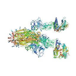
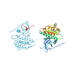
7V86
 | | Cryo-EM structure of SARS-CoV-2 S-Kappa variant (B.1.617.1) in complex with Angiotensin-converting enzyme 2 (ACE2) ectodomain, three ACE2-bound form | | 分子名称: | 2-acetamido-2-deoxy-beta-D-glucopyranose, 2-acetamido-2-deoxy-beta-D-glucopyranose-(1-4)-2-acetamido-2-deoxy-beta-D-glucopyranose, Angiotensin-converting enzyme 2,Green fluorescent protein, ... | | 著者 | Yang, T.J, Yu, P.Y, Chang, Y.C, Hsu, S.T.D. | | 登録日 | 2021-08-22 | | 公開日 | 2021-10-06 | | 最終更新日 | 2024-11-06 | | 実験手法 | ELECTRON MICROSCOPY (2.8 Å) | | 主引用文献 | Cryo-EM structure of SARS-CoV-2 S-Kappa variant (B.1.617.1) in complex with Angiotensin-converting enzyme 2 (ACE2) ectodomain, three ACE2-bound form
To Be Published
|
|
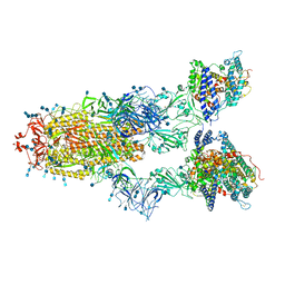
7V81
 | | Cryo-EM structure of SARS-CoV-2 S-Gamma variant (P.1) in complex with Angiotensin-converting enzyme 2 (ACE2) ectodomain, two ACE2-bound form | | 分子名称: | 2-acetamido-2-deoxy-beta-D-glucopyranose, 2-acetamido-2-deoxy-beta-D-glucopyranose-(1-4)-2-acetamido-2-deoxy-beta-D-glucopyranose, Angiotensin-converting enzyme 2,Green fluorescent protein, ... | | 著者 | Yang, T.J, Yu, P.Y, Chang, Y.C, Hsu, S.T.D. | | 登録日 | 2021-08-22 | | 公開日 | 2021-10-06 | | 最終更新日 | 2025-07-02 | | 実験手法 | ELECTRON MICROSCOPY (3.2 Å) | | 主引用文献 | Cryo-EM structure of SARS-CoV-2 S-Gamma variant (P.1) in complex with Angiotensin-converting enzyme 2 (ACE2) ectodomain, two ACE2-bound form
To Be Published
|
|
7V83
 | | Cryo-EM structure of SARS-CoV-2 S-Gamma variant (P.1) in complex with Angiotensin-converting enzyme 2 (ACE2) ectodomain, three ACE2-bound form conformation 2 | | 分子名称: | 2-acetamido-2-deoxy-beta-D-glucopyranose, 2-acetamido-2-deoxy-beta-D-glucopyranose-(1-4)-2-acetamido-2-deoxy-beta-D-glucopyranose, Angiotensin-converting enzyme 2,Green fluorescent protein, ... | | 著者 | Yang, T.J, Yu, P.Y, Chang, Y.C, Hsu, S.T.D. | | 登録日 | 2021-08-22 | | 公開日 | 2021-10-06 | | 最終更新日 | 2025-06-25 | | 実験手法 | ELECTRON MICROSCOPY (2.8 Å) | | 主引用文献 | Cryo-EM structure of SARS-CoV-2 S-Gamma variant (P.1) in complex with Angiotensin-converting enzyme 2 (ACE2) ectodomain, three ACE2-bound form conformation 2
To Be Published
|
|
7V84
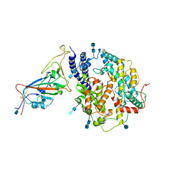 | | Local refinement of SARS-CoV-2 S-Gamma variant (P.1) RBD and Angiotensin-converting enzyme 2 (ACE2) ectodomain | | 分子名称: | 2-acetamido-2-deoxy-beta-D-glucopyranose, 2-acetamido-2-deoxy-beta-D-glucopyranose-(1-4)-2-acetamido-2-deoxy-beta-D-glucopyranose, Angiotensin-converting enzyme 2,Green fluorescent protein, ... | | 著者 | Yang, T.J, Yu, P.Y, Chang, Y.C, Hsu, S.T.D. | | 登録日 | 2021-08-22 | | 公開日 | 2021-10-06 | | 最終更新日 | 2025-07-02 | | 実験手法 | ELECTRON MICROSCOPY (3 Å) | | 主引用文献 | Local refinement of SARS-CoV-2 S-Gamma variant (P.1) RBD and Angiotensin-converting enzyme 2 (ACE2) ectodomain
To Be Published
|
|
2PSP
 | |
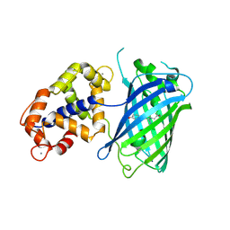
2P26
 | | Structure of the PHE2 and PHE3 fragments of the integrin beta2 subunit | | 分子名称: | 2-acetamido-2-deoxy-beta-D-glucopyranose, Integrin beta-2 | | 著者 | Shi, M, Foo, S.Y, Tan, S.M, Mitchell, E.P, Law, S.K.A, Lescar, J. | | 登録日 | 2007-03-06 | | 公開日 | 2007-08-14 | | 最終更新日 | 2024-11-13 | | 実験手法 | X-RAY DIFFRACTION (1.75 Å) | | 主引用文献 | A structural hypothesis for the transition between bent and extended conformations of the leukocyte beta2 integrins
J.Biol.Chem., 282, 2007
|
|
2PET
 | |
2PLK
 | |
6F5X
 | |
6GV1
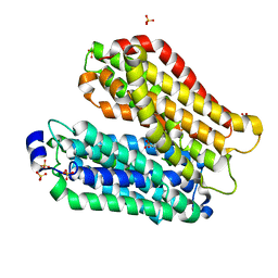
 | | Crystal structure of E.coli Multidrug/H+ antiporter MdfA in outward open conformation with bound Fab fragment | | 分子名称: | Fab fragment YN1074 heavy chain, Fab fragment YN1074 light chain, Major Facilitator Superfamily multidrug/H+ antiporter MdfA from E.coli, ... | | 著者 | Nagarathinam, K, Parthier, C, Stubbs, M.T, Tanabe, M. | | 登録日 | 2018-06-20 | | 公開日 | 2018-10-03 | | 最終更新日 | 2024-10-09 | | 実験手法 | X-RAY DIFFRACTION (3.4 Å) | | 主引用文献 | Outward open conformation of a Major Facilitator Superfamily multidrug/H+antiporter provides insights into switching mechanism.
Nat Commun, 9, 2018
|
|
4IKZ
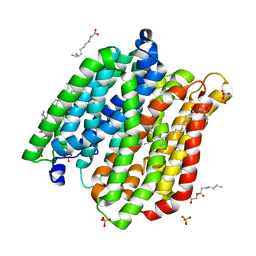
 | | Crystal structure of peptide transporter POT (E310Q mutant) in complex with alafosfalin | | 分子名称: | Di-tripeptide ABC transporter (Permease), N-[(1R)-1-phosphonoethyl]-L-alaninamide, SULFATE ION | | 著者 | Doki, S, Kato, H.E, Ishitani, R, Nureki, O. | | 登録日 | 2012-12-28 | | 公開日 | 2013-07-10 | | 最終更新日 | 2024-05-29 | | 実験手法 | X-RAY DIFFRACTION (2.4 Å) | | 主引用文献 | Structural basis for dynamic mechanism of proton-coupled symport by the peptide transporter POT.
Proc.Natl.Acad.Sci.USA, 110, 2013
|
|
4IKW
 | | Crystal structure of peptide transporter POT in complex with sulfate | | 分子名称: | (2R)-2,3-dihydroxypropyl (9Z)-octadec-9-enoate, Di-tripeptide ABC transporter (Permease), OLEIC ACID, ... | | 著者 | Doki, S, Kato, H.E, Ishitani, R, Nureki, O. | | 登録日 | 2012-12-28 | | 公開日 | 2013-07-10 | | 最終更新日 | 2024-05-29 | | 実験手法 | X-RAY DIFFRACTION (2.004 Å) | | 主引用文献 | Structural basis for dynamic mechanism of proton-coupled symport by the peptide transporter POT.
Proc.Natl.Acad.Sci.USA, 110, 2013
|
|
4IKY
 | | Crystal structure of peptide transporter POT (E310Q mutant) in complex with sulfate | | 分子名称: | (2R)-2,3-dihydroxypropyl (9Z)-octadec-9-enoate, Di-tripeptide ABC transporter (Permease), OLEIC ACID, ... | | 著者 | Doki, S, Kato, H.E, Ishitani, R, Nureki, O. | | 登録日 | 2012-12-28 | | 公開日 | 2013-07-10 | | 最終更新日 | 2024-05-29 | | 実験手法 | X-RAY DIFFRACTION (2.1 Å) | | 主引用文献 | Structural basis for dynamic mechanism of proton-coupled symport by the peptide transporter POT.
Proc.Natl.Acad.Sci.USA, 110, 2013
|
|
4IKX
 | | Crystal structure of peptide transporter POT (E310Q mutant) | | 分子名称: | Di-tripeptide ABC transporter (Permease), OLEIC ACID, SULFATE ION | | 著者 | Doki, S, Kato, H.E, Ishitani, R, Nureki, O. | | 登録日 | 2012-12-28 | | 公開日 | 2013-07-10 | | 最終更新日 | 2024-05-29 | | 実験手法 | X-RAY DIFFRACTION (2.3 Å) | | 主引用文献 | Structural basis for dynamic mechanism of proton-coupled symport by the peptide transporter POT.
Proc.Natl.Acad.Sci.USA, 110, 2013
|
|
4IKV
 | | Crystal structure of peptide transporter POT | | 分子名称: | (2S)-2,3-dihydroxypropyl (9Z)-octadec-9-enoate, Di-tripeptide ABC transporter (Permease), OLEIC ACID, ... | | 著者 | Doki, S, Kato, H.E, Ishitani, R, Nureki, O. | | 登録日 | 2012-12-28 | | 公開日 | 2013-07-10 | | 最終更新日 | 2024-04-03 | | 実験手法 | X-RAY DIFFRACTION (1.9 Å) | | 主引用文献 | Structural basis for dynamic mechanism of proton-coupled symport by the peptide transporter POT.
Proc.Natl.Acad.Sci.USA, 110, 2013
|
|
4JQ8
 | | Crystal structure of EGFR kinase domain in complex with compound 4b | | 分子名称: | Epidermal growth factor receptor, N-[3-(4-{[(1S)-2-hydroxy-1-phenylethyl]amino}-6-phenylfuro[2,3-d]pyrimidin-5-yl)phenyl]-N~3~,N~3~-dimethyl-beta-alaninamide | | 著者 | Peng, Y.H, Wu, J.S. | | 登録日 | 2013-03-20 | | 公開日 | 2013-06-19 | | 最終更新日 | 2023-11-08 | | 実験手法 | X-RAY DIFFRACTION (2.83 Å) | | 主引用文献 | Protein Kinase Inhibitor Design by Targeting the Asp-Phe-Gly (DFG) Motif: The Role of the DFG Motif in the Design of Epidermal Growth Factor Receptor Inhibitors
J.Med.Chem., 56, 2013
|
|
4JR3
 | | Crystal structure of EGFR kinase domain in complex with compound 3g | | 分子名称: | Epidermal growth factor receptor, N-[3-(4-{[(1S)-2-hydroxy-1-phenylethyl]amino}-6-phenylfuro[2,3-d]pyrimidin-5-yl)phenyl]acetamide | | 著者 | Peng, Y.H, Wu, J.S. | | 登録日 | 2013-03-21 | | 公開日 | 2013-06-19 | | 最終更新日 | 2023-11-08 | | 実験手法 | X-RAY DIFFRACTION (2.7 Å) | | 主引用文献 | Protein Kinase Inhibitor Design by Targeting the Asp-Phe-Gly (DFG) Motif: The Role of the DFG Motif in the Design of Epidermal Growth Factor Receptor Inhibitors
J.Med.Chem., 56, 2013
|
|
4JQ7
 | | Crystal structure of EGFR kinase domain in complex with compound 2a | | 分子名称: | (2S)-2-[(5,6-diphenylfuro[2,3-d]pyrimidin-4-yl)amino]-2-phenylethanol, Epidermal growth factor receptor | | 著者 | Peng, Y.H, Wu, J.S. | | 登録日 | 2013-03-20 | | 公開日 | 2013-06-19 | | 最終更新日 | 2023-11-08 | | 実験手法 | X-RAY DIFFRACTION (2.73 Å) | | 主引用文献 | Protein Kinase Inhibitor Design by Targeting the Asp-Phe-Gly (DFG) Motif: The Role of the DFG Motif in the Design of Epidermal Growth Factor Receptor Inhibitors
J.Med.Chem., 56, 2013
|
|
4JRV
 | | Crystal structure of EGFR kinase domain in complex with compound 4c | | 分子名称: | 4-(dimethylamino)-N-[3-(4-{[(1S)-2-hydroxy-1-phenylethyl]amino}-6-phenylfuro[2,3-d]pyrimidin-5-yl)phenyl]butanamide, Epidermal growth factor receptor | | 著者 | Peng, Y.H, Wu, J.S. | | 登録日 | 2013-03-22 | | 公開日 | 2013-06-19 | | 最終更新日 | 2023-11-08 | | 実験手法 | X-RAY DIFFRACTION (2.8 Å) | | 主引用文献 | Protein Kinase Inhibitor Design by Targeting the Asp-Phe-Gly (DFG) Motif: The Role of the DFG Motif in the Design of Epidermal Growth Factor Receptor Inhibitors
J.Med.Chem., 56, 2013
|
|
9K8X
 | | Crystal structure of the calcium indicator GCaMP6s-BrUS-145 in calcium-bounded state | | 分子名称: | 1,2-ETHANEDIOL, CALCIUM ION, Calcium indicator GCaMP6s-BrUS-145,Calmodulin-1, ... | | 著者 | Varfolomeeva, L.A, Simonyan, T.R, Mamontova, A.V, Popov, V.O, Bogdanov, A.M, Boyko, K.M. | | 登録日 | 2024-10-24 | | 公開日 | 2024-12-11 | | 最終更新日 | 2025-01-01 | | 実験手法 | X-RAY DIFFRACTION (2.05 Å) | | 主引用文献 | Calcium Indicators with Fluorescence Lifetime-Based Signal Readout: A Structure-Function Study.
Int J Mol Sci, 25, 2024
|
|
9K8W
 | | Crystal structure of the calcium indicator GCaMP6s-BrUS in calcium-bound state | | 分子名称: | CALCIUM ION, Calcium indicator GCaMP6s-BrUS,Calmodulin-1 | | 著者 | Varfolomeeva, L.A, Simonyan, T.R, Mamontova, A.V, Bogdanov, A.M, Popov, V.O, Boyko, K.M. | | 登録日 | 2024-10-24 | | 公開日 | 2024-12-11 | | 最終更新日 | 2025-01-01 | | 実験手法 | X-RAY DIFFRACTION (2.65 Å) | | 主引用文献 | Calcium Indicators with Fluorescence Lifetime-Based Signal Readout: A Structure-Function Study.
Int J Mol Sci, 25, 2024
|
|


