5I52
 
 | |
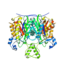
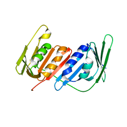
2GEY
 | | Crystal Structure of AclR a putative hydroxylase from Streptomyces galilaeus | | 分子名称: | AclR protein, DI(HYDROXYETHYL)ETHER, GLYCEROL, ... | | 著者 | Beinker, P, Lohkamp, B, Schneider, G. | | 登録日 | 2006-03-21 | | 公開日 | 2006-07-18 | | 最終更新日 | 2023-10-25 | | 実験手法 | X-RAY DIFFRACTION (1.8 Å) | | 主引用文献 | Crystal structures of SnoaL2 and AclR: two putative hydroxylases in the biosynthesis of aromatic polyketide antibiotics
J.Mol.Biol., 359, 2006
|
|
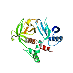
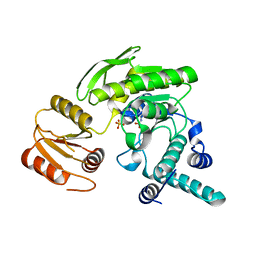
4EWD
 | | Study on structure and function relationships in human Pirin with Mn ion | | 分子名称: | MANGANESE (II) ION, Pirin | | 著者 | Liu, F, Rehmani, I, Chen, L, Fu, R, Serrano, V, Wilson, D.W, Liu, A. | | 登録日 | 2012-04-26 | | 公開日 | 2013-05-29 | | 最終更新日 | 2024-02-28 | | 実験手法 | X-RAY DIFFRACTION (2.15 Å) | | 主引用文献 | Pirin is an iron-dependent redox regulator of NF-kappa B.
Proc.Natl.Acad.Sci.USA, 110, 2013
|
|
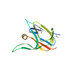
5I63
 | |
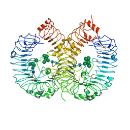
2G95
 | |
4EWP
 | | Crystal structure of FabH from Micrococcus luteus | | 分子名称: | 3-oxoacyl-[acyl-carrier-protein] synthase 3 | | 著者 | Pereira, J.H, Goh, E.-B, Keasling, J.D, Beller, H.R, Adams, P.D. | | 登録日 | 2012-04-27 | | 公開日 | 2012-10-03 | | 最終更新日 | 2024-02-28 | | 実験手法 | X-RAY DIFFRACTION (2.198 Å) | | 主引用文献 | Structure of FabH and factors affecting the distribution of branched fatty acids in Micrococcus luteus.
Acta Crystallogr.,Sect.D, 68, 2012
|
|
4GN1
 | |
2GC5
 | | G51S mutant of L. casei FPGS | | 分子名称: | Folylpolyglutamate synthase, SULFATE ION | | 著者 | Smith, C.A, Cross, J.A, Bognar, A.L, Sun, X. | | 登録日 | 2006-03-13 | | 公開日 | 2006-06-27 | | 最終更新日 | 2021-10-20 | | 実験手法 | X-RAY DIFFRACTION (1.85 Å) | | 主引用文献 | Mutation of Gly51 to serine in the P-loop of Lactobacillus casei folylpolyglutamate synthetase abolishes activity by altering the conformation of two adjacent loops.
Acta Crystallogr.,Sect.D, 62, 2006
|
|
2GCP
 | |
6MC4
 | |
2GCS
 | |
7Z3B
 | | Crystal structure of the cupredoxin AcoP from Acidithiobacillus ferrooxidans, reduced form | | 分子名称: | ACETATE ION, AcoP, COPPER (I) ION, ... | | 著者 | Leone, P, Sciara, G, Ilbert, M. | | 登録日 | 2022-03-02 | | 公開日 | 2023-09-13 | | 最終更新日 | 2024-02-28 | | 実験手法 | X-RAY DIFFRACTION (1.65 Å) | | 主引用文献 | Beyond the coupled distortion model: structural analysis of the single domain cupredoxin AcoP, a green mononuclear copper centre with original features.
Dalton Trans, 53, 2024
|
|
5I3V
 | |
7Z3F
 | | Crystal structure of the cupredoxin AcoP from Acidithiobacillus ferrooxidans, oxidized form | | 分子名称: | ACETATE ION, AcoP, CHLORIDE ION, ... | | 著者 | Leone, P, Sciara, G, Ilbert, M. | | 登録日 | 2022-03-02 | | 公開日 | 2023-09-13 | | 最終更新日 | 2024-02-28 | | 実験手法 | X-RAY DIFFRACTION (1.7 Å) | | 主引用文献 | Beyond the coupled distortion model: structural analysis of the single domain cupredoxin AcoP, a green mononuclear copper centre with original features.
Dalton Trans, 53, 2024
|
|
4GLF
 | |
7Z3G
 | | Crystal structure of the cupredoxin AcoP from Acidithiobacillus ferrooxidans, H166A mutant | | 分子名称: | AcoP, COPPER (I) ION, GLYCEROL | | 著者 | Leone, P, Sciara, G, Ilbert, M. | | 登録日 | 2022-03-02 | | 公開日 | 2023-09-13 | | 最終更新日 | 2024-02-28 | | 実験手法 | X-RAY DIFFRACTION (2.1 Å) | | 主引用文献 | Beyond the coupled distortion model: structural analysis of the single domain cupredoxin AcoP, a green mononuclear copper centre with original features.
Dalton Trans, 53, 2024
|
|
4GLS
 | | Crystal Structure of Chemically Synthesized Heterochiral {D-Protein Antagonist plus VEGF-A} Protein Complex in space group P21 | | 分子名称: | D- RFX001, D- Vascular endothelial growth factor-A, DI(HYDROXYETHYL)ETHER, ... | | 著者 | Mandal, K, Uppalapati, M, Ault-Riche, D, Kenney, J, Lowitz, J, Sidhu, S, Kent, S.B.H. | | 登録日 | 2012-08-14 | | 公開日 | 2012-09-05 | | 最終更新日 | 2023-12-06 | | 実験手法 | X-RAY DIFFRACTION (1.6 Å) | | 主引用文献 | Chemical synthesis and X-ray structure of a heterochiral {D-protein antagonist plus vascular endothelial growth factor} protein complex by racemic crystallography.
Proc.Natl.Acad.Sci.USA, 109, 2012
|
|
4L60
 | |
2GC6
 | | S73A mutant of L. casei FPGS | | 分子名称: | Folylpolyglutamate synthase, SULFATE ION | | 著者 | Smith, C.A, Cross, J.A, Bognar, A.L, Sun, X. | | 登録日 | 2006-03-13 | | 公開日 | 2006-06-27 | | 最終更新日 | 2021-10-20 | | 実験手法 | X-RAY DIFFRACTION (1.9 Å) | | 主引用文献 | Mutation of Gly51 to serine in the P-loop of Lactobacillus casei folylpolyglutamate synthetase abolishes activity by altering the conformation of two adjacent loops.
Acta Crystallogr.,Sect.D, 62, 2006
|
|
7Z3I
 | | Crystal structure of the cupredoxin AcoP from Acidithiobacillus ferrooxidans, M171A mutant | | 分子名称: | ACETATE ION, AcoP, COPPER (II) ION, ... | | 著者 | Leone, P, Sciara, G, Ilbert, M. | | 登録日 | 2022-03-02 | | 公開日 | 2023-09-13 | | 最終更新日 | 2024-02-28 | | 実験手法 | X-RAY DIFFRACTION (1.82 Å) | | 主引用文献 | Beyond the coupled distortion model: structural analysis of the single domain cupredoxin AcoP, a green mononuclear copper centre with original features.
Dalton Trans, 53, 2024
|
|
7YTX
 | | Crystal structure of TLR8 in complex with its antagonist | | 分子名称: | (2R,6R)-4-(8-cyanoquinolin-5-yl)-N-[(3S,4R)-4-fluoranylpyrrolidin-3-yl]-6-methyl-morpholine-2-carboxamide, 2-acetamido-2-deoxy-beta-D-glucopyranose, 2-acetamido-2-deoxy-beta-D-glucopyranose-(1-4)-2-acetamido-2-deoxy-beta-D-glucopyranose, ... | | 著者 | Shimizu, T, Sakaniwa, K. | | 登録日 | 2022-08-16 | | 公開日 | 2023-09-27 | | 実験手法 | X-RAY DIFFRACTION (2.9 Å) | | 主引用文献 | A novel Toll-like receptor 7/8-specific antagonist E6742 ameliorates clinically relevant disease parameters in murine models of lupus.
Eur.J.Pharmacol., 957, 2023
|
|
4GBQ
 | | SOLUTION NMR STRUCTURE OF THE GRB2 N-TERMINAL SH3 DOMAIN COMPLEXED WITH A TEN-RESIDUE PEPTIDE DERIVED FROM SOS DIRECT REFINEMENT AGAINST NOES, J-COUPLINGS, AND 1H AND 13C CHEMICAL SHIFTS, 15 STRUCTURES | | 分子名称: | GRB2, SOS-1 | | 著者 | Wittekind, M, Mapelli, C, Lee, V, Goldfarb, V, Friedrichs, M.S, Meyers, C.A, Mueller, L. | | 登録日 | 1996-12-23 | | 公開日 | 1997-09-04 | | 最終更新日 | 2022-03-16 | | 実験手法 | SOLUTION NMR | | 主引用文献 | Solution structure of the Grb2 N-terminal SH3 domain complexed with a ten-residue peptide derived from SOS: direct refinement against NOEs, J-couplings and 1H and 13C chemical shifts.
J.Mol.Biol., 267, 1997
|
|
2GCV
 | |
3NSE
 | | BOVINE ENOS, H4B-FREE, SEITU COMPLEX | | 分子名称: | ACETATE ION, ARGININE, CACODYLATE ION, ... | | 著者 | Raman, C.S, Li, H, Martasek, P, Kral, V, Masters, B.S.S, Poulos, T.L. | | 登録日 | 1998-09-23 | | 公開日 | 1999-05-18 | | 最終更新日 | 2024-02-21 | | 実験手法 | X-RAY DIFFRACTION (2.1 Å) | | 主引用文献 | Crystal structure of constitutive endothelial nitric oxide synthase: a paradigm for pterin function involving a novel metal center.
Cell(Cambridge,Mass.), 95, 1998
|
|
7YSP
 | |


