1ZH2
 
 | |
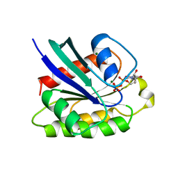
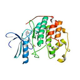
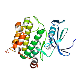
2X81
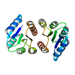
 | | STRUCTURE OF AURORA A IN COMPLEX WITH MLN8054 | | 分子名称: | 4-{[9-CHLORO-7-(2,6-DIFLUOROPHENYL)-5H-PYRIMIDO[5,4-D][2]BENZAZEPIN-2-YL]AMINO}BENZOIC ACID, SERINE/THREONINE-PROTEIN KINASE 6 | | 著者 | Savory, W, Mueller, I, Mason, C.S, Lamers, M, Williams, D.H, Eyers, P.A. | | 登録日 | 2010-03-05 | | 公開日 | 2010-05-05 | | 最終更新日 | 2023-12-20 | | 実験手法 | X-RAY DIFFRACTION (2.91 Å) | | 主引用文献 | Drug-Resistant Aurora a Mutants for Cellular Target Validation of the Small Molecule Kinase Inhibitors Mln8054 and Mln8237.
Acs Chem.Biol., 5, 2010
|
|
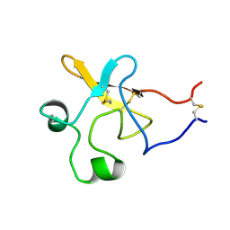
1KUN
 | | SOLUTION STRUCTURE OF THE HUMAN ALPHA3-CHAIN TYPE VI COLLAGEN C-TERMINAL KUNITZ DOMAIN, NMR, 20 STRUCTURES | | 分子名称: | ALPHA3-CHAIN TYPE VI COLLAGEN | | 著者 | Sorensen, M.D, Bjorn, S, Norris, K, Olsen, O, Petersen, L, James, T.L, Led, J.J. | | 登録日 | 1997-03-04 | | 公開日 | 1997-11-12 | | 最終更新日 | 2022-02-23 | | 実験手法 | SOLUTION NMR | | 主引用文献 | Solution structure and backbone dynamics of the human alpha3-chain type VI collagen C-terminal Kunitz domain,.
Biochemistry, 36, 1997
|
|
2B1J
 | |
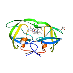
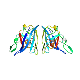
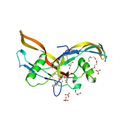
2ASM
 | | Structure of Rabbit Actin In Complex With Reidispongiolide A | | 分子名称: | 1,2-ETHANEDIOL, ADENOSINE-5'-TRIPHOSPHATE, Actin, ... | | 著者 | Allingham, J.S, Zampella, A, D'Auria, M.V, Rayment, I. | | 登録日 | 2005-08-23 | | 公開日 | 2005-10-11 | | 最終更新日 | 2023-10-25 | | 実験手法 | X-RAY DIFFRACTION (1.6 Å) | | 主引用文献 | Structures of microfilament destabilizing toxins bound to actin provide insight into toxin design and activity
Proc.Natl.Acad.Sci.Usa, 102, 2005
|
|
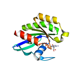
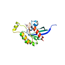
1KAO
 | | CRYSTAL STRUCTURE OF THE SMALL G PROTEIN RAP2A WITH GDP | | 分子名称: | GUANOSINE-5'-DIPHOSPHATE, MAGNESIUM ION, RAP2A | | 著者 | Cherfils, J, Menetrey, J, Le Bras, G. | | 登録日 | 1997-08-01 | | 公開日 | 1997-12-24 | | 最終更新日 | 2024-04-03 | | 実験手法 | X-RAY DIFFRACTION (1.7 Å) | | 主引用文献 | Crystal structures of the small G protein Rap2A in complex with its substrate GTP, with GDP and with GTPgammaS.
EMBO J., 16, 1997
|
|
2AVQ
 | | Kinetics, stability, and structural changes in high resolution crystal structures of HIV-1 protease with drug resistant mutations L24I, I50V, AND G73S | | 分子名称: | DIMETHYL SULFOXIDE, GLYCEROL, N-{(2S)-2-[(N-acetyl-L-threonyl-L-isoleucyl)amino]hexyl}-L-norleucyl-L-glutaminyl-N~5~-[amino(iminio)methyl]-L-ornithinamide, ... | | 著者 | Liu, F, Boross, P.I, Wang, Y.F, Tozser, J, Louis, J.M, Harrison, R.W, Weber, I.T. | | 登録日 | 2005-08-30 | | 公開日 | 2006-01-24 | | 最終更新日 | 2024-03-13 | | 実験手法 | X-RAY DIFFRACTION (1.3 Å) | | 主引用文献 | Kinetic, stability, and structural changes in high-resolution crystal structures of HIV-1 protease with drug-resistant mutations L24I, I50V, and G73S.
J.Mol.Biol., 354, 2005
|
|
2AI5
 | | Solution Structure of Cytochrome C552, determined by Distributed Computing Implementation for NMR data | | 分子名称: | Cytochrome c-552, HEME C | | 著者 | Nakamura, S, Ichiki, S.I, Takashima, H, Uchiyama, S, Hasegawa, J, Kobayashi, Y, Sambongi, Y, Ohkubo, T. | | 登録日 | 2005-07-29 | | 公開日 | 2006-05-23 | | 最終更新日 | 2022-03-09 | | 実験手法 | SOLUTION NMR | | 主引用文献 | Structure of Cytochrome c552 from a Moderate Thermophilic Bacterium, Hydrogenophilus thermoluteolus: Comparative Study on the Thermostability of Cytochrome c
Biochemistry, 45, 2006
|
|
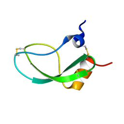
1KNT
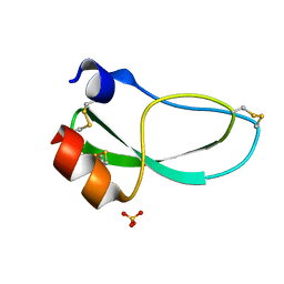 | | THE 1.6 ANGSTROMS STRUCTURE OF THE KUNITZ-TYPE DOMAIN FROM THE ALPHA3 CHAIN OF THE HUMAN TYPE VI COLLAGEN | | 分子名称: | COLLAGEN TYPE VI, SULFATE ION | | 著者 | Arnoux, B, Merigeau, K, Saludjian, P, Norris, F, Norris, K, Bjorn, S, Olsen, O, Petersen, L, Ducruix, A. | | 登録日 | 1994-08-18 | | 公開日 | 1994-11-01 | | 最終更新日 | 2024-06-05 | | 実験手法 | X-RAY DIFFRACTION (1.6 Å) | | 主引用文献 | The 1.6 A structure of Kunitz-type domain from the alpha 3 chain of human type VI collagen.
J.Mol.Biol., 246, 1995
|
|
1KIV
 | |
1ZIT
 | |
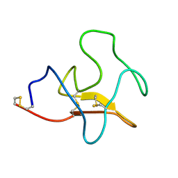
1KDU
 | | SEQUENTIAL 1H NMR ASSIGNMENTS AND SECONDARY STRUCTURE OF THE KRINGLE DOMAIN FROM UROKINASE | | 分子名称: | PLASMINOGEN ACTIVATOR | | 著者 | Li, X, Bokman, A.M, Llinas, M, Smith, R.A.G, Dobson, C.M. | | 登録日 | 1993-07-15 | | 公開日 | 1993-10-31 | | 最終更新日 | 2024-06-05 | | 実験手法 | SOLUTION NMR | | 主引用文献 | Solution structure of the kringle domain from urokinase-type plasminogen activator.
J.Mol.Biol., 235, 1994
|
|
1ZO4
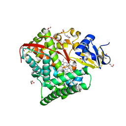 | | Crystal Structure Of A328S Mutant Of The Heme Domain Of P450BM-3 | | 分子名称: | 2-(N-MORPHOLINO)-ETHANESULFONIC ACID, Bifunctional P-450:NADPH-P450 reductase, GLYCEROL, ... | | 著者 | Hegda, A, Chen, B, Haines, D.C, Bondlela, M, Mullin, D, Graham, S.E, Tomchick, D.R, Machius, M, Peterson, J.A. | | 登録日 | 2005-05-12 | | 公開日 | 2006-08-01 | | 最終更新日 | 2023-08-23 | | 実験手法 | X-RAY DIFFRACTION (1.46 Å) | | 主引用文献 | A single active-site mutation of P450BM-3 dramatically enhances substrate binding and rate of product formation.
Biochemistry, 50, 2011
|
|
2AIU
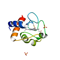 | | Crystal Structure of Mouse Testicular Cytochrome C at 1.6 Angstrom | | 分子名称: | Cytochrome c, testis-specific, PHOSPHATE ION, ... | | 著者 | Liu, Z, Ye, S, Lin, H, Rao, Z, Liu, X.J. | | 登録日 | 2005-08-01 | | 公開日 | 2006-07-18 | | 最終更新日 | 2011-07-13 | | 実験手法 | X-RAY DIFFRACTION (1.6 Å) | | 主引用文献 | Remarkably high activities of testicular cytochrome c in destroying reactive oxygen species and in triggering apoptosis
Proc.Natl.Acad.Sci.Usa, 103, 2006
|
|
1ZNG
 | | Strong Solute-Solute Dispersive Interactions in a Protein-Ligand Complex | | 分子名称: | CADMIUM ION, HEPTAN-1-OL, Major Urinary Protein | | 著者 | Malham, R, Johnstone, S, Bingham, R.J, Barratt, E, Phillips, S.E, Laughton, C.A, Homans, S.W. | | 登録日 | 2005-05-11 | | 公開日 | 2005-12-20 | | 最終更新日 | 2023-08-23 | | 実験手法 | X-RAY DIFFRACTION (1.6 Å) | | 主引用文献 | Strong Solute-Solute Dispersive Interactions in a Protein-Ligand Complex.
J.Am.Chem.Soc., 127, 2005
|
|
2ANG
 | |
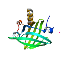
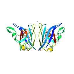
2AKQ
 | | The structure of bovine B-lactoglobulin A in crystals grown at very low ionic strength | | 分子名称: | Beta-lactoglobulin variant A | | 著者 | Adams, J.J, Anderson, B.F, Norris, G.E, Creamer, L.K, Jameson, G.B. | | 登録日 | 2005-08-03 | | 公開日 | 2005-08-16 | | 最終更新日 | 2023-10-25 | | 実験手法 | X-RAY DIFFRACTION (3 Å) | | 主引用文献 | Structure of bovine beta-lactoglobulin (variant A) at very low ionic strength
J.Struct.Biol., 154, 2006
|
|
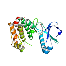
3UNJ
 | | CDK2 in complex with inhibitor YL1-038-31 | | 分子名称: | 4-{[4-(phenylamino)pyrimidin-2-yl]amino}benzoic acid, Cyclin-dependent kinase 2, PHOSPHATE ION | | 著者 | Zhu, J.-Y, Martin, M.P, Alam, R, Schonbrunn, E. | | 登録日 | 2011-11-15 | | 公開日 | 2012-01-25 | | 最終更新日 | 2023-09-13 | | 実験手法 | X-RAY DIFFRACTION (1.9001 Å) | | 主引用文献 | A Novel Mechanism by Which Small Molecule Inhibitors Induce the DFG Flip in Aurora A.
Acs Chem.Biol., 7, 2012
|
|
2APS
 | | CU/ZN SUPEROXIDE DISMUTASE FROM ACTINOBACILLUS PLEUROPNEUMONIAE | | 分子名称: | COPPER (II) ION, PROTEIN (CU,ZN SUPEROXIDE DISMUTASE), ZINC ION | | 著者 | Forest, K.T, Langford, P.R, Kroll, J.S, Getzoff, E.D. | | 登録日 | 1999-02-11 | | 公開日 | 1999-02-25 | | 最終更新日 | 2023-08-23 | | 実験手法 | X-RAY DIFFRACTION (1.9 Å) | | 主引用文献 | Cu,Zn superoxide dismutase structure from a microbial pathogen establishes a class with a conserved dimer interface.
J.Mol.Biol., 296, 2000
|
|
1Z22
 | | GDP-Bound Rab23 GTPase crystallized in C222(1) space group | | 分子名称: | GUANOSINE-5'-DIPHOSPHATE, MAGNESIUM ION, Ras-related protein Rab-23 | | 著者 | Eathiraj, S, Pan, X, Ritacco, C, Lambright, D.G. | | 登録日 | 2005-03-07 | | 公開日 | 2005-07-26 | | 最終更新日 | 2024-04-03 | | 実験手法 | X-RAY DIFFRACTION (2.06 Å) | | 主引用文献 | Structural basis of family-wide Rab GTPase recognition by rabenosyn-5.
Nature, 436, 2005
|
|
2AQN
 | | CU/ZN superoxide dismutase from neisseria meningitidis | | 分子名称: | COPPER (I) ION, COPPER (II) ION, SULFATE ION, ... | | 著者 | DiDonato, M, Kassmann, C.J, Bruns, C.K, Cabelli, D.E, Cao, Z, Tabatabai, L.B, Kroll, J.S, Getzoff, E.D. | | 登録日 | 2005-08-18 | | 公開日 | 2006-10-31 | | 最終更新日 | 2023-08-23 | | 実験手法 | X-RAY DIFFRACTION (1.4 Å) | | 主引用文献 | CU/ZN superoxide dismutase from neisseria meningitidis
To be Published
|
|
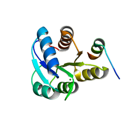
3BGP
 | | Human Pim-1 complexed with a benzoisoxazole inhibitor VX1 | | 分子名称: | 4-[3-(4-chlorophenyl)-2,1-benzisoxazol-5-yl]pyrimidin-2-amine, Proto-oncogene serine/threonine-protein kinase Pim-1 | | 著者 | Jacobs, M.D. | | 登録日 | 2007-11-27 | | 公開日 | 2007-12-11 | | 最終更新日 | 2011-07-13 | | 実験手法 | X-RAY DIFFRACTION (2.8 Å) | | 主引用文献 | Docking study yields four novel inhibitors of the protooncogene pim-1 kinase.
J.Med.Chem., 51, 2008
|
|
2ARV
 | | Structure of human Activin A | | 分子名称: | 2-(2-{2-[2-(2-METHOXY-ETHOXY)-ETHOXY]-ETHOXY}-ETHOXY)-ETHANOL, GLYCEROL, Inhibin beta A chain, ... | | 著者 | Harrington, A.E, Morris-Triggs, S.A, Ruotolo, B.T, Robinson, C.V, Ohnuma, S, Hyvonen, M. | | 登録日 | 2005-08-22 | | 公開日 | 2006-03-07 | | 最終更新日 | 2023-08-23 | | 実験手法 | X-RAY DIFFRACTION (2 Å) | | 主引用文献 | Structural basis for the inhibition of activin signalling by follistatin
Embo J., 25, 2006
|
|
1Z57
 | | Crystal structure of human CLK1 in complex with 10Z-Hymenialdisine | | 分子名称: | DEBROMOHYMENIALDISINE, Dual specificity protein kinase CLK1 | | 著者 | Debreczeni, J, Das, S, Knapp, S, Bullock, A, Guo, K, Amos, A, Fedorov, O, Edwards, A, Sundstrom, M, von Delft, F, Niesen, F.H, Ball, L, Sobott, F, Arrowsmith, C, Structural Genomics Consortium (SGC) | | 登録日 | 2005-03-17 | | 公開日 | 2005-04-12 | | 最終更新日 | 2023-08-23 | | 実験手法 | X-RAY DIFFRACTION (1.7 Å) | | 主引用文献 | Kinase domain insertions define distinct roles of CLK kinases in SR protein phosphorylation.
Structure, 17, 2009
|
|
2ATX
 | | Crystal Structure of the TC10 GppNHp complex | | 分子名称: | MAGNESIUM ION, PHOSPHOAMINOPHOSPHONIC ACID-GUANYLATE ESTER, small GTP binding protein TC10 | | 著者 | Hemsath, L, Dvorsky, R, Fiegen, D, Carlier, M.F, Ahmadian, M.R. | | 登録日 | 2005-08-26 | | 公開日 | 2005-09-13 | | 最終更新日 | 2024-04-03 | | 実験手法 | X-RAY DIFFRACTION (2.65 Å) | | 主引用文献 | An electrostatic steering mechanism of Cdc42 recognition by Wiskott-Aldrich syndrome proteins
Mol.Cell, 20, 2005
|
|


