1IWW
 
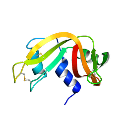
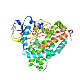
 | | Crystal Structure Analysis of Human lysozyme at 152K. | | 分子名称: | CHLORIDE ION, LYSOZYME C | | 著者 | Joti, Y, Nakasako, M, Kidera, A, Go, N. | | 登録日 | 2002-06-03 | | 公開日 | 2002-09-04 | | 最終更新日 | 2023-12-27 | | 実験手法 | X-RAY DIFFRACTION (1.4 Å) | | 主引用文献 | Nonlinear temperature dependence of the crystal structure of lysozyme: correlation between coordinate shifts and thermal factors.
Acta Crystallogr.,Sect.D, 58, 2002
|
|
1Z08
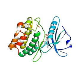
 | | GppNHp-Bound Rab21 Q53G mutant GTPase | | 分子名称: | MAGNESIUM ION, PHOSPHOAMINOPHOSPHONIC ACID-GUANYLATE ESTER, Ras-related protein Rab-21 | | 著者 | Eathiraj, S, Pan, X, Ritacco, C, Lambright, D.G. | | 登録日 | 2005-03-01 | | 公開日 | 2005-07-26 | | 最終更新日 | 2024-04-03 | | 実験手法 | X-RAY DIFFRACTION (1.8 Å) | | 主引用文献 | Structural basis of family-wide Rab GTPase recognition by rabenosyn-5.
Nature, 436, 2005
|
|
2BBT
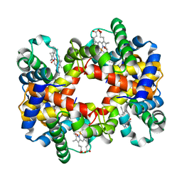
 | | Human deltaF508 NBD1 with two solublizing mutations. | | 分子名称: | ADENOSINE-5'-TRIPHOSPHATE, Cystic fibrosis transmembrane conductance regulator, MAGNESIUM ION | | 著者 | Lewis, H.A, Kearins, M.C, Conners, K, Zhao, X, Lu, F, Sauder, J.M, Emtage, S. | | 登録日 | 2005-10-17 | | 公開日 | 2005-11-01 | | 最終更新日 | 2023-08-23 | | 実験手法 | X-RAY DIFFRACTION (2.3 Å) | | 主引用文献 | Structure and dynamics of NBD1 from CFTR characterized using crystallography and hydrogen/deuterium exchange mass spectrometry.
J.Mol.Biol., 396, 2010
|
|
2B9I
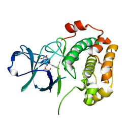
 | | Crystal structure of Fus3 with a docking motif from Msg5 | | 分子名称: | ADENOSINE-5'-DIPHOSPHATE, MAGNESIUM ION, Mitogen-activated protein kinase FUS3, ... | | 著者 | Remenyi, A, Good, M.C, Bhattacharyya, R.P, Lim, W.A. | | 登録日 | 2005-10-11 | | 公開日 | 2006-01-03 | | 最終更新日 | 2023-08-23 | | 実験手法 | X-RAY DIFFRACTION (2.5 Å) | | 主引用文献 | The role of docking interactions in mediating signaling input, output, and discrimination in the yeast MAPK network.
Mol.Cell, 20, 2005
|
|
1IVM
 | |
2A1E
 | | High resolution structure of HIV-1 PR with TS-126 | | 分子名称: | ACETATE ION, CHLORIDE ION, DIMETHYL SULFOXIDE, ... | | 著者 | Demitri, N, Geremia, S, Randaccio, L, Wuerges, J, Benedetti, F, Berti, F, Dinon, F, Campaner, P, Tell, G. | | 登録日 | 2005-06-20 | | 公開日 | 2006-02-21 | | 最終更新日 | 2023-10-25 | | 実験手法 | X-RAY DIFFRACTION (1.3 Å) | | 主引用文献 | A potent HIV protease inhibitor identified in an epimeric mixture by high-resolution protein crystallography.
Chemmedchem, 1, 2006
|
|
2A5J
 | | Crystal Structure of Human RAB2B | | 分子名称: | GUANOSINE-5'-DIPHOSPHATE, MAGNESIUM ION, Ras-related protein Rab-2B | | 著者 | Dong, A, Wang, J, Shen, Y, Arrowsmith, C.H, Edwards, A.M, Sundstrom, M, Bochkarev, A, Park, H.W, Structural Genomics Consortium (SGC) | | 登録日 | 2005-06-30 | | 公開日 | 2005-07-19 | | 最終更新日 | 2023-08-23 | | 実験手法 | X-RAY DIFFRACTION (1.501 Å) | | 主引用文献 | Crystal structure of human RAB2B
To be Published
|
|
2R3H
 | | Crystal Structure of Cyclin-Dependent Kinase 2 with inhibitor | | 分子名称: | 3-methyl-N-(pyridin-4-ylmethyl)imidazo[1,2-a]pyrazin-8-amine, Cell division protein kinase 2 | | 著者 | Fischmann, T.O, Hruza, A.W, Madison, V.M, Duca, J.S. | | 登録日 | 2007-08-29 | | 公開日 | 2008-01-22 | | 最終更新日 | 2011-07-13 | | 実験手法 | X-RAY DIFFRACTION (1.5 Å) | | 主引用文献 | Structure-guided discovery of cyclin-dependent kinase inhibitors.
Biopolymers, 89, 2008
|
|
2A31
 | | Trypsin in complex with borate | | 分子名称: | BORATE ION, CALCIUM ION, GUANIDINE-3-PROPANOL, ... | | 著者 | Transue, T.R, Gabel, S.A, London, R.E. | | 登録日 | 2005-06-23 | | 公開日 | 2006-07-04 | | 最終更新日 | 2023-08-23 | | 実験手法 | X-RAY DIFFRACTION (1.25 Å) | | 主引用文献 | NMR and crystallographic characterization of adventitious borate binding by trypsin.
Bioconjug.Chem., 17, 2006
|
|
1J3S
 | |
2AF2
 | | Solution structure of disulfide reduced and copper depleted Human Superoxide Dismutase | | 分子名称: | Superoxide dismutase [Cu-Zn], ZINC ION | | 著者 | Banci, L, Bertini, I, Cantini, F, D'Amelio, N, Gaggelli, E, Structural Proteomics in Europe (SPINE) | | 登録日 | 2005-07-25 | | 公開日 | 2005-11-15 | | 最終更新日 | 2024-05-29 | | 実験手法 | SOLUTION NMR | | 主引用文献 | Human SOD1 before harboring the catalytic metal: solution structure of copper-depleted, disulfide-reduced form
J.Biol.Chem., 281, 2006
|
|
1Y0W
 | |
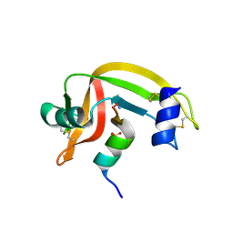
1IZQ
 | | F46V mutant of bovine pancreatic ribonuclease A | | 分子名称: | RIBONUCLEASE A | | 著者 | Kadonosono, T, Chatani, E, Hayashi, R, Moriyama, H, Ueki, T. | | 登録日 | 2002-10-11 | | 公開日 | 2003-11-25 | | 最終更新日 | 2023-10-25 | | 実験手法 | X-RAY DIFFRACTION (1.8 Å) | | 主引用文献 | Minimization of cavity size ensures protein stability and folding: structures of Phe46-replaced bovine pancreatic RNase A
Biochemistry, 42, 2003
|
|
1YDZ
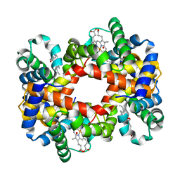 | | T-To-T(High) quaternary transitions in human hemoglobin: alphaY140F oxy (2MM IHP, 20% PEG) (1 test set) | | 分子名称: | Hemoglobin alpha chain, Hemoglobin beta chain, OXYGEN MOLECULE, ... | | 著者 | Kavanaugh, J.S, Rogers, P.H, Arnone, A. | | 登録日 | 2004-12-27 | | 公開日 | 2005-01-04 | | 最終更新日 | 2023-08-23 | | 実験手法 | X-RAY DIFFRACTION (3.3 Å) | | 主引用文献 | Crystallographic evidence for a new ensemble of ligand-induced allosteric transitions in hemoglobin: the T-to-T(high) quaternary transitions.
Biochemistry, 44, 2005
|
|
1YEA
 | |
1Y22
 | |
1J51
 | | CRYSTAL STRUCTURE OF CYTOCHROME P450CAM MUTANT (F87W/Y96F/V247L/C334A) WITH 1,3,5-TRICHLOROBENZENE | | 分子名称: | 1,3,5-TRICHLORO-BENZENE, CYTOCHROME P450CAM, POTASSIUM ION, ... | | 著者 | Chen, X, Christopher, A, Jones, J, Guo, Q, Xu, F, Cao, R, Wong, L.L, Rao, Z. | | 登録日 | 2002-01-05 | | 公開日 | 2002-01-23 | | 最終更新日 | 2023-12-27 | | 実験手法 | X-RAY DIFFRACTION (2.2 Å) | | 主引用文献 | Crystal structure of the F87W/Y96F/V247L mutant of cytochrome P-450cam with 1,3,5-trichlorobenzene bound and further protein engineering for the oxidation of pentachlorobenzene and hexachlorobenzene
J.BIOL.CHEM., 277, 2002
|
|
3GU6
 | |
1ZYX
 | | Crystal structure of the complex of a group IIA phospholipase A2 with a synthetic anti-inflammatory agent licofelone at 1.9A resolution | | 分子名称: | Phospholipase A2 VRV-PL-VIIIa, SULFATE ION, [6-(4-CHLOROPHENYL)-2,2-DIMETHYL-7-PHENYL-2,3-DIHYDRO-1H-PYRROLIZIN-5-YL]ACETIC ACID | | 著者 | Singh, N, Jabeen, T, Sharma, S, Bhushan, A, Singh, T.P. | | 登録日 | 2005-06-13 | | 公開日 | 2005-06-28 | | 最終更新日 | 2017-10-11 | | 実験手法 | X-RAY DIFFRACTION (1.95 Å) | | 主引用文献 | Crystal structure of the complex of a group IIA phospholipase A2 with a synthetic anti-inflammatory agent licofelone at 1.9A resolution
To be Published
|
|
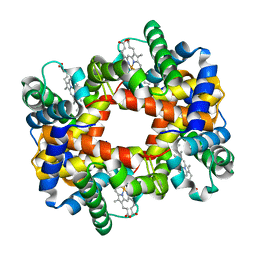
1YEN
 | | T-To-T(High) quaternary transitions in human hemoglobin: betaP36A oxy (2MM IHP, 20% PEG) (10 test sets) | | 分子名称: | Hemoglobin alpha chain, Hemoglobin beta chain, OXYGEN MOLECULE, ... | | 著者 | Kavanaugh, J.S, Rogers, P.H, Arnone, A. | | 登録日 | 2004-12-28 | | 公開日 | 2005-01-11 | | 最終更新日 | 2023-08-23 | | 実験手法 | X-RAY DIFFRACTION (2.8 Å) | | 主引用文献 | Crystallographic evidence for a new ensemble of ligand-induced allosteric transitions in hemoglobin: the T-to-T(high) quaternary transitions.
Biochemistry, 44, 2005
|
|
3UBD
 | | Structure of N-terminal domain of RSK2 kinase in complex with flavonoid glycoside SL0101 | | 分子名称: | 5,7-dihydroxy-2-(4-hydroxyphenyl)-4-oxo-4H-chromen-3-yl 3,4-di-O-acetyl-6-deoxy-alpha-L-mannopyranoside, Ribosomal protein S6 kinase alpha-3 | | 著者 | Utepbergenov, D, Derewenda, U, Derewenda, Z.S. | | 登録日 | 2011-10-24 | | 公開日 | 2012-09-05 | | 最終更新日 | 2023-09-13 | | 実験手法 | X-RAY DIFFRACTION (1.53 Å) | | 主引用文献 | Insights into the Inhibition of the p90 Ribosomal S6 Kinase (RSK) by the Flavonol Glycoside SL0101 from the 1.5 A Crystal Structure of the N-Terminal Domain of RSK2 with Bound Inhibitor.
Biochemistry, 51, 2012
|
|
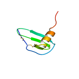
1J80
 | | Osmolyte Stabilization of RNase | | 分子名称: | RIBONUCLEASE PANCREATIC, SULFATE ION | | 著者 | Ratnaparkhi, G.S, Varadarajan, R. | | 登録日 | 2001-05-19 | | 公開日 | 2001-06-06 | | 最終更新日 | 2017-10-04 | | 実験手法 | X-RAY DIFFRACTION (2.1 Å) | | 主引用文献 | Osmolytes stabilize ribonuclease S by stabilizing its fragments S protein and S peptide to compact folding-competent states.
J.Biol.Chem., 276, 2001
|
|
3HA8
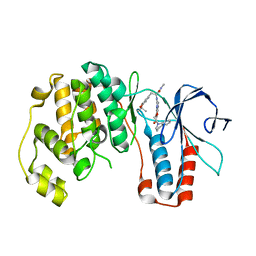 | | THE COMPLEX STRUCTURE OF THE MAP KINASE P38/Compound 14b | | 分子名称: | Mitogen-activated protein kinase 14, N~2~-{4-[6-(3,4-dihydroquinolin-1(2H)-ylcarbonyl)-1H-benzimidazol-1-yl]-6-ethoxy-1,3,5-triazin-2-yl}-3-(2,2-dimethyl-4H-1,3-benzodioxin-6-yl)-N-methyl-L-alaninamide | | 著者 | Zhao, B, Clark, M.A. | | 登録日 | 2009-05-01 | | 公開日 | 2009-08-04 | | 最終更新日 | 2023-09-06 | | 実験手法 | X-RAY DIFFRACTION (2.48 Å) | | 主引用文献 | Design, synthesis and selection of DNA-encoded small-molecule libraries.
Nat.Chem.Biol., 5, 2009
|
|
1J8I
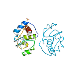
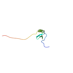 | | Solution Structure of Human Lymphotactin | | 分子名称: | Lymphotactin | | 著者 | Kuloglu, E.S, McCaslin, D.R, Markley, J.L, Pauza, C.D, Volkman, B.F. | | 登録日 | 2001-05-21 | | 公開日 | 2001-10-24 | | 最終更新日 | 2022-02-23 | | 実験手法 | SOLUTION NMR | | 主引用文献 | Monomeric solution structure of the prototypical 'C' chemokine lymphotactin.
Biochemistry, 40, 2001
|
|
1IKM
 | |


