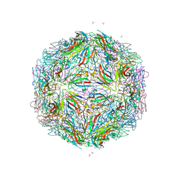
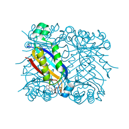
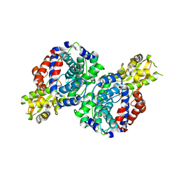
3ZJ5
 
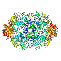 | | NEUROSPORA CRASSA CATALASE-3 EXPRESSED IN E. COLI, ORTHORHOMBIC FORM. | | 分子名称: | 1,2-ETHANEDIOL, 2-(2-ETHOXYETHOXY)ETHANOL, CATALASE-3, ... | | 著者 | Zarate-Romero, A, Rudino-Pinera, E. | | 登録日 | 2013-01-17 | | 公開日 | 2013-07-10 | | 最終更新日 | 2023-12-20 | | 実験手法 | X-RAY DIFFRACTION (1.95 Å) | | 主引用文献 | Conformational Stability and Crystal Packing: Polymorphism in Neurospora Crassa Cat-3
Acta Crystallogr.,Sect.F, 69, 2013
|
|
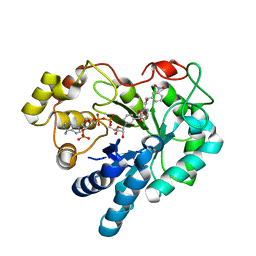
5BYY
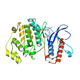 | | ERK5 IN COMPLEX WITH SMALL MOLECULE | | 分子名称: | 2-{[2-ethoxy-4-(4-hydroxypiperidin-1-yl)phenyl]amino}-5,11-dimethyl-5,11-dihydro-6H-pyrimido[4,5-b][1,4]benzodiazepin-6-one, Mitogen-activated protein kinase 7 | | 著者 | Chen, H, Tucker, J, Wang, X, Gavine, P.R, Philips, C, Augustin, M.A, Schreiner, P, Steinbacher, S, Preston, M, Ogg, D. | | 登録日 | 2015-06-11 | | 公開日 | 2016-05-04 | | 最終更新日 | 2024-05-08 | | 実験手法 | X-RAY DIFFRACTION (2.79 Å) | | 主引用文献 | Discovery of a novel allosteric inhibitor-binding site in ERK5: comparison with the canonical kinase hinge ATP-binding site.
Acta Crystallogr D Struct Biol, 72, 2016
|
|
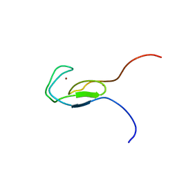
1J2O
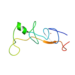 | | Structure of FLIN2, a complex containing the N-terminal LIM domain of LMO2 and ldb1-LID | | 分子名称: | Fusion of Rhombotin-2 and LIM domain-binding protein 1, ZINC ION | | 著者 | Deane, J.E, Mackay, J.P, Kwan, A.H, Sum, E.Y, Visvader, J.E, Matthews, J.M. | | 登録日 | 2003-01-08 | | 公開日 | 2003-05-13 | | 最終更新日 | 2023-12-27 | | 実験手法 | SOLUTION NMR | | 主引用文献 | Structural basis for the recognition of ldb1 by the N-terminal LIM domains of LMO2 and LMO4
EMBO J., 22, 2003
|
|
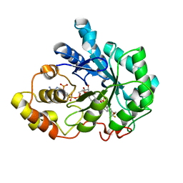
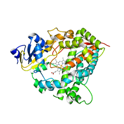
2C2S
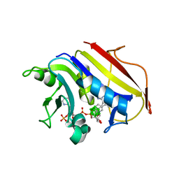 | | Human Dihydrofolate Reductase Complexed With NADPH and 2,4-Diamino-5-(1-o-carboranylmethyl)-6-methylpyrimidine, A novel boron containing, nonclassical Antifolate | | 分子名称: | 2,4-DIAMINO-5-(1-O-CARBORANYLMETHYL)-6-METHYLPYRIMIDINE, DIHYDROFOLATE REDUCTASE, GLYCEROL, ... | | 著者 | Leung, A.K.W, Reynolds, R.C, Riordan, J.M, Borhani, D.W. | | 登録日 | 2005-09-29 | | 公開日 | 2007-04-10 | | 最終更新日 | 2024-05-01 | | 実験手法 | X-RAY DIFFRACTION (1.4 Å) | | 主引用文献 | Novel Boron-Containing, Nonclassical Antifolates: Synthesis and Preliminary Biological and Structural Evaluation.
J.Med.Chem., 50, 2007
|
|
5BKQ
 | |
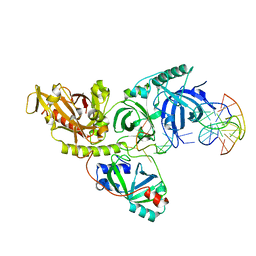
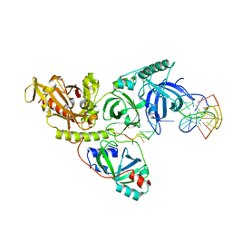
1JB1
 | | Lactobacillus casei HprK/P Bound to Phosphate | | 分子名称: | HPRK PROTEIN, PHOSPHATE ION | | 著者 | Fieulaine, S, Morera, S, Poncet, S, Monedero, V, Gueguen-Chaignon, V, Galinier, A, Janin, J, Deutscher, J, Nessler, S. | | 登録日 | 2001-06-01 | | 公開日 | 2001-08-08 | | 最終更新日 | 2024-11-06 | | 実験手法 | X-RAY DIFFRACTION (2.8 Å) | | 主引用文献 | X-ray structure of HPr kinase: a bacterial protein kinase with a P-loop nucleotide-binding domain.
EMBO J., 20, 2001
|
|
2NC7
 | |
1K4U
 | | Solution structure of the C-terminal SH3 domain of p67phox complexed with the C-terminal tail region of p47phox | | 分子名称: | PHAGOCYTE NADPH OXIDASE SUBUNIT P47PHOX, PHAGOCYTE NADPH OXIDASE SUBUNIT P67PHOX | | 著者 | Kami, K, Takeya, R, Sumimoto, H, Kohda, D. | | 登録日 | 2001-10-08 | | 公開日 | 2002-04-08 | | 最終更新日 | 2024-05-29 | | 実験手法 | SOLUTION NMR | | 主引用文献 | Diverse recognition of non-PxxP peptide ligands by the SH3 domains from p67(phox), Grb2 and Pex13p.
EMBO J., 21, 2002
|
|
1TBN
 | | NMR STRUCTURE OF A PROTEIN KINASE C-G PHORBOL-BINDING DOMAIN, MINIMIZED AVERAGE STRUCTURE | | 分子名称: | PROTEIN KINASE C, GAMMA TYPE, ZINC ION | | 著者 | Xu, R.X, Pawelczyk, T, Xia, T, Brown, S.C. | | 登録日 | 1997-04-15 | | 公開日 | 1998-04-29 | | 最終更新日 | 2024-05-22 | | 実験手法 | SOLUTION NMR | | 主引用文献 | NMR structure of a protein kinase C-gamma phorbol-binding domain and study of protein-lipid micelle interactions.
Biochemistry, 36, 1997
|
|
2PDL
 | |
2PDX
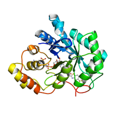 | | Human aldose reductase double mutant S302R-C303D complexed with zopolrestat. | | 分子名称: | 3,4-DIHYDRO-4-OXO-3-((5-TRIFLUOROMETHYL-2-BENZOTHIAZOLYL)METHYL)-1-PHTHALAZINE ACETIC ACID, Aldose reductase, NADP NICOTINAMIDE-ADENINE-DINUCLEOTIDE PHOSPHATE | | 著者 | Steuber, H, Heine, A, Klebe, G. | | 登録日 | 2007-04-01 | | 公開日 | 2008-04-01 | | 最終更新日 | 2023-08-30 | | 実験手法 | X-RAY DIFFRACTION (1.65 Å) | | 主引用文献 | Merging the binding sites of aldose and aldehyde reductase for detection of inhibitor selectivity-determining features.
J.Mol.Biol., 379, 2008
|
|
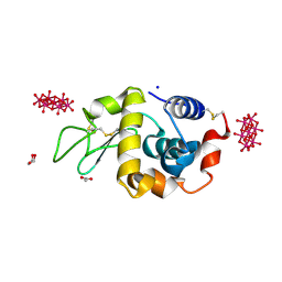
2NM3
 | | Crystal structure of dihydroneopterin aldolase from S. aureus in complex with (1S,2S)-monapterin at 1.68 angstrom resolution | | 分子名称: | ACETATE ION, D-MONAPTERIN, Dihydroneopterin aldolase | | 著者 | Blaszczyk, J, Ji, X, Yan, H. | | 登録日 | 2006-10-20 | | 公開日 | 2007-09-04 | | 最終更新日 | 2023-08-30 | | 実験手法 | X-RAY DIFFRACTION (1.68 Å) | | 主引用文献 | Structural basis for the aldolase and epimerase activities of Staphylococcus aureus dihydroneopterin aldolase.
J.Mol.Biol., 368, 2007
|
|
2PDP
 | | Human aldose reductase mutant S302R complexed with IDD 393. | | 分子名称: | (5-CHLORO-2-{[(3-NITROBENZYL)AMINO]CARBONYL}PHENOXY)ACETIC ACID, Aldose reductase, NADP NICOTINAMIDE-ADENINE-DINUCLEOTIDE PHOSPHATE | | 著者 | Steuber, H, Heine, A, Klebe, G. | | 登録日 | 2007-04-01 | | 公開日 | 2008-04-01 | | 最終更新日 | 2023-08-30 | | 実験手法 | X-RAY DIFFRACTION (1.65 Å) | | 主引用文献 | Merging the binding sites of aldose and aldehyde reductase for detection of inhibitor selectivity-determining features.
J.Mol.Biol., 379, 2008
|
|
1PH1
 | |
1PH2
 | |
1PH4
 | |
1PH3
 | |
1PH5
 | |
1PH7
 | |
1TBO
 | | NMR STRUCTURE OF A PROTEIN KINASE C-G PHORBOL-BINDING DOMAIN, 30 STRUCTURES | | 分子名称: | PROTEIN KINASE C, GAMMA TYPE, ZINC ION | | 著者 | Xu, R.X, Pawelczyk, T, Xia, T, Brown, S.C. | | 登録日 | 1997-04-15 | | 公開日 | 1998-04-29 | | 最終更新日 | 2024-05-22 | | 実験手法 | SOLUTION NMR | | 主引用文献 | NMR structure of a protein kinase C-gamma phorbol-binding domain and study of protein-lipid micelle interactions.
Biochemistry, 36, 1997
|
|
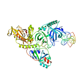
1TQN
 | | Crystal Structure of Human Microsomal P450 3A4 | | 分子名称: | PROTOPORPHYRIN IX CONTAINING FE, cytochrome P450 3A4 | | 著者 | Yano, J.K, Wester, M.R, Schoch, G.A, Griffin, K.J, Stout, C.D, Johnson, E.F. | | 登録日 | 2004-06-17 | | 公開日 | 2004-07-27 | | 最終更新日 | 2024-02-14 | | 実験手法 | X-RAY DIFFRACTION (2.05 Å) | | 主引用文献 | The Structure of Human Microsomal Cytochrome P450 3A4 Determined by X-ray Crystallography to 2.05-A Resolution
J.Biol.Chem., 279, 2004
|
|
2NM2
 | |
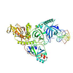
4PHI
 | | Crystal structure of HEWL with hexatungstotellurate(VI) | | 分子名称: | 6-tungstotellurate(VI), ACETATE ION, GLYCEROL, ... | | 著者 | Bijelic, A, Molitor, C, Mauracher, S.G, Al-Oweini, R, Kortz, U, Rompel, A. | | 登録日 | 2014-05-06 | | 公開日 | 2015-01-14 | | 最終更新日 | 2024-11-06 | | 実験手法 | X-RAY DIFFRACTION (1.811 Å) | | 主引用文献 | Hen Egg-White Lysozyme Crystallisation: Protein Stacking and Structure Stability Enhanced by a Tellurium(VI)-Centred Polyoxotungstate.
Chembiochem, 16, 2015
|
|
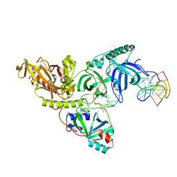
7BR9
 | | Crystal structure of mus musculus IRG1 | | 分子名称: | Cis-aconitate decarboxylase | | 著者 | Park, H.H, Chun, H.L. | | 登録日 | 2020-03-27 | | 公開日 | 2021-02-03 | | 最終更新日 | 2023-11-29 | | 実験手法 | X-RAY DIFFRACTION (3.3 Å) | | 主引用文献 | The crystal structure of mouse IRG1 suggests that cis-aconitate decarboxylase has an open and closed conformation.
Plos One, 15, 2020
|
|
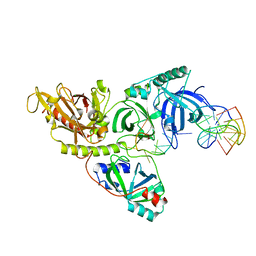
6IEV
 | | Crystal structure of a designed protein | | 分子名称: | Designed protein | | 著者 | Han, M, Liao, S, Chen, Q, Liu, H. | | 登録日 | 2018-09-17 | | 公開日 | 2019-09-18 | | 最終更新日 | 2023-11-22 | | 実験手法 | X-RAY DIFFRACTION (2.25 Å) | | 主引用文献 | Selection and analyses of variants of a designed protein suggest importance of hydrophobicity of partially buried sidechains for protein stability at high temperatures.
Protein Sci., 28, 2019
|
|


