1KCY
 
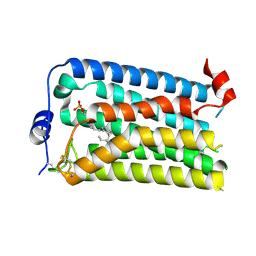
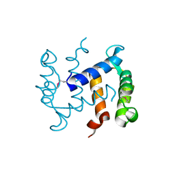
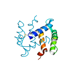
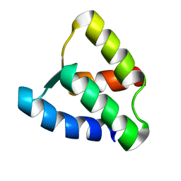 | | NMR solution structure of apo calbindin D9k (F36G + P43M mutant) | | 分子名称: | calbindin D9k | | 著者 | Nelson, M.R, Thulin, E, Fagan, P.A, Forsen, S, Chazin, W.J. | | 登録日 | 2001-11-12 | | 公開日 | 2001-11-21 | | 最終更新日 | 2024-05-22 | | 実験手法 | SOLUTION NMR | | 主引用文献 | The EF-hand domain: a globally cooperative structural unit.
Protein Sci., 11, 2002
|
|
7YU5
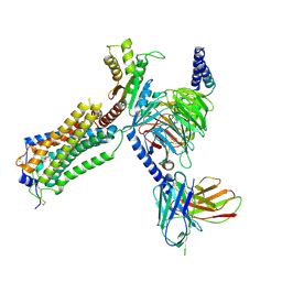
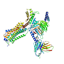 | | Human Lysophosphatidic Acid Receptor 1-Gi complex bound to ONO-0740556, state1 | | 分子名称: | Guanine nucleotide-binding protein G(I)/G(S)/G(O) subunit gamma-2, Guanine nucleotide-binding protein G(I)/G(S)/G(T) subunit beta-1, Guanine nucleotide-binding protein G(i) subunit alpha-1, ... | | 著者 | Akasaka, H, Shihoya, W, Nureki, O. | | 登録日 | 2022-08-16 | | 公開日 | 2022-10-05 | | 実験手法 | ELECTRON MICROSCOPY (3.3 Å) | | 主引用文献 | Structure of the active G i -coupled human lysophosphatidic acid receptor 1 complexed with a potent agonist.
Nat Commun, 13, 2022
|
|
7YU3
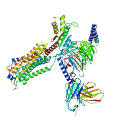 | | Human Lysophosphatidic Acid Receptor 1-Gi complex bound to ONO-0740556 | | 分子名称: | Guanine nucleotide-binding protein G(I)/G(S)/G(O) subunit gamma-2, Guanine nucleotide-binding protein G(I)/G(S)/G(T) subunit beta-1, Guanine nucleotide-binding protein G(i) subunit alpha-1, ... | | 著者 | Akasaka, H, Shihoya, W, Nureki, O. | | 登録日 | 2022-08-16 | | 公開日 | 2022-10-05 | | 実験手法 | ELECTRON MICROSCOPY (3.4 Å) | | 主引用文献 | Structure of the active G i -coupled human lysophosphatidic acid receptor 1 complexed with a potent agonist.
Nat Commun, 13, 2022
|
|
7YU8
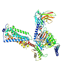 | | Human Lysophosphatidic Acid Receptor 1-Gi complex bound to ONO-0740556, state4 | | 分子名称: | Guanine nucleotide-binding protein G(I)/G(S)/G(O) subunit gamma-2, Guanine nucleotide-binding protein G(I)/G(S)/G(T) subunit beta-1, Guanine nucleotide-binding protein G(i) subunit alpha-1, ... | | 著者 | Akasaka, H, Shihoya, W, Nureki, O. | | 登録日 | 2022-08-16 | | 公開日 | 2022-10-05 | | 実験手法 | ELECTRON MICROSCOPY (4.5 Å) | | 主引用文献 | Structure of the active G i -coupled human lysophosphatidic acid receptor 1 complexed with a potent agonist.
Nat Commun, 13, 2022
|
|
7YU6
 | | Human Lysophosphatidic Acid Receptor 1-Gi complex bound to ONO-0740556, state2 | | 分子名称: | Guanine nucleotide-binding protein G(I)/G(S)/G(O) subunit gamma-2, Guanine nucleotide-binding protein G(I)/G(S)/G(T) subunit beta-1, Guanine nucleotide-binding protein G(i) subunit alpha-1, ... | | 著者 | Akasaka, H, Shihoya, W, Nureki, O. | | 登録日 | 2022-08-16 | | 公開日 | 2022-10-05 | | 実験手法 | ELECTRON MICROSCOPY (3.5 Å) | | 主引用文献 | Structure of the active G i -coupled human lysophosphatidic acid receptor 1 complexed with a potent agonist.
Nat Commun, 13, 2022
|
|
7YU4
 | |
7YU7
 | | Human Lysophosphatidic Acid Receptor 1-Gi complex bound to ONO-0740556, state3 | | 分子名称: | Guanine nucleotide-binding protein G(I)/G(S)/G(O) subunit gamma-2, Guanine nucleotide-binding protein G(I)/G(S)/G(T) subunit beta-1, Guanine nucleotide-binding protein G(i) subunit alpha-1, ... | | 著者 | Akasaka, H, Shihoya, W, Nureki, O. | | 登録日 | 2022-08-16 | | 公開日 | 2022-10-05 | | 実験手法 | ELECTRON MICROSCOPY (3.8 Å) | | 主引用文献 | Structure of the active G i -coupled human lysophosphatidic acid receptor 1 complexed with a potent agonist.
Nat Commun, 13, 2022
|
|
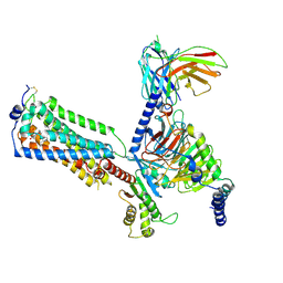
7YTJ
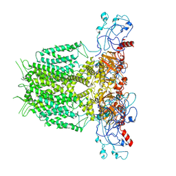
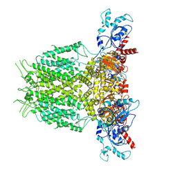
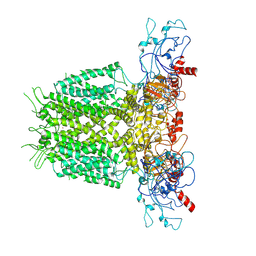
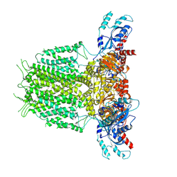
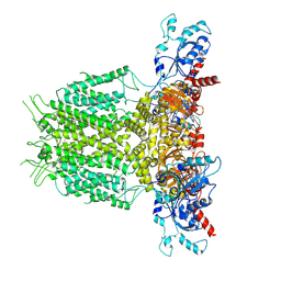
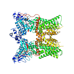
 | | Cryo-EM structure of VTC complex | | 分子名称: | 1,2-DIACYL-SN-GLYCERO-3-PHOSPHOCHOLINE, INOSITOL HEXAKISPHOSPHATE, PHOSPHATE ION, ... | | 著者 | Guan, Z.Y, Chen, J, Liu, R.W, Chen, Y.K, Xing, Q, Du, Z.M, Liu, Z. | | 登録日 | 2022-08-15 | | 公開日 | 2023-02-22 | | 最終更新日 | 2024-07-03 | | 実験手法 | ELECTRON MICROSCOPY (3 Å) | | 主引用文献 | The cytoplasmic synthesis and coupled membrane translocation of eukaryotic polyphosphate by signal-activated VTC complex.
Nat Commun, 14, 2023
|
|
7YID
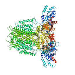 | | Human KCNH5 closed state 1 | | 分子名称: | POTASSIUM ION, Potassium voltage-gated channel subfamily H member 5 | | 著者 | Zhang, M.F. | | 登録日 | 2022-07-16 | | 公開日 | 2023-04-05 | | 最終更新日 | 2024-07-03 | | 実験手法 | ELECTRON MICROSCOPY (3.4 Å) | | 主引用文献 | Mechanism underlying delayed rectifying in human voltage-mediated activation Eag2 channel.
Nat Commun, 14, 2023
|
|
7YIJ
 | | Human KCNH5 pore dilation but the non-conducting state | | 分子名称: | POTASSIUM ION, Potassium voltage-gated channel subfamily H member 5 | | 著者 | Zhang, M.F. | | 登録日 | 2022-07-16 | | 公開日 | 2023-04-05 | | 最終更新日 | 2024-07-03 | | 実験手法 | ELECTRON MICROSCOPY (3.8 Å) | | 主引用文献 | Mechanism underlying delayed rectifying in human voltage-mediated activation Eag2 channel.
Nat Commun, 14, 2023
|
|
7YIG
 | | Human KCNH5 pre-open state 2 | | 分子名称: | POTASSIUM ION, Potassium voltage-gated channel subfamily H member 5 | | 著者 | Zhang, M.F. | | 登録日 | 2022-07-16 | | 公開日 | 2023-04-05 | | 最終更新日 | 2024-07-03 | | 実験手法 | ELECTRON MICROSCOPY (3.6 Å) | | 主引用文献 | Mechanism underlying delayed rectifying in human voltage-mediated activation Eag2 channel.
Nat Commun, 14, 2023
|
|
7YIH
 | | Human KCNH5 open state | | 分子名称: | POTASSIUM ION, Potassium voltage-gated channel subfamily H member 5 | | 著者 | Zhang, M.F. | | 登録日 | 2022-07-16 | | 公開日 | 2023-04-05 | | 最終更新日 | 2024-07-03 | | 実験手法 | ELECTRON MICROSCOPY (3.5 Å) | | 主引用文献 | Mechanism underlying delayed rectifying in human voltage-mediated activation Eag2 channel.
Nat Commun, 14, 2023
|
|
7YIE
 | | Human KCNH5-closed state 2 | | 分子名称: | POTASSIUM ION, Potassium voltage-gated channel subfamily H member 5 | | 著者 | Zhang, M.F. | | 登録日 | 2022-07-16 | | 公開日 | 2023-04-05 | | 最終更新日 | 2024-07-03 | | 実験手法 | ELECTRON MICROSCOPY (3.4 Å) | | 主引用文献 | Mechanism underlying delayed rectifying in human voltage-mediated activation Eag2 channel.
Nat Commun, 14, 2023
|
|
7YIF
 | | Human KCNH5 pre-open state 1 | | 分子名称: | POTASSIUM ION, Potassium voltage-gated channel subfamily H member 5 | | 著者 | Zhang, M.F. | | 登録日 | 2022-07-16 | | 公開日 | 2023-04-05 | | 最終更新日 | 2024-07-03 | | 実験手法 | ELECTRON MICROSCOPY (3.5 Å) | | 主引用文献 | Mechanism underlying delayed rectifying in human voltage-mediated activation Eag2 channel.
Nat Commun, 14, 2023
|
|
1N2D
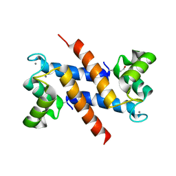
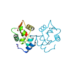
 | | Ternary complex of MLC1P bound to IQ2 and IQ3 of Myo2p, a class V myosin | | 分子名称: | IQ2 AND IQ3 MOTIFS FROM MYO2P, A CLASS V MYOSIN, Myosin Light Chain | | 著者 | Terrak, M, Wu, G, Stafford, W.F, Lu, R.C, Dominguez, R. | | 登録日 | 2002-10-22 | | 公開日 | 2003-11-04 | | 最終更新日 | 2024-02-14 | | 実験手法 | X-RAY DIFFRACTION (2 Å) | | 主引用文献 | Structure of the light chain-binding domain of myosin V.
Proc.Natl.Acad.Sci.USA, 102, 2005
|
|
1K8U
 | |
1K96
 | |
1K9K
 | |
1K9P
 | |
1K9U
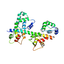
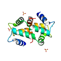 | | Crystal Structure of the Calcium-Binding Pollen Allergen Phl p 7 (Polcalcin) at 1.75 Angstroem | | 分子名称: | CALCIUM ION, Polcalcin Phl p 7, SULFATE ION | | 著者 | Verdino, P, Westritschnig, K, Valenta, R, Keller, W. | | 登録日 | 2001-10-30 | | 公開日 | 2003-04-30 | | 最終更新日 | 2024-03-13 | | 実験手法 | X-RAY DIFFRACTION (1.75 Å) | | 主引用文献 | The cross-reactive calcium-binding pollen allergen, Phl p 7, reveals a novel dimer assembly
EMBO J., 21, 2002
|
|
1CDP
 | |
7L2O
 | |
7L2K
 | |
7L2H
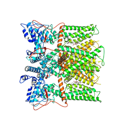
 | | Cryo-EM structure of unliganded full-length TRPV1 at neutral pH | | 分子名称: | (10R,13S)-16-amino-13-hydroxy-7,13-dioxo-8,12,14-trioxa-13lambda~5~-phosphahexadecan-10-yl tridecanoate, (2S)-1-(butanoyloxy)-3-{[(R)-hydroxy{[(1r,2R,3S,4S,5R,6S)-2,3,4,5,6-pentahydroxycyclohexyl]oxy}phosphoryl]oxy}propan-2-yl tridecanoate, SODIUM ION, ... | | 著者 | Zhang, K, Julius, D, Cheng, Y. | | 登録日 | 2020-12-17 | | 公開日 | 2021-09-22 | | 最終更新日 | 2021-10-13 | | 実験手法 | ELECTRON MICROSCOPY (2.63 Å) | | 主引用文献 | Structural snapshots of TRPV1 reveal mechanism of polymodal functionality.
Cell, 184, 2021
|
|
7L2J
 | | Cryo-EM structure of full-length TRPV1 at pH6c state | | 分子名称: | (2S)-1-(butanoyloxy)-3-{[(R)-hydroxy{[(1r,2R,3S,4S,5R,6S)-2,3,4,5,6-pentahydroxycyclohexyl]oxy}phosphoryl]oxy}propan-2-yl tridecanoate, Transient receptor potential cation channel subfamily V member 1 | | 著者 | Zhang, K, Julius, D, Cheng, Y. | | 登録日 | 2020-12-17 | | 公開日 | 2021-09-22 | | 最終更新日 | 2021-10-13 | | 実験手法 | ELECTRON MICROSCOPY (3.66 Å) | | 主引用文献 | Structural snapshots of TRPV1 reveal mechanism of polymodal functionality.
Cell, 184, 2021
|
|


