3GUB
 
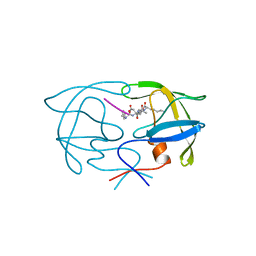
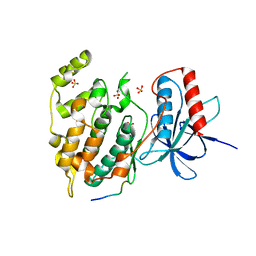
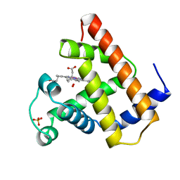
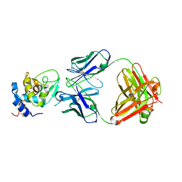
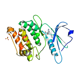 | | Crystal structure of DAPKL93G complexed with N6-(2-Phenylethyl)adenosine | | 分子名称: | 9-alpha-L-lyxofuranosyl-N-(2-phenylethyl)-9H-purin-6-amine, Death-associated protein kinase 1, SULFATE ION | | 著者 | McNamara, L.K, Schumacher, A.M, Schavocky, J.S, Watterson, D.M, Brunzelle, J.S. | | 登録日 | 2009-03-29 | | 公開日 | 2010-03-09 | | 最終更新日 | 2023-09-06 | | 実験手法 | X-RAY DIFFRACTION (1.71 Å) | | 主引用文献 | Crystal structures of the DAPK gatekeeper mutant complexed with N6-modified adenosine analogs.
To be Published
|
|
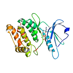
3GU7
 | |
1YT9
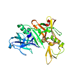
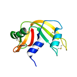
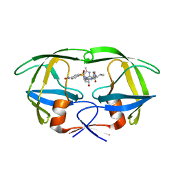 | | HIV Protease with oximinoarylsulfonamide bound | | 分子名称: | (S)-N-((2S,3R)-3-HYDROXY-4-(4-((E)-(HYDROXYIMINO)METHYL)-N-ISOBUTYLPHENYLSULFONAMIDO)-1-PHENYLBUTAN-2-YL)-3-METHYL-2-(3 -((2-METHYLTHIAZOL-4-YL)METHYL)-2-OXOIMIDAZOLIDIN-1-YL)BUTANAMIDE, Pol polyprotein | | 著者 | Yeung, C.M, Klein, L.L, Flentge, C.A, Randolph, J.T, Zhao, C, Sun, M, Dekhtyar, T, Stoll, V.S, Kempf, D.J. | | 登録日 | 2005-02-10 | | 公開日 | 2005-04-12 | | 最終更新日 | 2024-02-14 | | 実験手法 | X-RAY DIFFRACTION (3 Å) | | 主引用文献 | Oximinoarylsulfonamides as potent HIV protease inhibitors.
Bioorg.Med.Chem.Lett., 15, 2005
|
|
2DWB
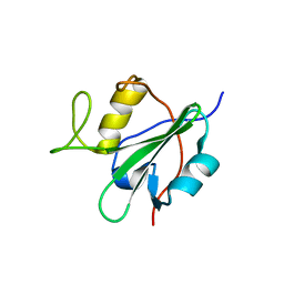
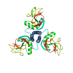
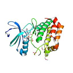 | | Aurora-A kinase complexed with AMPPNP | | 分子名称: | PHOSPHOAMINOPHOSPHONIC ACID-ADENYLATE ESTER, SULFATE ION, Serine/threonine-protein kinase 6 | | 著者 | Kukimoto-Niino, M, Murayama, K, Shirouzu, S, Yokoyama, S, RIKEN Structural Genomics/Proteomics Initiative (RSGI) | | 登録日 | 2006-08-10 | | 公開日 | 2007-07-31 | | 最終更新日 | 2023-11-15 | | 実験手法 | X-RAY DIFFRACTION (2.5 Å) | | 主引用文献 | Aurora-A kinase complexed with AMPPNP
To be Published
|
|
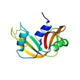
1YTI
 | | SIV PROTEASE CRYSTALLIZED WITH PEPTIDE PRODUCT | | 分子名称: | PEPTIDE PRODUCT, SIV PROTEASE | | 著者 | Rose, R.B, Craik, C.S, Douglas, N.L, Stroud, R.M. | | 登録日 | 1996-08-01 | | 公開日 | 1997-03-12 | | 最終更新日 | 2024-02-14 | | 実験手法 | X-RAY DIFFRACTION (2.2 Å) | | 主引用文献 | Three-dimensional structures of HIV-1 and SIV protease product complexes.
Biochemistry, 35, 1996
|
|
1YM2
 | | Crystal structure of human beta secretase complexed with NVP-AUR200 | | 分子名称: | Beta-secretase 1, NVP-AUR200 INHIBITOR | | 著者 | Hanessian, S, Yun, H, Hou, Y, Yang, G, Bayrakdarian, M, Therrien, E, Moitessier, N, Roggo, S, Veenstra, S. | | 登録日 | 2005-01-20 | | 公開日 | 2006-01-17 | | 最終更新日 | 2023-11-15 | | 実験手法 | X-RAY DIFFRACTION (2.05 Å) | | 主引用文献 | Structure-based design, synthesis, and memapsin 2 (BACE) inhibitory activity of carbocyclic and heterocyclic peptidomimetics
J.Med.Chem., 48, 2005
|
|
2DCR
 | |
3A7G
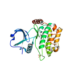 | | Human MST3 kinase | | 分子名称: | Serine/threonine kinase 24 (STE20 homolog, yeast) | | 著者 | Ko, T.P, Jeng, W.Y, Liu, C.I, Lai, M.D, Wang, A.H.J. | | 登録日 | 2009-09-26 | | 公開日 | 2010-02-02 | | 最終更新日 | 2023-11-01 | | 実験手法 | X-RAY DIFFRACTION (2 Å) | | 主引用文献 | Structures of human MST3 kinase in complex with adenine, ADP and Mn2+.
Acta Crystallogr.,Sect.D, 66, 2010
|
|
3VUI
 | | Crystal structure of a cysteine-deficient mutant M2 in MAP kinase JNK1 | | 分子名称: | Mitogen-activated protein kinase 8, Peptide from C-Jun-amino-terminal kinase-interacting protein 1, SULFATE ION | | 著者 | Nakaniwa, T, Kinoshita, T, Inoue, T. | | 登録日 | 2012-06-28 | | 公開日 | 2013-02-13 | | 最終更新日 | 2024-03-20 | | 実験手法 | X-RAY DIFFRACTION (2.8 Å) | | 主引用文献 | Seven cysteine-deficient mutants depict the interplay between thermal and chemical stabilities of individual cysteine residues in mitogen-activated protein kinase c-Jun N-terminal kinase 1
Biochemistry, 51, 2012
|
|
2DLQ
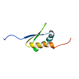
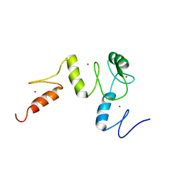 | | Solution structure of the tandem four zf-C2H2 domain repeats of murine GLI-Kruppel family member HKR3 | | 分子名称: | GLI-Kruppel family member HKR3, ZINC ION | | 著者 | Inoue, K, Hayashi, F, Izumi, K, Yoshida, M, Yokoyama, S, RIKEN Structural Genomics/Proteomics Initiative (RSGI) | | 登録日 | 2006-04-20 | | 公開日 | 2006-10-20 | | 最終更新日 | 2024-05-29 | | 実験手法 | SOLUTION NMR | | 主引用文献 | Solution structure of the tandem four zf-C2H2 domain repeats of murine GLI-Kruppel family member HKR3
To be published
|
|
2DMS
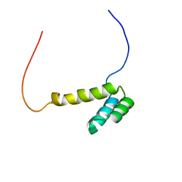 | | Solution structure of the homeobox domain of Homeobox protein OTX2 | | 分子名称: | Homeobox protein OTX2 | | 著者 | Ohnishi, S, Saito, K, Koshiba, S, Inoue, M, Kigawa, T, Yokoyama, S, RIKEN Structural Genomics/Proteomics Initiative (RSGI) | | 登録日 | 2006-04-24 | | 公開日 | 2006-10-24 | | 最終更新日 | 2024-05-29 | | 実験手法 | SOLUTION NMR | | 主引用文献 | Solution structure of the homeobox domain of Homeobox protein OTX2
To be Published
|
|
1KVZ
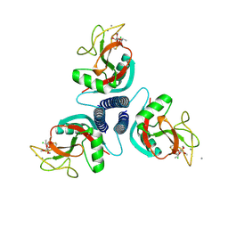
 | | Solution Structure of Cytotoxic RC-RNase4 | | 分子名称: | RC-RNase4 | | 著者 | Hsu, C.-H, Liao, Y.-D, Chen, L.-W, Wu, S.-H, Chen, C. | | 登録日 | 2002-01-28 | | 公開日 | 2002-07-28 | | 最終更新日 | 2022-12-21 | | 実験手法 | SOLUTION NMR | | 主引用文献 | Solution Structure of the Cytotoxic RNase 4 from the Oocytes of Bullfrog Rana Catesbeiana
J.MOL.BIOL., 326, 2003
|
|
1KWW
 | | Rat mannose protein A complexed with a-Me-Fuc. | | 分子名称: | CALCIUM ION, CHLORIDE ION, MANNOSE-BINDING PROTEIN A, ... | | 著者 | Ng, K.K, Kolatkar, A.R, Park-Snyder, S, Feinberg, H, Clark, D.A, Drickamer, K, Weis, W.I. | | 登録日 | 2002-01-30 | | 公開日 | 2002-07-05 | | 最終更新日 | 2020-07-29 | | 実験手法 | X-RAY DIFFRACTION (1.9 Å) | | 主引用文献 | Orientation of bound ligands in mannose-binding proteins. Implications for multivalent ligand recognition.
J.Biol.Chem., 277, 2002
|
|
1KX1
 | | Rat mannose protein A complexed with Man6-GlcNAc2-Asn | | 分子名称: | CALCIUM ION, MANNOSE-BINDING PROTEIN A, alpha-D-mannopyranose, ... | | 著者 | Ng, K.K, Kolatkar, A.R, Park-Snyder, S, Feinberg, H, Clark, D.A, Drickamer, K, Weis, W.I. | | 登録日 | 2002-01-30 | | 公開日 | 2002-07-05 | | 最終更新日 | 2020-07-29 | | 実験手法 | X-RAY DIFFRACTION (2.8 Å) | | 主引用文献 | Orientation of bound ligands in mannose-binding proteins. Implications for multivalent ligand recognition.
J.Biol.Chem., 277, 2002
|
|
1YOI
 | | COBALT MYOGLOBIN (OXY) | | 分子名称: | MYOGLOBIN, OXYGEN MOLECULE, PROTOPORPHYRIN IX CONTAINING CO, ... | | 著者 | Brucker, E.A, Phillips Jr, G.N. | | 登録日 | 1996-06-14 | | 公開日 | 1996-12-07 | | 最終更新日 | 2024-02-14 | | 実験手法 | X-RAY DIFFRACTION (1.65 Å) | | 主引用文献 | High resolution crystal structures of the deoxy, oxy, and aquomet forms of cobalt myoglobin.
J.Biol.Chem., 271, 1996
|
|
2DGR
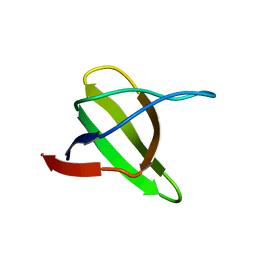
 | | Solution structure of the second KH domain in ring finger and KH domain containing protein 1 | | 分子名称: | RING finger and KH domain-containing protein 1 | | 著者 | Abe, C, Muto, Y, Inoue, M, Kigawa, T, Terada, T, Shirouzu, M, Yokoyama, S, RIKEN Structural Genomics/Proteomics Initiative (RSGI) | | 登録日 | 2006-03-15 | | 公開日 | 2006-09-15 | | 最終更新日 | 2024-05-29 | | 実験手法 | SOLUTION NMR | | 主引用文献 | Solution structure of the second KH domain in ring finger and KH domain containing protein 1
To be Published
|
|
1YP5
 | | Yeast Myo5 SH3 domain, tetragonal crystal form | | 分子名称: | Myosin-5 isoform | | 著者 | Gonfloni, S, Kursula, P, Sacco, R, Cesareni, G, Wilmanns, M. | | 登録日 | 2005-01-29 | | 公開日 | 2006-01-17 | | 最終更新日 | 2024-03-13 | | 実験手法 | X-RAY DIFFRACTION (1.68 Å) | | 主引用文献 | Yeast Myo5 SH3 domain, tetragonal crystal form
To be Published
|
|
1KVW
 | |
1YP9
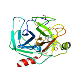 | | Trypsin Inhibitor Complex | | 分子名称: | (1R,3AS,4R,8AS,8BR)-4-(2-BENZO[1,3]DIOXOL-5-YLMETHYL)-1-BENZYL-3-OXO-DECAHYDRO-PYRROLO[3,4-A]PYRROZILIN-4-YL-BENZAMIDINE, CALCIUM ION, Cationic trypsin, ... | | 著者 | Fokkens, J, Obst-Sander, U, Heine, A, Diederich, F, Klebe, G. | | 登録日 | 2005-01-31 | | 公開日 | 2006-01-17 | | 最終更新日 | 2023-10-25 | | 実験手法 | X-RAY DIFFRACTION (2.1 Å) | | 主引用文献 | A simple protocol to estimate differences in protein binding affinity for enantiomers without prior resolution of racemates
Angew.Chem.Int.Ed.Engl., 45, 2006
|
|
1YQV
 | | The crystal structure of the antibody Fab HyHEL5 complex with lysozyme at 1.7A resolution | | 分子名称: | Hen Egg White Lysozyme, HyHEL-5 Antibody Heavy Chain, HyHEL-5 Antibody Light Chain | | 著者 | Cohen, G.H, Silverton, E.W, Padlan, E.A, Dyda, F, Wibbenmeyer, J.A, Wilson, R.C, Davies, D.R. | | 登録日 | 2005-02-02 | | 公開日 | 2005-04-26 | | 最終更新日 | 2023-08-23 | | 実験手法 | X-RAY DIFFRACTION (1.7 Å) | | 主引用文献 | Water molecules in the antibody-antigen interface of the structure of the Fab HyHEL-5-lysozyme complex at 1.7 A resolution: comparison with results from isothermal titration calorimetry.
Acta Crystallogr.,Sect.D, 61, 2005
|
|
1KF4
 | | Atomic Resolution Structure of RNase A at pH 6.3 | | 分子名称: | SULFATE ION, pancreatic ribonuclease | | 著者 | Berisio, R, Sica, F, Lamzin, V.S, Wilson, K.S, Zagari, A, Mazzarella, L. | | 登録日 | 2001-11-19 | | 公開日 | 2001-12-19 | | 最終更新日 | 2023-08-16 | | 実験手法 | X-RAY DIFFRACTION (1.1 Å) | | 主引用文献 | Atomic resolution structures of ribonuclease A at six pH values.
Acta Crystallogr.,Sect.D, 58, 2002
|
|
1KWU
 | | Rat mannose binding protein A complexed with a-Me-Man | | 分子名称: | CALCIUM ION, CHLORIDE ION, MANNOSE-BINDING PROTEIN A, ... | | 著者 | Ng, K.K, Kolatkar, A.R, Park-Snyder, S, Feinberg, H, Clark, D.A, Drickamer, K, Weis, W.I. | | 登録日 | 2002-01-30 | | 公開日 | 2002-07-05 | | 最終更新日 | 2020-07-29 | | 実験手法 | X-RAY DIFFRACTION (1.95 Å) | | 主引用文献 | Orientation of bound ligands in mannose-binding proteins. Implications for multivalent ligand recognition.
J.Biol.Chem., 277, 2002
|
|
1YMR
 | | The study of reductive unfolding pathways of RNase A (Y92A mutant) | | 分子名称: | Ribonuclease pancreatic | | 著者 | Xu, G, Narayan, M, Kurinov, I, Ripoll, D.R, Welker, E, Khalili, M, Ealick, S.E, Scheraga, H.A. | | 登録日 | 2005-01-21 | | 公開日 | 2006-01-31 | | 最終更新日 | 2021-10-20 | | 実験手法 | X-RAY DIFFRACTION (1.5 Å) | | 主引用文献 | A localized specific interaction alters the unfolding pathways of structural homologues.
J.Am.Chem.Soc., 128, 2006
|
|
2DIX
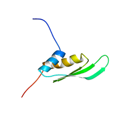 | | Solution structure of the DSRM domain of Protein activator of the interferon-induced protein kinase | | 分子名称: | Interferon-inducible double stranded RNA-dependent protein kinase activator A | | 著者 | Dang, W, Muto, Y, Inoue, M, Kigawa, T, Shirouzu, M, Terada, T, Yokoyama, S, RIKEN Structural Genomics/Proteomics Initiative (RSGI) | | 登録日 | 2006-03-30 | | 公開日 | 2006-09-30 | | 最終更新日 | 2024-05-29 | | 実験手法 | SOLUTION NMR | | 主引用文献 | Solution structure of the DSRM domain of Protein activator of the interferon-induced protein kinase
To be published
|
|
1YPQ
 | |


