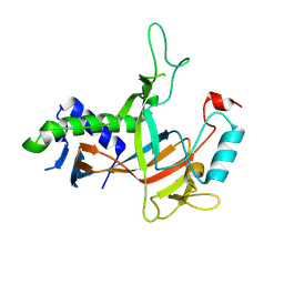
6IET
 
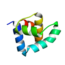
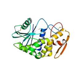
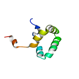
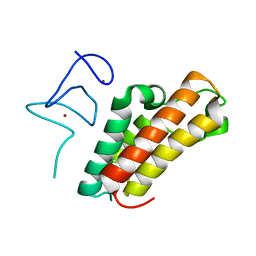 | | The crystal structure of TRIM66 PHD-Bromo domain | | 分子名称: | Tripartite motif-containing protein 66, ZINC ION | | 著者 | Chen, J. | | 登録日 | 2018-09-17 | | 公開日 | 2019-09-18 | | 最終更新日 | 2023-11-22 | | 実験手法 | X-RAY DIFFRACTION (2.101 Å) | | 主引用文献 | TRIM66 reads unmodified H3R2K4 and H3K56ac to respond to DNA damage in embryonic stem cells.
Nat Commun, 10, 2019
|
|
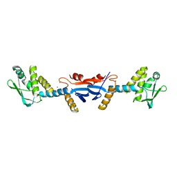
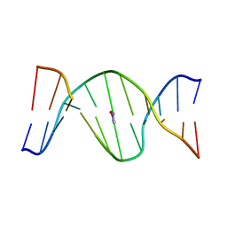
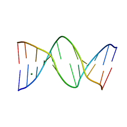
3ZHM
 | | N-terminal domain of the CI repressor from bacteriophage TP901-1 in complex with the OL2 operator half-site | | 分子名称: | 5'-D(*AP*CP*GP*TP*GP*AP*AP*CP*TP*TP*GP*CP*AP*CP *TP*TP*GP*A)-3', 5'-D(*AP*GP*TP*TP*CP*AP*CP*GP*TP*TP*CP*AP*AP*GP *TP*GP*CP*A)-3', CI | | 著者 | Frandsen, K.H, Rasmussen, K.K, Poulsen, J.N, Lo Leggio, L. | | 登録日 | 2012-12-22 | | 公開日 | 2013-12-25 | | 最終更新日 | 2023-12-20 | | 実験手法 | X-RAY DIFFRACTION (2.6 Å) | | 主引用文献 | Binding of the N-Terminal Domain of the Lactococcal Bacteriophage Tp901-1 Ci Repressor to its Target DNA: A Crystallography, Small Angle Scattering, and Nuclear Magnetic Resonance Study.
Biochemistry, 52, 2013
|
|
2GK4
 | |
1KIJ
 | | Crystal structure of the 43K ATPase domain of Thermus thermophilus gyrase B in complex with novobiocin | | 分子名称: | DNA GYRASE SUBUNIT B, FORMIC ACID, NOVOBIOCIN | | 著者 | Lamour, V, Hoermann, L, Jeltsch, J.-M, Oudet, P, Moras, D. | | 登録日 | 2001-12-03 | | 公開日 | 2002-06-03 | | 最終更新日 | 2024-02-14 | | 実験手法 | X-RAY DIFFRACTION (2.3 Å) | | 主引用文献 | An open conformation of the Thermus thermophilus gyrase B ATP-binding domain.
J.Biol.Chem., 277, 2002
|
|
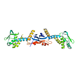
3ZHI
 | | N-terminal domain of the CI repressor from bacteriophage TP901-1 | | 分子名称: | CI | | 著者 | Frandsen, K.H, Rasmussen, K.K, Poulsen, J.N, Lo Leggio, L. | | 登録日 | 2012-12-21 | | 公開日 | 2013-12-25 | | 最終更新日 | 2024-05-08 | | 実験手法 | X-RAY DIFFRACTION (1.6 Å) | | 主引用文献 | Binding of the N-Terminal Domain of the Lactococcal Bacteriophage Tp901-1 Ci Repressor to its Target DNA: A Crystallography, Small Angle Scattering, and Nuclear Magnetic Resonance Study.
Biochemistry, 52, 2013
|
|
6H09
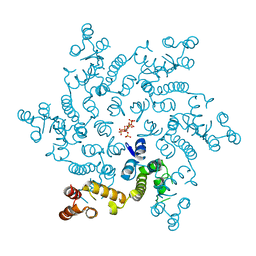 | | HIV capsid hexamer with IP6 ligand | | 分子名称: | Gag polyprotein, INOSITOL HEXAKISPHOSPHATE | | 著者 | James, L.C. | | 登録日 | 2018-07-06 | | 公開日 | 2018-08-15 | | 最終更新日 | 2020-10-07 | | 実験手法 | X-RAY DIFFRACTION (2 Å) | | 主引用文献 | IP6 is an HIV pocket factor that prevents capsid collapse and promotes DNA synthesis.
Elife, 7, 2018
|
|
2KFP
 | | Solution NMR structure of PSPTO_3016 from Pseudomonas syringae. Northeast Structural Genomics Consortium target PsR293. | | 分子名称: | PSPTO_3016 protein | | 著者 | Feldmann, E.A, Ramelot, T.A, Zhao, L, Hamilton, K, Ciccosanti, C, Xiao, R, Nair, R, Everett, J.K, Swapna, G, Acton, T.B, Rost, B, Montelione, G.T, Kennedy, M.A, Northeast Structural Genomics Consortium (NESG) | | 登録日 | 2009-02-24 | | 公開日 | 2009-03-24 | | 最終更新日 | 2024-05-08 | | 実験手法 | SOLUTION NMR | | 主引用文献 | Solution NMR and X-ray crystal structures of Pseudomonas syringae Pspto_3016 from protein domain family PF04237 (DUF419) adopt a "double wing" DNA binding motif.
J.Struct.Funct.Genom., 13, 2012
|
|
427D
 | |
3H5K
 | | Crystal structure of the ribosome inactivating protein PDL1 | | 分子名称: | 1,2-ETHANEDIOL, 2-acetamido-2-deoxy-beta-D-glucopyranose, Ribosome-inactivating protein PD-L1/PD-L2 | | 著者 | Ruggiero, A, Di Maro, A, Severino, V, Chambery, A, Berisio, R. | | 登録日 | 2009-04-22 | | 公開日 | 2009-10-13 | | 最終更新日 | 2020-07-29 | | 実験手法 | X-RAY DIFFRACTION (1.45 Å) | | 主引用文献 | Crystal structure of PD-L1, a ribosome inactivating protein from Phytolacca dioica L. Leaves with the property to induce DNA cleavage
Biopolymers, 91, 2009
|
|
3KDK
 | |
3KDG
 | |
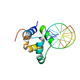
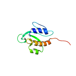
2LQ6
 | | Solution structure of BRD1 PHD2 finger | | 分子名称: | Bromodomain-containing protein 1, ZINC ION | | 著者 | Liu, L, Wu, J. | | 登録日 | 2012-02-25 | | 公開日 | 2012-10-24 | | 最終更新日 | 2024-05-01 | | 実験手法 | SOLUTION NMR | | 主引用文献 | Solution structure of an atypical PHD finger in BRPF2 and its interaction with DNA
J.Struct.Biol., 180, 2012
|
|
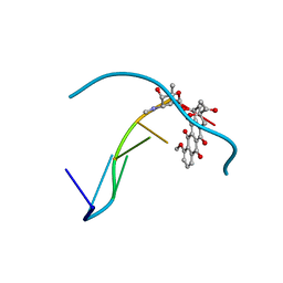
1U6O
 | | Mispairing of a Site-Specific Major Groove (2S,3S)-N6-(2,3,4-Trihydroxybutyl)-2-deoxyadenosyl DNA Adduct of Butadiene Diol Epoxide with Deoxyguanosine: Formation of a dA(anti)dG(anti) Pairing Interaction | | 分子名称: | 5'-D(*CP*GP*GP*AP*CP*(2BU)P*AP*GP*AP*AP*G)-3', 5'-D(*CP*TP*TP*CP*TP*GP*GP*TP*CP*CP*G)-3' | | 著者 | Scholdberg, T.A, Nechev, L.N, Merritt, W.K, Harris, T.M, Harris, C.M, Lloyd, R.S, Stone, M.P. | | 登録日 | 2004-07-30 | | 公開日 | 2004-08-10 | | 最終更新日 | 2024-05-22 | | 実験手法 | SOLUTION NMR | | 主引用文献 | Mispairing of a Site Specific Major Groove (2S,3S)-N6-(2,3,4-Trihydroxybutyl)-2'-deoxyadenosyl DNA Adduct of Butadiene Diol Epoxide with Deoxyguanosine: Formation of a dA(anti)dG(anti) Pairing Interaction.
CHEM.RES.TOXICOL., 18, 2005
|
|
7YRD
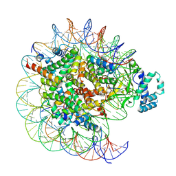 | | histone methyltransferase | | 分子名称: | DNA (146-MER), Histone H2A.Z, Histone H2B 1.1, ... | | 著者 | Li, H, Wang, W.Y. | | 登録日 | 2022-08-09 | | 公開日 | 2023-08-16 | | 最終更新日 | 2023-12-13 | | 実験手法 | ELECTRON MICROSCOPY (3.2 Å) | | 主引用文献 | Structural insight into H4K20 methylation on H2A.Z-nucleosome by SUV420H1.
Mol.Cell, 83, 2023
|
|
3RIQ
 | | Siphovirus 9NA tailspike receptor binding domain | | 分子名称: | GLYCEROL, Tailspike protein | | 著者 | Andres, D, Roske, Y, Doering, C, Heinemann, U, Seckler, R, Barbirz, S. | | 登録日 | 2011-04-14 | | 公開日 | 2012-02-29 | | 最終更新日 | 2023-09-13 | | 実験手法 | X-RAY DIFFRACTION (1.5 Å) | | 主引用文献 | Tail morphology controls DNA release in two Salmonella phages with one lipopolysaccharide receptor recognition system.
Mol.Microbiol., 83, 2012
|
|
7YRG
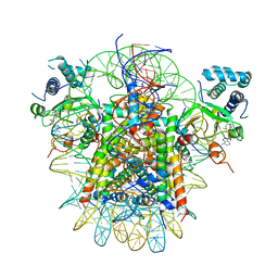 | | histone methyltransferase | | 分子名称: | DNA (146-MER), Histone H2A.Z, Histone H2B 1.1, ... | | 著者 | Li, H, Wang, W.Y. | | 登録日 | 2022-08-09 | | 公開日 | 2023-12-13 | | 実験手法 | ELECTRON MICROSCOPY (4.2 Å) | | 主引用文献 | Structural insight into H4K20 methylation on H2A.Z-nucleosome by SUV420H1.
Mol.Cell, 83, 2023
|
|
1NGT
 | | The Role of Minor Groove Functional Groups in DNA Hydration | | 分子名称: | 5'-D(*CP*GP*CP*GP*AP*AP*(MTR)P*TP*CP*GP*CP*G)-3', MAGNESIUM ION | | 著者 | Woods, K.K, Lan, T, McLaughlin, L.W, Williams, L.D. | | 登録日 | 2002-12-17 | | 公開日 | 2003-03-04 | | 最終更新日 | 2024-02-14 | | 実験手法 | X-RAY DIFFRACTION (2.04 Å) | | 主引用文献 | The Role of Minor Groove Functional Groups in DNA Hydration
Nucleic Acids Res., 31, 2003
|
|
1PVE
 | |
1SSF
 | | Solution structure of the mouse 53BP1 fragment (residues 1463-1617) | | 分子名称: | Transformation related protein 53 binding protein 1 | | 著者 | Charier, G, Couprie, J, Alpha-Bazin, B, Meyer, V, Quemeneur, E, Guerois, R, Callebaut, I, Gilquin, B, Zinn-Justin, S. | | 登録日 | 2004-03-24 | | 公開日 | 2004-09-14 | | 最終更新日 | 2024-05-22 | | 実験手法 | SOLUTION NMR | | 主引用文献 | The Tudor Tandem of 53BP1; A New Structural Motif Involved in DNA and RG-Rich Peptide Binding
Structure, 12, 2004
|
|
1S14
 | |
7YEQ
 | |
1ZUJ
 | | The crystal structure of the Lactococcus lactis MG1363 DpsA protein | | 分子名称: | hypothetical protein Llacc01001955 | | 著者 | Stillman, T.J, Upadhyay, M, Norte, V.A, Sedelnikova, S.E, Carradus, M, Tzokov, S, Bullough, P.A, Shearman, C.A, Gasson, M.J, Williams, C.H, Artymiuk, P.J, Green, J. | | 登録日 | 2005-05-31 | | 公開日 | 2005-08-30 | | 最終更新日 | 2023-08-23 | | 実験手法 | X-RAY DIFFRACTION (2.9 Å) | | 主引用文献 | The crystal structures of Lactococcus lactis MG1363 Dps proteins reveal the presence of an N-terminal helix that is required for DNA binding.
Mol.Microbiol., 57, 2005
|
|
1ZS3
 | | The crystal structure of the Lactococcus lactis MG1363 DpsB protein | | 分子名称: | Lactococcus lactis MG1363 DpsA | | 著者 | Stillman, T.J, Upadhyay, M, Norte, V.A, Sedelnikova, S.E, Carradus, M, Tzokov, S, Bullough, P.A, Shearman, C.A, Gasson, M.J, Williams, C.H, Artymiuk, P.J, Green, J. | | 登録日 | 2005-05-23 | | 公開日 | 2005-08-30 | | 最終更新日 | 2023-08-23 | | 実験手法 | X-RAY DIFFRACTION (2.7 Å) | | 主引用文献 | The crystal structures of Lactococcus lactis MG1363 Dps proteins reveal the presence of an N-terminal helix that is required for DNA binding.
Mol.Microbiol., 57, 2005
|
|
1ZVU
 | |
1T6F
 | | Crystal Structure of the Coiled-coil Dimerization Motif of Geminin | | 分子名称: | Geminin | | 著者 | Thepaut, M, Maiorano, D, Guichou, J.-F, Auge, M.-T, Dumas, C, Mechali, M, Padilla, A. | | 登録日 | 2004-05-06 | | 公開日 | 2004-07-27 | | 最終更新日 | 2023-08-23 | | 実験手法 | X-RAY DIFFRACTION (1.47 Å) | | 主引用文献 | Crystal Structure of the Coiled-coil Dimerization Motif of Geminin: Structural and Functional Insights on DNA Replication Regulation
J.Mol.Biol., 342, 2004
|
|


