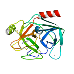
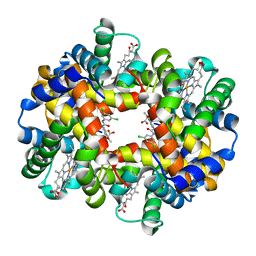
1XZ2
 
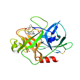
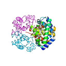
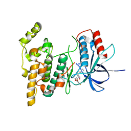
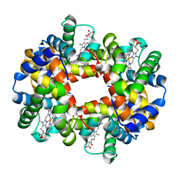 | | wild-type hemoglobin deoxy no-salt | | 分子名称: | Hemoglobin alpha chain, Hemoglobin beta chain, PROTOPORPHYRIN IX CONTAINING FE | | 著者 | Kavanaugh, J.S, Rogers, P.H, Arnone, A, Hui, H.L, Wierzba, A, DeYoung, A, Kwiatkowski, L.D, Noble, R.W, Juszczak, L.J, Peterson, E.S, Friedman, J.M. | | 登録日 | 2004-11-11 | | 公開日 | 2004-11-30 | | 最終更新日 | 2023-08-23 | | 実験手法 | X-RAY DIFFRACTION (1.9 Å) | | 主引用文献 | Intersubunit interactions associated with tyr42alpha stabilize the quaternary-T tetramer but are not major quaternary constraints in deoxyhemoglobin
Biochemistry, 44, 2005
|
|
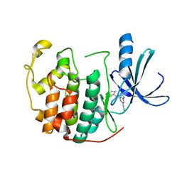
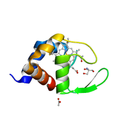
1HF4
 | | STRUCTURAL EFFECTS OF MONOVALENT ANIONS ON POLYMORPHIC LYSOZYME CRYSTALS | | 分子名称: | LYSOZYME, NITRATE ION, SODIUM ION | | 著者 | Vaney, M.C, Broutin, I, Ries-Kautt, M, Ducruix, A. | | 登録日 | 2000-11-29 | | 公開日 | 2001-01-07 | | 最終更新日 | 2023-12-13 | | 実験手法 | X-RAY DIFFRACTION (1.45 Å) | | 主引用文献 | Structural Effects of Monovalent Anions on Polymorphic Lysozyme Crystals
Acta Crystallogr.,Sect.D, 57, 2001
|
|
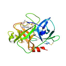
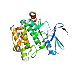
2D8W
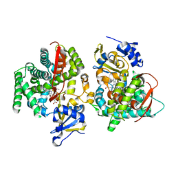
 | | Structure of HYPER-VIL-trypsin | | 分子名称: | CALCIUM ION, Cationic trypsin, IODIDE ION | | 著者 | Miyatake, H, Hasegawa, T, Yamano, A. | | 登録日 | 2005-12-08 | | 公開日 | 2006-07-11 | | 最終更新日 | 2011-07-13 | | 実験手法 | X-RAY DIFFRACTION (2 Å) | | 主引用文献 | New methods to prepare iodinated derivatives by vaporizing iodine labelling (VIL) and hydrogen peroxide VIL (HYPER-VIL)
ACTA CRYSTALLOGR.,SECT.D, 62, 2006
|
|
2D93
 | |
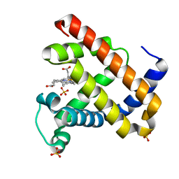
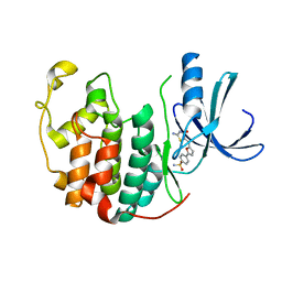
1H1X
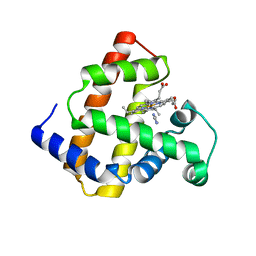
 | | Sperm whale Myoglobin mutant T67R S92D | | 分子名称: | CYANIDE ION, MYOGLOBIN, PROTOPORPHYRIN IX CONTAINING FE, ... | | 著者 | Zuccotti, S, Bolognesi, M. | | 登録日 | 2002-07-25 | | 公開日 | 2003-10-23 | | 最終更新日 | 2023-12-13 | | 実験手法 | X-RAY DIFFRACTION (1.4 Å) | | 主引用文献 | Engineering Peroxidase Activity in Myoglobin: The Haem Cavity Structure and Peroxide Activation in the T67R/S92D Mutant and its Derivative Reconstituted with Protohaemin-L-Histidine.
Biochem.J., 377, 2004
|
|
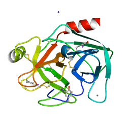
1H4W
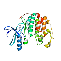
 | | Structure of human trypsin IV (brain trypsin) | | 分子名称: | BENZAMIDINE, CALCIUM ION, TRYPSIN IVA | | 著者 | Katona, G, Berglund, G.I, Hajdu, J, Graf, L, Szilagyi, L. | | 登録日 | 2001-05-15 | | 公開日 | 2002-02-11 | | 最終更新日 | 2023-12-13 | | 実験手法 | X-RAY DIFFRACTION (1.7 Å) | | 主引用文献 | Crystal structure reveals basis for the inhibitor resistance of human brain trypsin.
J. Mol. Biol., 315, 2002
|
|
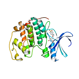
1H0W
 | | Human cyclin dependent protein kinase 2 in complex with the inhibitor 2-Amino-6-[cyclohex-3-enyl]methoxypurine | | 分子名称: | 1-AMINO-6-CYCLOHEX-3-ENYLMETHYLOXYPURINE, CELL DIVISION PROTEIN KINASE 2 | | 著者 | Gibson, A.E, Arris, C.E, Bentley, J, Boyle, F.T, Curtin, N.J, Davies, T.G, Endicott, J.A, Golding, B.T, Grant, S, Griffin, R.J, Jewsbury, P, Johnson, L.N, Mesguiche, V, Newell, D.R, Noble, M.E.M, Tucker, J.A, Whitfield, H.J. | | 登録日 | 2002-06-27 | | 公開日 | 2003-06-27 | | 最終更新日 | 2023-12-13 | | 実験手法 | X-RAY DIFFRACTION (2.1 Å) | | 主引用文献 | Probing the ATP Ribose-Binding Domain of Cyclin-Dependent Kinases 1 and 2 with O(6)-Substituted Guanine Derivatives
J.Med.Chem., 45, 2002
|
|
1W10
 | | Urokinase type plasminogen activator | | 分子名称: | N-(ISOBUTOXYCARBONYL)-D-SERYL-N-((1S)-4-{[AMINO(IMINO)METHYL]AMINO}-1-FORMYLBUTYL)-L-ALANINAMIDE, SULFATE ION, UROKINASE-TYPE PLASMINOGEN ACTIVATOR | | 著者 | Jacob, U. | | 登録日 | 2004-06-15 | | 公開日 | 2008-05-20 | | 最終更新日 | 2019-09-18 | | 実験手法 | X-RAY DIFFRACTION (2 Å) | | 主引用文献 | Crystals of urokinase type plasminogen activator complexes reveal the binding mode of peptidomimetic inhibitors.
J.Mol.Biol., 328, 2003
|
|
1RXP
 | |
1JP8
 | |
1W13
 | | UROKINASE TYPE PLASMINOGEN ACTIVATOR | | 分子名称: | N-(BENZYLSULFONYL)-D-SERYL-N-(4-{[AMINO(IMINO)METHYL]AMINO}BENZYL)-L-ALANINAMIDE, SULFATE ION, UROKINASE-TYPE PLASMINOGEN ACTIVATOR | | 著者 | Jacob, U. | | 登録日 | 2004-06-15 | | 公開日 | 2008-05-20 | | 最終更新日 | 2019-05-22 | | 実験手法 | X-RAY DIFFRACTION (2 Å) | | 主引用文献 | Crystals of Urokinase Type Plasminogen Activator Complexes Reveal the Binding Mode of Peptidomimetic Inhibitors.
J.Mol.Biol., 328, 2003
|
|
1W2L
 | | Cytochrome c domain of caa3 oxygen oxidoreductase | | 分子名称: | 2-AMINO-2-HYDROXYMETHYL-PROPANE-1,3-DIOL, ACETATE ION, CYTOCHROME OXIDASE SUBUNIT II, ... | | 著者 | Srinivasan, V, Rajendran, C, Sousa, F.L, Melo, A.M.P, Saraiva, L.M, Pereira, M.M, Santana, M, Teixeira, M, Michel, H. | | 登録日 | 2004-07-06 | | 公開日 | 2005-01-19 | | 最終更新日 | 2019-05-22 | | 実験手法 | X-RAY DIFFRACTION (1.3 Å) | | 主引用文献 | Structure at 1.3 A resolution of Rhodothermus marinus caa(3) cytochrome c domain.
J. Mol. Biol., 345, 2005
|
|
3VBX
 | |
2UZN
 | | Crystal structure of human CDK2 complexed with a thiazolidinone inhibitor | | 分子名称: | 4-{5-[(1Z)-1-(2-IMINO-4-OXO-1,3-THIAZOLIDIN-5-YLIDENE)ETHYL]-2-FURYL}BENZENESULFONAMIDE, CELL DIVISION PROTEIN KINASE 2 | | 著者 | Richardson, C.M, Dokurno, P, Murray, J.B, Surgenor, A.E. | | 登録日 | 2007-04-30 | | 公開日 | 2007-06-26 | | 最終更新日 | 2023-12-13 | | 実験手法 | X-RAY DIFFRACTION (2.3 Å) | | 主引用文献 | Discovery of a Potent Cdk2 Inhibitor with a Novel Binding Mode, Using Virtual Screening and Initial, Structure-Guided Lead Scoping.
Bioorg.Med.Chem.Lett., 17, 2007
|
|
2D5X
 | | Crystal structure of carbonmonoxy horse hemoglobin complexed with L35 | | 分子名称: | 2-[4-({[(3,5-DICHLOROPHENYL)AMINO]CARBONYL}AMINO)PHENOXY]-2-METHYLPROPANOIC ACID, CARBON MONOXIDE, Hemoglobin alpha subunit, ... | | 著者 | Yokoyama, T, Neya, S, Tsuneshige, A, Yonetani, T, Park, S.Y, Tame, J.R. | | 登録日 | 2005-11-08 | | 公開日 | 2006-03-07 | | 最終更新日 | 2024-03-13 | | 実験手法 | X-RAY DIFFRACTION (1.45 Å) | | 主引用文献 | R-state haemoglobin with low oxygen affinity: crystal structures of deoxy human and carbonmonoxy horse haemoglobin bound to the effector molecule L35
J.Mol.Biol., 356, 2006
|
|
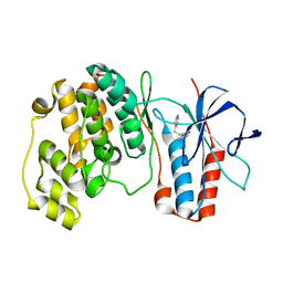
1JME
 | | Crystal Structure of Phe393His Cytochrome P450 BM3 | | 分子名称: | BIFUNCTIONAL P-450:NADPH-P450 REDUCTASE, PROTOPORPHYRIN IX CONTAINING FE | | 著者 | Ost, T.W.B, Munro, A.W, Mowat, C.G, Pesseguiero, A, Fulco, A.J, Cho, A.K, Cheesman, M.A, Walkinshaw, M.D, Chapman, S.K. | | 登録日 | 2001-07-18 | | 公開日 | 2001-11-23 | | 最終更新日 | 2023-08-16 | | 実験手法 | X-RAY DIFFRACTION (2 Å) | | 主引用文献 | Structural and spectroscopic analysis of the F393H mutant of flavocytochrome P450 BM3.
Biochemistry, 40, 2001
|
|
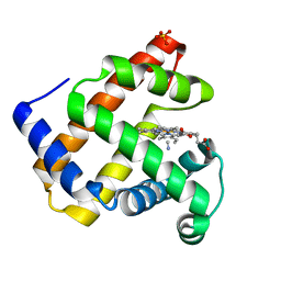
2D6C
 | | Crystal structure of myoglobin reconstituted with iron porphycene | | 分子名称: | IMIDAZOLE, Myoglobin, PORPHYCENE CONTAINING FE | | 著者 | Hayashi, T, Murata, D, Makino, M, Sugimoto, H, Matsuo, T, Sato, H, Shiro, Y, Hisaeda, Y, RIKEN Structural Genomics/Proteomics Initiative (RSGI) | | 登録日 | 2005-11-11 | | 公開日 | 2006-10-31 | | 最終更新日 | 2023-10-25 | | 実験手法 | X-RAY DIFFRACTION (2.26 Å) | | 主引用文献 | Crystal structure and peroxidase activity of myoglobin reconstituted with iron porphycene
Inorg.Chem., 45, 2006
|
|
1W0X
 | |
2UZO
 | | Crystal structure of human CDK2 complexed with a thiazolidinone inhibitor | | 分子名称: | 4-{5-[(Z)-(2,4-DIOXO-1,3-THIAZOLIDIN-5-YLIDENE)METHYL]FURAN-2-YL}BENZENESULFONAMIDE, CELL DIVISION PROTEIN KINASE 2 | | 著者 | Richardson, C.M, Dokurno, P, Murray, J.B, Surgenor, A.E. | | 登録日 | 2007-04-30 | | 公開日 | 2007-06-26 | | 最終更新日 | 2023-12-13 | | 実験手法 | X-RAY DIFFRACTION (2.3 Å) | | 主引用文献 | Discovery of a Potent Cdk2 Inhibitor with a Novel Binding Mode, Using Virtual Screening and Initial, Structure-Guided Lead Scoping.
Bioorg.Med.Chem.Lett., 17, 2007
|
|
1JN4
 | | The Crystal Structure of Ribonuclease A in complex with 2'-deoxyuridine 3'-pyrophosphate (P'-5') adenosine | | 分子名称: | ADENOSINE-5'-[TRIHYDROGEN DIPHOSPHATE] P'-3'-ESTER WITH 2'-DEOXYURIDINE, Pancreatic Ribonuclease A | | 著者 | Jardine, A.M, Leonidas, D.D, Jenkins, J.L, Park, C, Raines, R.T, Acharya, K.R, Shapiro, R. | | 登録日 | 2001-07-23 | | 公開日 | 2003-06-03 | | 最終更新日 | 2023-08-16 | | 実験手法 | X-RAY DIFFRACTION (1.8 Å) | | 主引用文献 | Cleavage of 3',5'-Pyrophosphate-Linked Dinucleotides by Ribonuclease A and Angiogenin
Biochemistry, 40, 2001
|
|
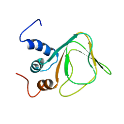
1JQQ
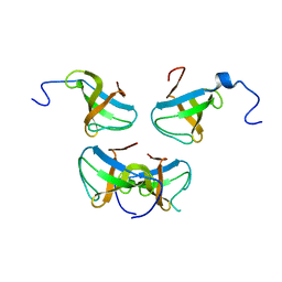 | | Crystal structure of Pex13p(301-386) SH3 domain | | 分子名称: | PEROXISOMAL MEMBRANE PROTEIN PAS20 | | 著者 | Douangamath, A, Mayans, O, Barnett, P, Distel, B, Wilmanns, M. | | 登録日 | 2001-08-08 | | 公開日 | 2002-12-06 | | 最終更新日 | 2024-02-07 | | 実験手法 | X-RAY DIFFRACTION (2.65 Å) | | 主引用文献 | Topography for Independent Binding of alpha-Helical and PPII-Helical Ligands to a Peroxisomal SH3 Domain
Mol.Cell, 10, 2002
|
|
2D60
 | | Crystal structure of deoxy human hemoglobin complexed with two L35 molecules | | 分子名称: | 2-[4-({[(3,5-DICHLOROPHENYL)AMINO]CARBONYL}AMINO)PHENOXY]-2-METHYLPROPANOIC ACID, Hemoglobin alpha subunit, Hemoglobin beta subunit, ... | | 著者 | Yokoyama, T, Neya, S, Tsuneshige, A, Yonetani, T, Park, S.Y, Tame, J.R. | | 登録日 | 2005-11-08 | | 公開日 | 2006-03-07 | | 最終更新日 | 2024-03-13 | | 実験手法 | X-RAY DIFFRACTION (1.7 Å) | | 主引用文献 | R-state haemoglobin with low oxygen affinity: crystal structures of deoxy human and carbonmonoxy horse haemoglobin bound to the effector molecule L35
J.Mol.Biol., 356, 2006
|
|
1W84
 | | p38 Kinase crystal structure in complex with small molecule inhibitor | | 分子名称: | 3-(2-PYRIDIN-4-YLETHYL)-1H-INDOLE, MITOGEN-ACTIVATED PROTEIN KINASE 14 | | 著者 | Tickle, J, Jhoti, H, Cleasby, A, Devine, L. | | 登録日 | 2004-09-16 | | 公開日 | 2005-02-08 | | 最終更新日 | 2024-05-08 | | 実験手法 | X-RAY DIFFRACTION (2.2 Å) | | 主引用文献 | Identification of novel p38alpha MAP kinase inhibitors using fragment-based lead generation.
J. Med. Chem., 48, 2005
|
|
1W11
 | | UROKINASE TYPE PLASMINOGEN ACTIVATOR | | 分子名称: | N-(BENZYLSULFONYL)-D-SERYL-N-{4-[AMINO(IMINO)METHYL]BENZYL}-L-ALANINAMIDE, SULFATE ION, UROKINASE-TYPE PLASMINOGEN ACTIVATOR | | 著者 | Jacob, U. | | 登録日 | 2004-06-15 | | 公開日 | 2008-05-20 | | 最終更新日 | 2019-10-09 | | 実験手法 | X-RAY DIFFRACTION (2 Å) | | 主引用文献 | Crystals of Urokinase Type Plasminogen Activator Complexes Reveal the Binding Mode of Peptidomimetic Inhibitors.
J.Mol.Biol., 328, 2003
|
|
3G9N
 | |


