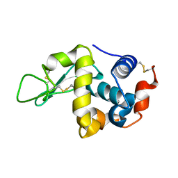
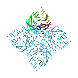
1IO3
 
 | |
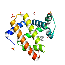
2CQ4
 | | solution structure of RNA binding domain in RNA binding motif protein 23 | | 分子名称: | RNA binding motif protein 23 | | 著者 | Tsuda, K, Muto, Y, Inoue, M, Kigawa, T, Terada, T, Shirouzu, M, Yokoyama, S, RIKEN Structural Genomics/Proteomics Initiative (RSGI) | | 登録日 | 2005-05-19 | | 公開日 | 2005-11-19 | | 最終更新日 | 2024-05-29 | | 実験手法 | SOLUTION NMR | | 主引用文献 | solution structure of RNA binding domain in RNA binding motif protein 23
To be Published
|
|
2CQP
 | | Solution structure of the RNA binding domain of RNA-binding protein 12 | | 分子名称: | RNA-binding protein 12 | | 著者 | Suzuki, S, Muto, Y, Inoue, M, Kigawa, T, Terada, T, Shirouzu, M, Yokoyama, S, RIKEN Structural Genomics/Proteomics Initiative (RSGI) | | 登録日 | 2005-05-20 | | 公開日 | 2005-11-20 | | 最終更新日 | 2024-05-29 | | 実験手法 | SOLUTION NMR | | 主引用文献 | Solution structure of the RNA binding domain of RNA-binding protein 12
To be Published
|
|
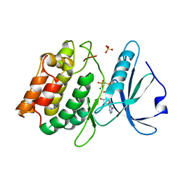
1IOC
 | | CRYSTAL STRUCTURE OF MUTANT HUMAN LYSOZYME, EAEA-I56T | | 分子名称: | LYSOZYME C, SODIUM ION | | 著者 | Goda, S, Takano, K, Yamagata, Y, Yutani, K. | | 登録日 | 2001-02-27 | | 公開日 | 2002-10-09 | | 最終更新日 | 2024-04-03 | | 実験手法 | X-RAY DIFFRACTION (2.4 Å) | | 主引用文献 | Elongation in a beta-structure promotes amyloid-like fibril formation of human lysozyme.
J.Biochem., 132, 2002
|
|
2CSH
 | | Solution structure of tandem repeat of the zf-C2H2 domains of human zinc finger protein 297B | | 分子名称: | ZINC ION, Zinc finger protein 297B | | 著者 | Inoue, K, Saitoh, K, Hayashi, F, Kigawa, T, Yokoyama, S, RIKEN Structural Genomics/Proteomics Initiative (RSGI) | | 登録日 | 2005-05-21 | | 公開日 | 2005-11-21 | | 最終更新日 | 2024-05-29 | | 実験手法 | SOLUTION NMR | | 主引用文献 | Solution structure of tandem repeat of the zf-C2H2 domains of human zinc finger protein 297B
To be Published
|
|
3FC2
 | | PLK1 in complex with BI6727 | | 分子名称: | 1,2-ETHANEDIOL, ACETATE ION, DI(HYDROXYETHYL)ETHER, ... | | 著者 | Bader, G. | | 登録日 | 2008-11-21 | | 公開日 | 2009-05-12 | | 最終更新日 | 2023-12-27 | | 実験手法 | X-RAY DIFFRACTION (2.45 Å) | | 主引用文献 | BI 6727, A Polo-like Kinase Inhibitor with Improved Pharmacokinetic Profile and Broad Antitumor Activity.
Clin.Cancer Res., 15, 2009
|
|
2CTK
 | | Solution structure of the 12th KH type I domain from human Vigilin | | 分子名称: | Vigilin | | 著者 | Tomizawa, T, Kigawa, T, Koshiba, S, Inoue, M, Yokoyama, S, RIKEN Structural Genomics/Proteomics Initiative (RSGI) | | 登録日 | 2005-05-24 | | 公開日 | 2005-11-24 | | 最終更新日 | 2024-05-29 | | 実験手法 | SOLUTION NMR | | 主引用文献 | Solution structure of the 12th KH type I domain from human Vigilin
To be Published
|
|
1SMF
 | | Studies on an artificial trypsin inhibitor peptide derived from the mung bean inhibitor | | 分子名称: | BOWMAN-BIRK TYPE TRYPSIN INHIBITOR, CALCIUM ION, TRYPSIN | | 著者 | Huang, Q, Li, Y, Zhang, S, Liu, S, Tang, Y, Qi, C. | | 登録日 | 1992-10-24 | | 公開日 | 1994-07-31 | | 最終更新日 | 2017-06-28 | | 実験手法 | X-RAY DIFFRACTION (2.1 Å) | | 主引用文献 | Studies on an artificial trypsin inhibitor peptide derived from the mung bean trypsin inhibitor: chemical synthesis, refolding, and crystallographic analysis of its complex with trypsin.
J.Biochem.(Tokyo), 116, 1994
|
|
2CUD
 | | Solution structure of the SH3 domain of the human SRC-like adopter protein (SLAP) | | 分子名称: | SRC-like-adapter | | 著者 | Ohnishi, S, Kigawa, T, Tochio, N, Sato, M, Nameki, N, Koshiba, S, Inoue, M, Yokoyama, S, RIKEN Structural Genomics/Proteomics Initiative (RSGI) | | 登録日 | 2005-05-26 | | 公開日 | 2005-11-26 | | 最終更新日 | 2024-05-29 | | 実験手法 | SOLUTION NMR | | 主引用文献 | Solution structure of the SH3 domain of the human SRC-like adopter protein (SLAP)
To be Published
|
|
2CQ3
 | | solution structure of RNA binding domain in RNA binding motif protein 9 | | 分子名称: | RNA-binding protein 9 | | 著者 | Tsuda, K, Muto, Y, Inoue, M, Kigawa, T, Terada, T, Shirouzu, M, Yokoyama, S, RIKEN Structural Genomics/Proteomics Initiative (RSGI) | | 登録日 | 2005-05-19 | | 公開日 | 2005-11-19 | | 最終更新日 | 2024-05-29 | | 実験手法 | SOLUTION NMR | | 主引用文献 | solution structure of RNA binding domain in RNA binding motif protein 9
To be Published
|
|
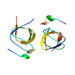
1IR8
 | |
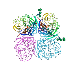
3F5U
 | | Crystal structure of the death associated protein kinase in complex with AMPPNP and Mg2+ | | 分子名称: | Death-associated protein kinase 1, MAGNESIUM ION, PHOSPHOAMINOPHOSPHONIC ACID-ADENYLATE ESTER, ... | | 著者 | McNamara, L.K, Watterson, D.M, Brunzelle, J.S. | | 登録日 | 2008-11-04 | | 公開日 | 2009-03-24 | | 最終更新日 | 2023-09-06 | | 実験手法 | X-RAY DIFFRACTION (2 Å) | | 主引用文献 | Structural insight into nucleotide recognition by human death-associated protein kinase.
Acta Crystallogr.,Sect.D, 65, 2009
|
|
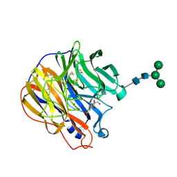
1IVD
 | |
2CTJ
 | | Solution structure of the 8th KH type I domain from human Vigilin | | 分子名称: | Vigilin | | 著者 | Tomizawa, T, Kigawa, T, Koshiba, S, Inoue, M, Yokoyama, S, RIKEN Structural Genomics/Proteomics Initiative (RSGI) | | 登録日 | 2005-05-24 | | 公開日 | 2005-11-24 | | 最終更新日 | 2024-05-29 | | 実験手法 | SOLUTION NMR | | 主引用文献 | Solution structure of the 8th KH type I domain from human Vigilin
To be Published
|
|
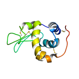
1INF
 | |
2D0N
 | |
1INW
 | | A SIALIC ACID DERIVED PHOSPHONATE ANALOG INHIBITS DIFFERENT STRAINS OF INFLUENZA VIRUS NEURAMINIDASE WITH DIFFERENT EFFICIENCIES | | 分子名称: | (1S)-4-acetamido-1,5-anhydro-2,4-dideoxy-1-phosphono-D-glycero-D-galacto-octitol, 2-acetamido-2-deoxy-beta-D-glucopyranose, CALCIUM ION, ... | | 著者 | White, C.L, Janakiraman, M.N, Laver, W.G, Philippon, C, Vasella, A, Air, G.M, Luo, M. | | 登録日 | 1994-09-26 | | 公開日 | 1995-02-07 | | 最終更新日 | 2020-07-29 | | 実験手法 | X-RAY DIFFRACTION (2.4 Å) | | 主引用文献 | A sialic acid-derived phosphonate analog inhibits different strains of influenza virus neuraminidase with different efficiencies.
J.Mol.Biol., 245, 1995
|
|
1IY4
 | | Solution structure of the human lysozyme at 35 degree C | | 分子名称: | Lysozyme | | 著者 | Kumeta, H, Miura, A, Kobashigawa, Y, Miura, K, Oka, C, Nitta, K, Nemoto, N, Tsuda, S. | | 登録日 | 2002-07-15 | | 公開日 | 2002-07-31 | | 最終更新日 | 2023-12-27 | | 実験手法 | SOLUTION NMR | | 主引用文献 | Low-temperature-induced structural changes in human lysozyme elucidated by three-dimensional NMR spectroscopy
Biochemistry, 42, 2003
|
|
1WSD
 | | Alkaline M-protease form I crystal structure | | 分子名称: | CALCIUM ION, M-protease, SULFATE ION | | 著者 | Shirai, T, Suzuki, A, Yamane, T, Ashida, T, Kobayashi, T, Hitomi, J, Ito, S. | | 登録日 | 2004-11-05 | | 公開日 | 2004-11-16 | | 最終更新日 | 2024-03-13 | | 実験手法 | X-RAY DIFFRACTION (1.5 Å) | | 主引用文献 | High-resolution crystal structure of M-protease: phylogeny aided analysis of the high-alkaline adaptation mechanism
Protein Eng., 10, 1997
|
|
2CR4
 | | Solution structure of the SH2 domain of human SH3BP2 protein | | 分子名称: | SH3 domain-binding protein 2 | | 著者 | Hatta, R, Tomizawa, T, Hayashi, F, Yoshida, M, Yokoyama, S, RIKEN Structural Genomics/Proteomics Initiative (RSGI) | | 登録日 | 2005-05-20 | | 公開日 | 2005-11-20 | | 最終更新日 | 2024-05-29 | | 実験手法 | SOLUTION NMR | | 主引用文献 | Solution structure of the SH2 domain of human SH3BP2 protein
To be Published
|
|
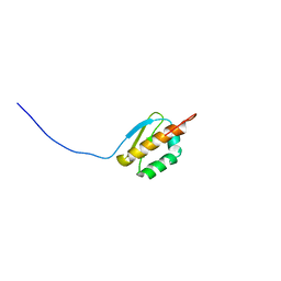
1J16
 | |
1J1B
 | | Binary complex structure of human tau protein kinase I with AMPPNP | | 分子名称: | Glycogen synthase kinase-3 beta, PHOSPHOAMINOPHOSPHONIC ACID-ADENYLATE ESTER | | 著者 | Aoki, M, Yokota, T, Sugiura, I, Sasaki, C, Hasegawa, T, Okumura, C, Kohno, T, Sugio, S, Matsuzaki, T. | | 登録日 | 2002-12-03 | | 公開日 | 2003-12-03 | | 最終更新日 | 2023-12-27 | | 実験手法 | X-RAY DIFFRACTION (1.8 Å) | | 主引用文献 | Structural insight into nucleotide recognition in tau-protein kinase I/glycogen synthase kinase 3 beta.
Acta Crystallogr.,Sect.D, 60, 2004
|
|
2CUI
 | | Solution structure of the 31st fibronectin type III domain of the human tenascin X | | 分子名称: | Tenascin-X | | 著者 | Ohnishi, S, Kigawa, T, Tochio, N, Koshiba, S, Inoue, M, Yokoyama, S, RIKEN Structural Genomics/Proteomics Initiative (RSGI) | | 登録日 | 2005-05-26 | | 公開日 | 2005-11-26 | | 最終更新日 | 2024-05-29 | | 実験手法 | SOLUTION NMR | | 主引用文献 | Solution structure of the 31st fibronectin type III domain of the human tenascin X
To be Published
|
|
1WSG
 | | Co-crystal structure of E.coli RNase HI active site mutant (E48A/D134N*) with Mn2+ | | 分子名称: | MANGANESE (II) ION, Ribonuclease HI | | 著者 | Tsunaka, Y, Takano, K, Matsumura, H, Yamagata, Y, Kanaya, S. | | 登録日 | 2004-11-05 | | 公開日 | 2005-02-08 | | 最終更新日 | 2024-05-29 | | 実験手法 | X-RAY DIFFRACTION (2.2 Å) | | 主引用文献 | Identification of Single Mn(2+) Binding Sites Required for Activation of the Mutant Proteins of E.coli RNase HI at Glu48 and/or Asp134 by X-ray Crystallography
J.Mol.Biol., 345, 2005
|
|
1J3F
 | | Crystal Structure of an Artificial Metalloprotein:Cr(III)(3,3'-Me2-salophen)/apo-A71G Myoglobin | | 分子名称: | 'N,N'-BIS-(2-HYDROXY-3-METHYL-BENZYLIDENE)-BENZENE-1,2-DIAMINE', CHROMIUM ION, Myoglobin, ... | | 著者 | Koshiyama, T, Kono, M, Ohashi, M, Ueno, T, Suzuki, A, Yamane, T, Watanabe, Y. | | 登録日 | 2003-01-24 | | 公開日 | 2004-05-18 | | 最終更新日 | 2023-10-25 | | 実験手法 | X-RAY DIFFRACTION (1.45 Å) | | 主引用文献 | Coordinated Design of Cofactor and Active Site Structures in Development of New Protein Catalysts
J.Am.Chem.Soc., 127, 2005
|
|


