6D39
 
 | |
6D38
 | |
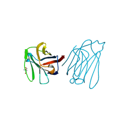
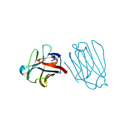
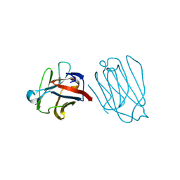
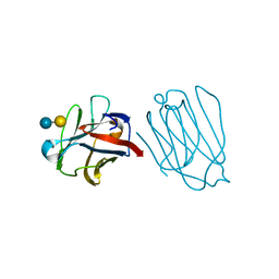
6BQN
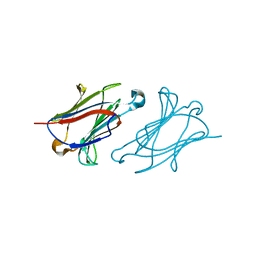
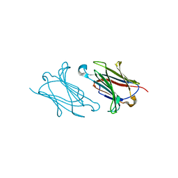
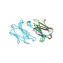
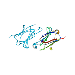
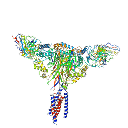 | | Cryo-EM structure of ENaC | | 分子名称: | 10D4 fab, 7B1 fab, EGFP-SCNN1G chimera, ... | | 著者 | Noreng, S, Bharadwaj, A, Posert, R, Yoshioka, C, Baconguis, I. | | 登録日 | 2017-11-28 | | 公開日 | 2018-10-10 | | 最終更新日 | 2019-12-18 | | 実験手法 | ELECTRON MICROSCOPY (3.9 Å) | | 主引用文献 | Structure of the human epithelial sodium channel by cryo-electron microscopy.
Elife, 7, 2018
|
|
6B9C
 | | Superfolder Green Fluorescent Protein with 4-nitro-L-phenylalanine at the chromophore (position 66) | | 分子名称: | CARBON DIOXIDE, Green fluorescent protein | | 著者 | Phillips-Piro, C.M, Brewer, S.H, Olenginski, G.M, Piacentini, J. | | 登録日 | 2017-10-10 | | 公開日 | 2018-10-17 | | 最終更新日 | 2023-11-15 | | 実験手法 | X-RAY DIFFRACTION (1.695 Å) | | 主引用文献 | Structural and spectrophotometric investigation of two unnatural amino-acid altered chromophores in the superfolder green fluorescent protein
Acta Crystallogr.,Sect.D, 2021
|
|
6B7T
 | | Truncated strand 10-less green fluorescent protein | | 分子名称: | Green fluorescent protein,Green fluorescent protein | | 著者 | Deng, A, Boxer, S.G. | | 登録日 | 2017-10-05 | | 公開日 | 2017-12-27 | | 最終更新日 | 2023-11-15 | | 実験手法 | X-RAY DIFFRACTION (1.91 Å) | | 主引用文献 | Structural Insight into the Photochemistry of Split Green Fluorescent Proteins: A Unique Role for a His-Tag.
J. Am. Chem. Soc., 140, 2018
|
|
6B7R
 | | Truncated strand 11-less green fluorescent protein | | 分子名称: | 2-[N-CYCLOHEXYLAMINO]ETHANE SULFONIC ACID, Green fluorescent protein | | 著者 | Deng, A, Boxer, S.G. | | 登録日 | 2017-10-05 | | 公開日 | 2017-12-27 | | 最終更新日 | 2023-11-15 | | 実験手法 | X-RAY DIFFRACTION (1.73 Å) | | 主引用文献 | Structural Insight into the Photochemistry of Split Green Fluorescent Proteins: A Unique Role for a His-Tag.
J. Am. Chem. Soc., 140, 2018
|
|
6AZ1
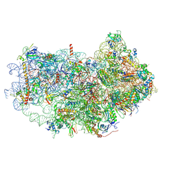 | | Cryo-EM structure of the small subunit of Leishmania ribosome bound to paromomycin | | 分子名称: | E-site tRNA, LACK1, MAGNESIUM ION, ... | | 著者 | Shalev-Benami, M, Zhang, Y, Rozenberg, H, Matzov, D, Zimmerman, E, Bashan, A, Jaffe, C.L, Yonath, A, Skiniotis, G. | | 登録日 | 2017-09-09 | | 公開日 | 2017-12-06 | | 最終更新日 | 2019-07-03 | | 実験手法 | ELECTRON MICROSCOPY (2.7 Å) | | 主引用文献 | Atomic resolution snapshot of Leishmania ribosome inhibition by the aminoglycoside paromomycin.
Nat Commun, 8, 2017
|
|
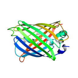
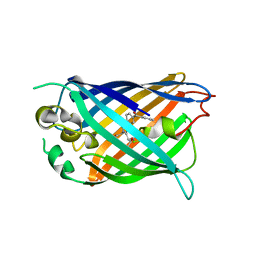
6AS9
 | | Filamentous Assembly of Green Fluorescent Protein Supported by a C-terminal fusion of 18-residues, viewed in space group P212121 form 2 | | 分子名称: | (4S)-2-METHYL-2,4-PENTANEDIOL, ACETATE ION, Green fluorescent protein | | 著者 | Sawaya, M.R, Heller, D.M, McPartland, L, Hochschild, A, Eisenberg, D.S. | | 登録日 | 2017-08-23 | | 公開日 | 2018-05-30 | | 最終更新日 | 2019-11-20 | | 実験手法 | X-RAY DIFFRACTION (1.75 Å) | | 主引用文献 | Atomic insights into the genesis of cellular filaments by globular proteins.
Nat. Struct. Mol. Biol., 25, 2018
|
|
6AA6
 | |
6AA2
 | | X-ray structure of ReQy1 (oxidized form) | | 分子名称: | Green fluorescent protein | | 著者 | Sugiura, K, Yasuda, A, Tabushi, N, Tanaka, H, Kurisu, G, Hisabori, T. | | 登録日 | 2018-07-17 | | 公開日 | 2019-05-29 | | 最終更新日 | 2023-11-22 | | 実験手法 | X-RAY DIFFRACTION (2.3 Å) | | 主引用文献 | Multicolor redox sensor proteins can visualize redox changes in various compartments of the living cell.
Biochim Biophys Acta Gen Subj, 1863, 2019
|
|
6A66
 | | Placental protein 13/galectin-13 variant R53H with Tris | | 分子名称: | 2-AMINO-2-HYDROXYMETHYL-PROPANE-1,3-DIOL, Galactoside-binding soluble lectin 13 | | 著者 | Su, J.Y. | | 登録日 | 2018-06-26 | | 公開日 | 2018-12-26 | | 実験手法 | X-RAY DIFFRACTION (1.4 Å) | | 主引用文献 | Resetting the ligand binding site of placental protein 13/galectin-13 recovers its ability to bind lactose
Biosci. Rep., 38, 2018
|
|
6A65
 | | Placental protein 13/galectin-13 variant R53HR55N with Tris | | 分子名称: | 2-AMINO-2-HYDROXYMETHYL-PROPANE-1,3-DIOL, Galactoside-binding soluble lectin 13 | | 著者 | Su, J. | | 登録日 | 2018-06-26 | | 公開日 | 2018-12-26 | | 実験手法 | X-RAY DIFFRACTION (1.771 Å) | | 主引用文献 | Resetting the ligand binding site of placental protein 13/galectin-13 recovers its ability to bind lactose
Biosci. Rep., 38, 2018
|
|
6A64
 | |
6A63
 | | Placental protein 13/galectin-13 variant R53HH57R with Lactose | | 分子名称: | Galactoside-binding soluble lectin 13, beta-D-galactopyranose-(1-4)-beta-D-glucopyranose | | 著者 | Su, J. | | 登録日 | 2018-06-26 | | 公開日 | 2018-12-26 | | 最終更新日 | 2020-07-29 | | 実験手法 | X-RAY DIFFRACTION (1.63 Å) | | 主引用文献 | Resetting the ligand binding site of placental protein 13/galectin-13 recovers its ability to bind lactose
Biosci. Rep., 38, 2018
|
|
6A62
 | | Placental protein 13/galectin-13 variant R53HH57RD33G with Lactose | | 分子名称: | Galactoside-binding soluble lectin 13, beta-D-galactopyranose-(1-4)-beta-D-glucopyranose | | 著者 | Su, J. | | 登録日 | 2018-06-26 | | 公開日 | 2018-12-26 | | 最終更新日 | 2020-07-29 | | 実験手法 | X-RAY DIFFRACTION (2.03 Å) | | 主引用文献 | Resetting the ligand binding site of placental protein 13/galectin-13 recovers its ability to bind lactose
Biosci. Rep., 38, 2018
|
|
6A1Y
 | | Charcot-Leyden crystal protein/Galectin-10 variant Y35A | | 分子名称: | Galectin-10 | | 著者 | Su, J. | | 登録日 | 2018-06-08 | | 公開日 | 2018-12-26 | | 最終更新日 | 2024-03-27 | | 実験手法 | X-RAY DIFFRACTION (1.63 Å) | | 主引用文献 | Identification of key amino acid residues determining ligand binding specificity, homodimerization and cellular distribution of human galectin-10
Glycobiology, 29, 2019
|
|
6A1X
 | | Charcot-Leyden crystal protein/Galectin-10 variant W127A | | 分子名称: | Galectin-10 | | 著者 | Su, J. | | 登録日 | 2018-06-08 | | 公開日 | 2018-12-26 | | 最終更新日 | 2024-03-27 | | 実験手法 | X-RAY DIFFRACTION (1.99 Å) | | 主引用文献 | Identification of key amino acid residues determining ligand binding specificity, homodimerization and cellular distribution of human galectin-10
Glycobiology, 29, 2019
|
|
6A1V
 | | Charcot-Leyden crystal protein/Galectin-10 variant E33Q | | 分子名称: | Galectin-10 | | 著者 | Su, J. | | 登録日 | 2018-06-08 | | 公開日 | 2018-12-26 | | 最終更新日 | 2024-03-27 | | 実験手法 | X-RAY DIFFRACTION (1.984 Å) | | 主引用文献 | Identification of key amino acid residues determining ligand binding specificity, homodimerization and cellular distribution of human galectin-10
Glycobiology, 29, 2019
|
|
6A1U
 | | Charcot-Leyden crystal protein/Galectin-10 variant E33D | | 分子名称: | Galectin-10 | | 著者 | Su, J. | | 登録日 | 2018-06-08 | | 公開日 | 2018-12-26 | | 最終更新日 | 2024-03-27 | | 実験手法 | X-RAY DIFFRACTION (1.62 Å) | | 主引用文献 | Identification of key amino acid residues determining ligand binding specificity, homodimerization and cellular distribution of human galectin-10
Glycobiology, 29, 2019
|
|
6A1T
 | | Charcot-Leyden crystal protein/Galectin-10 variant E33A with lactose | | 分子名称: | Galectin-10, beta-D-galactopyranose-(1-4)-beta-D-glucopyranose | | 著者 | Su, J. | | 登録日 | 2018-06-08 | | 公開日 | 2018-12-26 | | 最終更新日 | 2024-03-27 | | 実験手法 | X-RAY DIFFRACTION (1.97 Å) | | 主引用文献 | Identification of key amino acid residues determining ligand binding specificity, homodimerization and cellular distribution of human galectin-10
Glycobiology, 29, 2019
|
|
6A1S
 | | Charcot-Leyden crystal protein/Galectin-10 variant E33A | | 分子名称: | Galectin-10 | | 著者 | Su, J. | | 登録日 | 2018-06-08 | | 公開日 | 2018-12-26 | | 最終更新日 | 2024-03-27 | | 実験手法 | X-RAY DIFFRACTION (1.63 Å) | | 主引用文献 | Identification of key amino acid residues determining ligand binding specificity, homodimerization and cellular distribution of human galectin-10
Glycobiology, 29, 2019
|
|
5ZMD
 | | Crystal structure of FTO in complex with m6dA modified ssDNA | | 分子名称: | Alpha-ketoglutarate-dependent dioxygenase FTO, DNA (5'-D(P*TP*CP*TP*(6MA)P*TP*AP*TP*CP*G)-3'), MANGANESE (II) ION, ... | | 著者 | Zhang, X, Wei, L.H, Luo, J, Xiao, Y, Liu, J, Zhang, W, Zhang, L, Jia, G.F. | | 登録日 | 2018-04-02 | | 公開日 | 2019-04-10 | | 最終更新日 | 2023-11-22 | | 実験手法 | X-RAY DIFFRACTION (3.3 Å) | | 主引用文献 | Structural insights into FTO's catalytic mechanism for the demethylation of multiple RNA substrates.
Proc. Natl. Acad. Sci. U.S.A., 116, 2019
|
|
5Z6Y
 | |
5YR2
 | |
5Y8R
 | | ZsYellow at pH 3.5 | | 分子名称: | GFP-like fluorescent chromoprotein FP538 | | 著者 | Bae, J.E, Kim, I.J, Nam, K.H. | | 登録日 | 2017-08-21 | | 公開日 | 2017-09-13 | | 最終更新日 | 2023-11-22 | | 実験手法 | X-RAY DIFFRACTION (2.3 Å) | | 主引用文献 | Disruption of the hydrogen bonding network determines the pH-induced non-fluorescent state of the fluorescent protein ZsYellow by protonation of Glu221.
Biochem. Biophys. Res. Commun., 493, 2017
|
|


