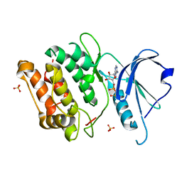
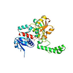
1IMX
 
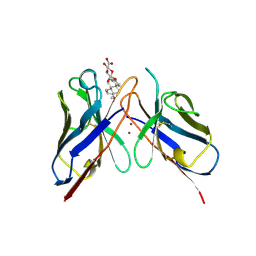
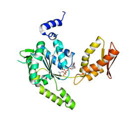
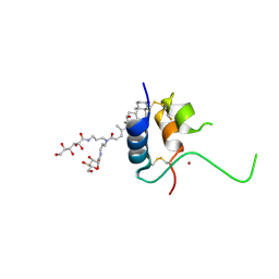 | | 1.8 Angstrom crystal structure of IGF-1 | | 分子名称: | BROMIDE ION, Insulin-like Growth Factor 1A, N,N-BIS(3-D-GLUCONAMIDOPROPYL)DEOXYCHOLAMIDE | | 著者 | Vajdos, F.F, Ultsch, M, Schaffer, M.L, Deshayes, K.D, Liu, J, Skelton, N.J, de Vos, A.M. | | 登録日 | 2001-05-11 | | 公開日 | 2001-09-05 | | 最終更新日 | 2011-07-13 | | 実験手法 | X-RAY DIFFRACTION (1.82 Å) | | 主引用文献 | Crystal structure of human insulin-like growth factor-1: detergent binding inhibits binding protein interactions.
Biochemistry, 40, 2001
|
|
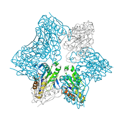
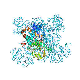
1SAY
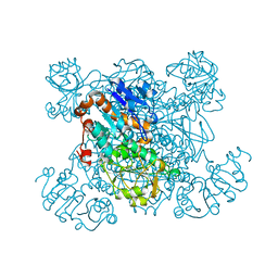 | | L-ALANINE DEHYDROGENASE COMPLEXED WITH PYRUVATE | | 分子名称: | L-ALANINE DEHYDROGENASE, PYRUVIC ACID | | 著者 | Baker, P.J, Sawa, Y, Shibata, H, Sedelnikova, S.E, Rice, D.W. | | 登録日 | 1998-06-05 | | 公開日 | 1999-06-08 | | 最終更新日 | 2023-11-15 | | 実験手法 | X-RAY DIFFRACTION (2.1 Å) | | 主引用文献 | Analysis of the structure and substrate binding of Phormidium lapideum alanine dehydrogenase.
Nat.Struct.Biol., 5, 1998
|
|
4LYX
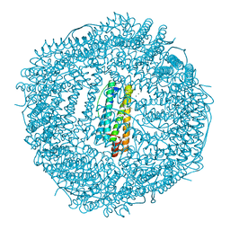 | | five minutes iron loaded frog M ferritin | | 分子名称: | CHLORIDE ION, FE (II) ION, Ferritin, ... | | 著者 | Mangani, S, Di Pisa, F, Pozzi, C, Turano, P, Lalli, D. | | 登録日 | 2013-07-31 | | 公開日 | 2014-08-06 | | 最終更新日 | 2023-09-20 | | 実験手法 | X-RAY DIFFRACTION (1.23 Å) | | 主引用文献 | Time-lapse anomalous X-ray diffraction shows how Fe(2+) substrate ions move through ferritin protein nanocages to oxidoreductase sites.
Acta Crystallogr.,Sect.D, 71, 2015
|
|
4LYU
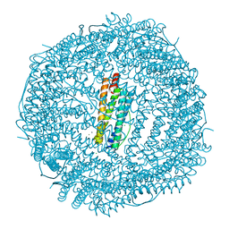 | | Fifteen minutes iron loaded frog M ferritin | | 分子名称: | CHLORIDE ION, FE (II) ION, Ferritin, ... | | 著者 | Mangani, S, Di Pisa, F, Pozzi, C, Turano, P, Lalli, D. | | 登録日 | 2013-07-31 | | 公開日 | 2014-08-06 | | 最終更新日 | 2023-09-20 | | 実験手法 | X-RAY DIFFRACTION (1.75 Å) | | 主引用文献 | Time-lapse anomalous X-ray diffraction shows how Fe(2+) substrate ions move through ferritin protein nanocages to oxidoreductase sites.
Acta Crystallogr.,Sect.D, 71, 2015
|
|
3EH9
 | | Crystal structure of death associated protein kinase complexed with ADP | | 分子名称: | ADENOSINE-5'-DIPHOSPHATE, Death-associated protein kinase 1, SULFATE ION | | 著者 | McNamara, L.K, Watterson, D.M, Brunzelle, J.S. | | 登録日 | 2008-09-11 | | 公開日 | 2009-04-28 | | 最終更新日 | 2023-08-30 | | 実験手法 | X-RAY DIFFRACTION (1.7 Å) | | 主引用文献 | Structural insight into nucleotide recognition by human death-associated protein kinase.
Acta Crystallogr.,Sect.D, 65, 2009
|
|
1WQY
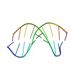 | | X-RAY structural analysis of B-DNA decamer D(CCATTAATGG)2 crystal grown in D2O solution | | 分子名称: | 5'-D(*CP*CP*AP*TP*TP*AP*AP*TP*GP*G)-3' | | 著者 | Arai, S, Chatake, T, Ohhara, T, Kurihara, K, Tanaka, I, Suzuki, N, Fujimoto, Z, Mizuno, H, Niimura, N. | | 登録日 | 2004-10-07 | | 公開日 | 2005-06-21 | | 最終更新日 | 2023-10-25 | | 実験手法 | X-RAY DIFFRACTION (2 Å) | | 主引用文献 | Complicated water orientations in the minor groove of the B-DNA decamer d(CCATTAATGG)2 observed by neutron diffraction measurements
Nucleic Acids Res., 33, 2005
|
|
3SSF
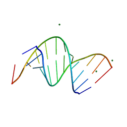 | | Crystal structure of RNA:DNA dodecamer corresponding to HIV-1 polypurine tract, at 1.6 A resolution. | | 分子名称: | 5'-D(*CP*CP*TP*TP*TP*TP*CP*TP*TP*TP*TP*A)-3', 5'-R(*UP*AP*AP*AP*AP*GP*AP*AP*AP*AP*GP*G)-3', MAGNESIUM ION | | 著者 | Drozdzal, P, Michalska, K, Kierzek, R, Lomozik, L, Jaskolski, M. | | 登録日 | 2011-07-08 | | 公開日 | 2012-02-08 | | 最終更新日 | 2023-09-13 | | 実験手法 | X-RAY DIFFRACTION (1.6 Å) | | 主引用文献 | Structure of an RNA/DNA dodecamer corresponding to the HIV-1 polypurine tract at 1.6 Angstrom resolution
Acta Crystallogr.,Sect.D, 68, 2012
|
|
3EE8
 | |
1JJW
 | |
154D
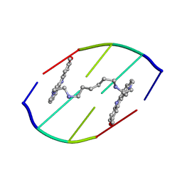 | | DNA DISTORTION IN BIS-INTERCALATED COMPLEXES | | 分子名称: | BIS-(N-ETHYLPYRIDINIUM-(3-METHOXYCARBAZOLE))HEXANE-1,6-DIAMINE, DNA (5'-D(*(CBR)P*GP*CP*G)-3') | | 著者 | Peek, M.E, Lipscomb, L.A, Bertrand, J.A, Gao, Q, Roques, B.P, Garbay-Jaureguiberry, C, Williams, L.D. | | 登録日 | 1993-12-14 | | 公開日 | 1994-08-31 | | 最終更新日 | 2024-02-07 | | 実験手法 | X-RAY DIFFRACTION (2.5 Å) | | 主引用文献 | DNA distortion in bis-intercalated complexes.
Biochemistry, 33, 1994
|
|
3QUT
 | | Crystal structure of pyrophosphatase from bacteroides thetaiotaomicron, asp13asn mutant, an open cap conformation | | 分子名称: | CHLORIDE ION, D-MALATE, INORGANIC PYROPHOSPHATASE, ... | | 著者 | Patskovsky, Y, Huang, H, Toro, R, Gerlt, J.A, Burley, S.K, Dunaway-Mariano, D, Almo, S.C, New York SGX Research Center for Structural Genomics (NYSGXRC), Enzyme Function Initiative (EFI) | | 登録日 | 2011-02-24 | | 公開日 | 2011-04-13 | | 最終更新日 | 2023-09-13 | | 実験手法 | X-RAY DIFFRACTION (1.5 Å) | | 主引用文献 | Divergence of Structure and Function in the Haloacid Dehalogenase Enzyme Superfamily: Bacteroides thetaiotaomicron BT2127 Is an Inorganic Pyrophosphatase.
Biochemistry, 50, 2011
|
|
1PJB
 | | L-ALANINE DEHYDROGENASE | | 分子名称: | L-ALANINE DEHYDROGENASE | | 著者 | Baker, P.J, Sawa, Y, Shibata, H, Sedelnikova, S.E, Rice, D.W. | | 登録日 | 1998-06-05 | | 公開日 | 1999-06-08 | | 最終更新日 | 2024-02-14 | | 実験手法 | X-RAY DIFFRACTION (2.1 Å) | | 主引用文献 | Analysis of the structure and substrate binding of Phormidium lapideum alanine dehydrogenase.
Nat.Struct.Biol., 5, 1998
|
|
1PJC
 | | L-ALANINE DEHYDROGENASE COMPLEXED WITH NAD | | 分子名称: | NICOTINAMIDE-ADENINE-DINUCLEOTIDE, PROTEIN (L-ALANINE DEHYDROGENASE) | | 著者 | Baker, P.J, Sawa, Y, Shibata, H, Sedelnikova, S.E, Rice, D.W. | | 登録日 | 1998-06-05 | | 公開日 | 1999-07-15 | | 最終更新日 | 2023-12-27 | | 実験手法 | X-RAY DIFFRACTION (2 Å) | | 主引用文献 | Analysis of the structure and substrate binding of Phormidium lapideum alanine dehydrogenase.
Nat.Struct.Biol., 5, 1998
|
|
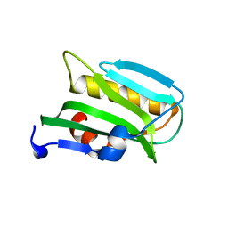
1F3S
 | | Solution Structure of DNA Sequence GGGTTCAGG Forms GGGG Tetrade and G(C-A) Triad. | | 分子名称: | DNA (5'-D(*GP*GP*GP*TP*TP*CP*AP*GP*G)-3') | | 著者 | Kettani, A, Basu, G, Gorin, A, Majumdar, A, Skripkin, E, Patel, D.J. | | 登録日 | 2000-06-06 | | 公開日 | 2000-11-13 | | 最終更新日 | 2024-05-22 | | 実験手法 | SOLUTION NMR | | 主引用文献 | A two-stranded template-based approach to G.(C-A) triad formation: designing novel structural elements into an existing DNA framework.
J.Mol.Biol., 301, 2000
|
|
3EQP
 | | Crystal Structure of Ack1 with compound T95 | | 分子名称: | Activated CDC42 kinase 1, CHLORIDE ION, N-(2,6-dimethylphenyl)-4-(2-ethoxyphenoxy)-2-({4-[4-(2-hydroxyethyl)piperazin-1-yl]phenyl}amino)pyrimidine-5-carboxamide | | 著者 | Liu, J, Wang, Z, Walker, N.P.C. | | 登録日 | 2008-10-01 | | 公開日 | 2008-12-02 | | 最終更新日 | 2023-12-27 | | 実験手法 | X-RAY DIFFRACTION (2.3 Å) | | 主引用文献 | Identification and optimization of N3,N6-diaryl-1H-pyrazolo[3,4-d]pyrimidine-3,6-diamines as a novel class of ACK1 inhibitors.
Bioorg.Med.Chem.Lett., 18, 2008
|
|
1PKZ
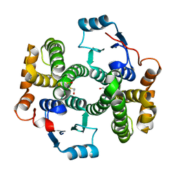 | | Crystal structure of human glutathione transferase (GST) A1-1 | | 分子名称: | 2-HYDROXYETHYL DISULFIDE, Glutathione S-transferase A1 | | 著者 | Grahn, E, Jakobsson, E, Gustafsson, A, Grehn, L, Olin, B, Wahlberg, M, Madsen, D, Kleywegt, G.J, Mannervik, B. | | 登録日 | 2003-06-06 | | 公開日 | 2004-06-22 | | 最終更新日 | 2018-03-07 | | 実験手法 | X-RAY DIFFRACTION (2.1 Å) | | 主引用文献 | New crystal structures of human glutathione transferase A1-1 shed light on glutathione binding and the conformation of the C-terminal helix.
Acta Crystallogr.,Sect.D, 62, 2006
|
|
1PR3
 | | Crystal Structure of the R103K Mutant of Aspartate Semialdehyde dehydrogenase from Haemophilus influenzae | | 分子名称: | Aspartate semialdehyde dehydrogenase, PHOSPHATE ION | | 著者 | Blanco, J, Moore, R.A, Faehnle, C.R, Coe, D.M, Viola, R.E. | | 登録日 | 2003-06-19 | | 公開日 | 2004-07-27 | | 最終更新日 | 2023-08-16 | | 実験手法 | X-RAY DIFFRACTION (2.15 Å) | | 主引用文献 | The role of substrate-binding groups in the mechanism of aspartate-beta-semialdehyde dehydrogenase.
Acta Crystallogr.,Sect.D, 60, 2004
|
|
4MY7
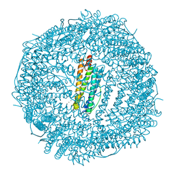 | | frog M ferritin iron-loaded under anaerobic environment | | 分子名称: | CHLORIDE ION, FE (II) ION, Ferritin, ... | | 著者 | Mangani, S, Di Pisa, F, Pozzi, C, Turano, P, Lalli, D. | | 登録日 | 2013-09-27 | | 公開日 | 2014-09-17 | | 最終更新日 | 2023-09-20 | | 実験手法 | X-RAY DIFFRACTION (1.48 Å) | | 主引用文献 | Time-lapse anomalous X-ray diffraction shows how Fe(2+) substrate ions move through ferritin protein nanocages to oxidoreductase sites.
Acta Crystallogr.,Sect.D, 71, 2015
|
|
1FCA
 | |
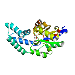
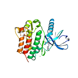
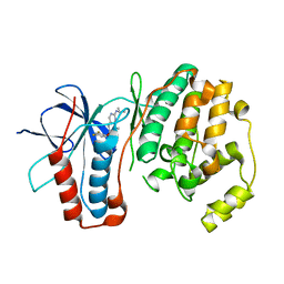
1KOU
 | | Crystal Structure of the Photoactive Yellow Protein Reconstituted with Caffeic Acid at 1.16 A Resolution | | 分子名称: | CAFFEIC ACID, N-BUTANE, PHOTOACTIVE YELLOW PROTEIN | | 著者 | van Aalten, D.M.F, Crielaard, W, Hellingwerf, K.J, Joshua-Tor, L. | | 登録日 | 2001-12-22 | | 公開日 | 2002-04-03 | | 最終更新日 | 2023-08-16 | | 実験手法 | X-RAY DIFFRACTION (1.16 Å) | | 主引用文献 | Structure of the photoactive yellow protein reconstituted with caffeic acid at 1.16 A resolution.
Acta Crystallogr.,Sect.D, 58, 2002
|
|
1BFV
 | |
3FLY
 | |
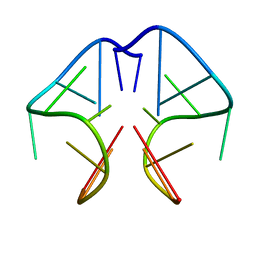
352D
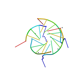 | | THE CRYSTAL STRUCTURE OF A PARALLEL-STRANDED PARALLEL-STRANDED GUANINE TETRAPLEX AT 0.95 ANGSTROM RESOLUTION | | 分子名称: | CALCIUM ION, DNA (5'-D(*TP*GP*GP*GP*GP*T)-3'), SODIUM ION | | 著者 | Phillips, K, Dauter, Z, Murchie, A.I.H, Lilley, D.M.J, Luisi, B. | | 登録日 | 1997-09-04 | | 公開日 | 1997-11-10 | | 最終更新日 | 2023-08-02 | | 実験手法 | X-RAY DIFFRACTION (0.95 Å) | | 主引用文献 | The crystal structure of a parallel-stranded guanine tetraplex at 0.95 A resolution.
J.Mol.Biol., 273, 1997
|
|
1L0W
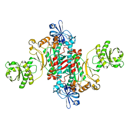 | | Aspartyl-tRNA synthetase-1 from space-grown crystals | | 分子名称: | Aspartyl-tRNA synthetase | | 著者 | Ng, J.D, Sauter, C, Lorber, B, Kirkland, N, Arnez, J, Giege, R. | | 登録日 | 2002-02-14 | | 公開日 | 2002-03-20 | | 最終更新日 | 2024-02-14 | | 実験手法 | X-RAY DIFFRACTION (2.01 Å) | | 主引用文献 | Comparative analysis of space-grown and earth-grown crystals of an aminoacyl-tRNA synthetase: space-grown crystals are more useful for structural determination.
Acta Crystallogr.,Sect.D, 58, 2002
|
|
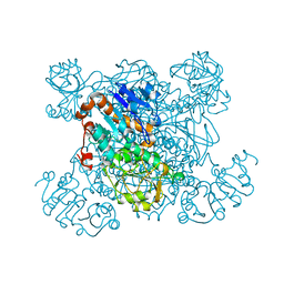
4LJA
 | | ClpB NBD2 R621Q from T. thermophilus in complex with AMPPCP and guanidinium chloride | | 分子名称: | Chaperone protein ClpB, GUANIDINE, MAGNESIUM ION, ... | | 著者 | Zeymer, C, Barends, T.R.M, Werbeck, N.D, Schlichting, I, Reinstein, J. | | 登録日 | 2013-07-04 | | 公開日 | 2014-02-12 | | 最終更新日 | 2023-11-08 | | 実験手法 | X-RAY DIFFRACTION (2 Å) | | 主引用文献 | Elements in nucleotide sensing and hydrolysis of the AAA+ disaggregation machine ClpB: a structure-based mechanistic dissection of a molecular motor
Acta Crystallogr.,Sect.D, 70, 2014
|
|


