8DLF
 
 | |
4NE3
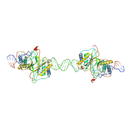
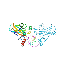
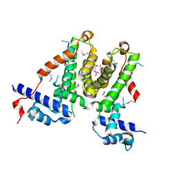
 | | Human MHF1-MHF2 complex | | 分子名称: | Centromere protein S, Centromere protein X | | 著者 | Zhao, Q, Saro, D, Sachpatzidis, A, Sung, P, Xiong, Y. | | 登録日 | 2013-10-28 | | 公開日 | 2013-12-25 | | 最終更新日 | 2014-02-12 | | 実験手法 | X-RAY DIFFRACTION (1.8007 Å) | | 主引用文献 | The MHF complex senses branched DNA by binding a pair of crossover DNA duplexes.
Nat Commun, 5, 2014
|
|
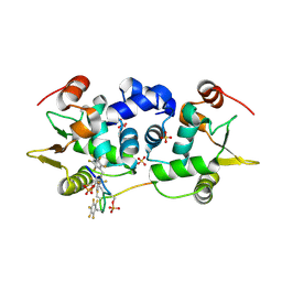
5VPC
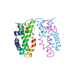
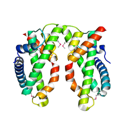
 | | Transcription factor FosB/JunD bZIP domain in its oxidized form, type-II crystal | | 分子名称: | CHLORIDE ION, Protein fosB, SODIUM ION, ... | | 著者 | Yin, Z, Machius, M.C, Rudenko, G. | | 登録日 | 2017-05-04 | | 公開日 | 2017-09-06 | | 最終更新日 | 2023-10-04 | | 実験手法 | X-RAY DIFFRACTION (2.498 Å) | | 主引用文献 | Activator Protein-1: redox switch controlling structure and DNA-binding.
Nucleic Acids Res., 45, 2017
|
|
5VPB
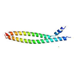
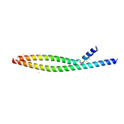 | | Transcription factor FosB/JunD bZIP domain in its oxidized form, type-I crystal | | 分子名称: | CHLORIDE ION, Protein fosB, Transcription factor jun-D | | 著者 | Yin, Z, Machius, M, Rudenko, G. | | 登録日 | 2017-05-04 | | 公開日 | 2017-09-06 | | 最終更新日 | 2023-10-04 | | 実験手法 | X-RAY DIFFRACTION (2.691 Å) | | 主引用文献 | Activator Protein-1: redox switch controlling structure and DNA-binding.
Nucleic Acids Res., 45, 2017
|
|
5VPD
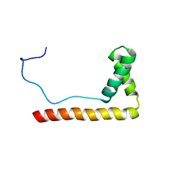
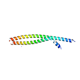 | | Transcription factor FosB/JunD bZIP domain in its oxidized form, type-III crystal | | 分子名称: | CHLORIDE ION, Protein fosB, SODIUM ION, ... | | 著者 | Yin, Z, Machius, M, Rudenko, G. | | 登録日 | 2017-05-04 | | 公開日 | 2017-09-06 | | 最終更新日 | 2023-10-04 | | 実験手法 | X-RAY DIFFRACTION (2.79 Å) | | 主引用文献 | Activator Protein-1: redox switch controlling structure and DNA-binding.
Nucleic Acids Res., 45, 2017
|
|
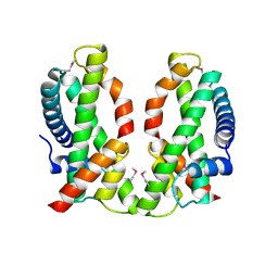
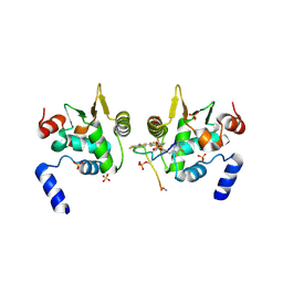
4NE6
 | | Human MHF1-MHF2 complex | | 分子名称: | Centromere protein S, Centromere protein X | | 著者 | Zhao, Q, Saro, D, Sachpatzidis, A, Sung, P, Xiong, Y. | | 登録日 | 2013-10-28 | | 公開日 | 2013-12-25 | | 最終更新日 | 2014-02-12 | | 実験手法 | X-RAY DIFFRACTION (2.1001 Å) | | 主引用文献 | The MHF complex senses branched DNA by binding a pair of crossover DNA duplexes.
Nat Commun, 5, 2014
|
|
5VPA
 | | Transcription factor FosB/JunD bZIP domain | | 分子名称: | CHLORIDE ION, Protein fosB, SODIUM ION, ... | | 著者 | Yin, Z, Machius, M, Rudenko, G. | | 登録日 | 2017-05-04 | | 公開日 | 2017-09-06 | | 最終更新日 | 2023-10-04 | | 実験手法 | X-RAY DIFFRACTION (2.83 Å) | | 主引用文献 | Activator Protein-1: redox switch controlling structure and DNA-binding.
Nucleic Acids Res., 45, 2017
|
|
1LWM
 | | Solution Structure of the Sequence-Non-Specific HMGB protein NHP6A | | 分子名称: | NONHISTONE CHROMOSOMAL PROTEIN 6A | | 著者 | Masse, J.E, Wong, B, Yen, Y.-M, Allain, F.H.-T, Johnson, R.C, Feigon, J. | | 登録日 | 2002-05-31 | | 公開日 | 2002-10-16 | | 最終更新日 | 2024-05-22 | | 実験手法 | SOLUTION NMR | | 主引用文献 | The S. cerevisiae architectural HMGB protein NHP6A complexed with DNA: DNA and protein conformational changes upon binding
J.Mol.Biol., 323, 2002
|
|
4NE5
 | | Human MHF1-MHF2 complex | | 分子名称: | Centromere protein S, Centromere protein X | | 著者 | Zhao, Q, Saro, D, Sachpatzidis, A, Sung, P, Xiong, Y. | | 登録日 | 2013-10-28 | | 公開日 | 2013-12-25 | | 最終更新日 | 2014-02-12 | | 実験手法 | X-RAY DIFFRACTION (2.5 Å) | | 主引用文献 | The MHF complex senses branched DNA by binding a pair of crossover DNA duplexes.
Nat Commun, 5, 2014
|
|
2D9A
 | | Solution Structure of RSGI RUH-050, a myb DNA-binding domain in mouse cDNA | | 分子名称: | Myb-related protein B | | 著者 | Doi-Katayama, Y, Hirota, H, Hayashi, F, Yokoyama, S, RIKEN Structural Genomics/Proteomics Initiative (RSGI) | | 登録日 | 2005-12-09 | | 公開日 | 2006-06-09 | | 最終更新日 | 2024-05-29 | | 実験手法 | SOLUTION NMR | | 主引用文献 | Solution Structure of RSGI RUH-050, a myb DNA-binding domain in mouse cDNA
To be Published
|
|
8X7U
 | | MCM in complex with dsDNA in presence of ATP. | | 分子名称: | ADENOSINE-5'-TRIPHOSPHATE, MAGNESIUM ION, mini-chromosome maintenance complex 3 | | 著者 | Ma, J, Yi, G, Ye, M, MacGregor-Chatwin, C, Sheng, Y, Lu, Y, Li, M, Gilbert, R.J.C, Zhang, P. | | 登録日 | 2023-11-25 | | 公開日 | 2024-01-17 | | 実験手法 | ELECTRON MICROSCOPY (3.57 Å) | | 主引用文献 | MCM in complex with dsDNA in presence of ATP
To Be Published
|
|
5DWC
 | |
5LOI
 | |
7B4F
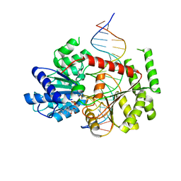
 | | Structural basis of reactivation of oncogenic p53 mutants by a small molecule: methylene quinuclidinone (MQ). Human p53DBD-R282W mutant bound to DNA: R282W-MQ (I) | | 分子名称: | Cellular tumor antigen p53, DNA target, FORMIC ACID, ... | | 著者 | Rozenberg, H, Degtjarik, O, Shakked, Z. | | 登録日 | 2020-12-02 | | 公開日 | 2021-12-08 | | 最終更新日 | 2024-01-31 | | 実験手法 | X-RAY DIFFRACTION (1.78 Å) | | 主引用文献 | Structural basis of reactivation of oncogenic p53 mutants by a small molecule: methylene quinuclidinone (MQ).
Nat Commun, 12, 2021
|
|
5FQ5
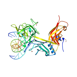
 | | Crystal structure of Cas9-sgRNA-DNA complex solved by native SAD phasing | | 分子名称: | CRISPR-ASSOCIATED ENDONUCLEASE CAS9/CSN1, MAGNESIUM ION, NON-TARGET DNA STRAND, ... | | 著者 | Olieric, V, Weinert, T, Finke, A, Anders, C, Jinek, M, Wang, M. | | 登録日 | 2015-12-07 | | 公開日 | 2016-03-23 | | 最終更新日 | 2024-05-08 | | 実験手法 | X-RAY DIFFRACTION (2.136 Å) | | 主引用文献 | Data-Collection Strategy for Challenging Native Sad Phasing.
Acta Crystallogr.,Sect.D, 72, 2016
|
|
3IAG
 | | CSL (RBP-Jk) bound to HES-1 nonconsensus site | | 分子名称: | 1,2-ETHANEDIOL, 5'-D(*AP*AP*TP*CP*TP*TP*TP*CP*AP*CP*AP*CP*GP*AP*T)-3', 5'-D(*TP*TP*AP*TP*CP*GP*TP*GP*TP*GP*AP*AP*AP*GP*A)-3', ... | | 著者 | Friedmann, D.R, Kovall, R.A. | | 登録日 | 2009-07-13 | | 公開日 | 2009-11-10 | | 最終更新日 | 2024-02-21 | | 実験手法 | X-RAY DIFFRACTION (2 Å) | | 主引用文献 | Thermodynamic and structural insights into CSL-DNA complexes.
Protein Sci., 19, 2010
|
|
8OO9
 | | CryoEM Structure INO80core Hexasome complex ATPase-DNA refinement state1 | | 分子名称: | ADENOSINE-5'-DIPHOSPHATE, Chromatin-remodeling ATPase INO80, DNA strand 1, ... | | 著者 | Zhang, M, Jungblut, A, Hoffmann, T, Eustermann, S. | | 登録日 | 2023-04-04 | | 公開日 | 2023-07-26 | | 最終更新日 | 2024-07-24 | | 実験手法 | ELECTRON MICROSCOPY (3.2 Å) | | 主引用文献 | Hexasome-INO80 complex reveals structural basis of noncanonical nucleosome remodeling.
Science, 381, 2023
|
|
5XAY
 | |
3M89
 | | Structure of TubZ-GTP-g-S | | 分子名称: | 5'-GUANOSINE-DIPHOSPHATE-MONOTHIOPHOSPHATE, FtsZ/tubulin-related protein | | 著者 | Ni, L, Xu, W, Schumacher, M.A. | | 登録日 | 2010-03-17 | | 公開日 | 2010-07-07 | | 最終更新日 | 2023-11-22 | | 実験手法 | X-RAY DIFFRACTION (2 Å) | | 主引用文献 | From the Cover: Plasmid protein TubR uses a distinct mode of HTH-DNA binding and recruits the prokaryotic tubulin homolog TubZ to effect DNA partition.
Proc.Natl.Acad.Sci.USA, 107, 2010
|
|
6DA1
 | | ETS1 in complex with synthetic SRR mimic | | 分子名称: | Protein C-ets-1, SULFATE ION, serine-rich region (SRR) peptide | | 著者 | Perez-Borrajero, C, Okon, M, Lin, C.S, Scheu, K, Murphy, M.E.P, Graves, B.J, McIntosh, L.P. | | 登録日 | 2018-05-01 | | 公開日 | 2019-01-16 | | 最終更新日 | 2023-10-04 | | 実験手法 | X-RAY DIFFRACTION (2.000127 Å) | | 主引用文献 | The Biophysical Basis for Phosphorylation-Enhanced DNA-Binding Autoinhibition of the ETS1 Transcription Factor.
J. Mol. Biol., 431, 2019
|
|
6DAT
 | | ETS1 in complex with synthetic SRR mimic | | 分子名称: | Protein C-ets-1, SULFATE ION, serine-rich region (SRR) peptide | | 著者 | Perez-Borrajero, C, Okon, M, Lin, C.S, Scheu, K, Murphy, M.E.P, Graves, B.J, McIntosh, L.P. | | 登録日 | 2018-05-02 | | 公開日 | 2019-01-16 | | 最終更新日 | 2023-10-04 | | 実験手法 | X-RAY DIFFRACTION (2.35002637 Å) | | 主引用文献 | The Biophysical Basis for Phosphorylation-Enhanced DNA-Binding Autoinhibition of the ETS1 Transcription Factor.
J. Mol. Biol., 431, 2019
|
|
5XAZ
 | |
2CKX
 | |
7NMB
 | |
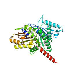
5IHE
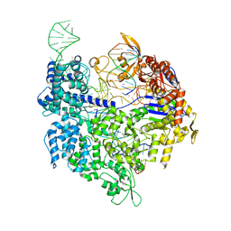
 | | D-family DNA polymerase - DP1 subunit (3'-5' proof-reading exonuclease) | | 分子名称: | 1,2-ETHANEDIOL, 2'-DEOXYADENOSINE-5'-MONOPHOSPHATE, ACETATE ION, ... | | 著者 | Sauguet, L, Raia, P, De Larue, M. | | 登録日 | 2016-02-29 | | 公開日 | 2016-08-31 | | 最終更新日 | 2024-05-08 | | 実験手法 | X-RAY DIFFRACTION (2.5 Å) | | 主引用文献 | Shared active site architecture between archaeal PolD and multi-subunit RNA polymerases revealed by X-ray crystallography.
Nat Commun, 7, 2016
|
|


