8CEB
 
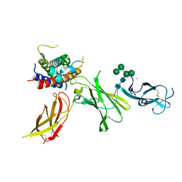
 | | Type2 alpha-synuclein filament assembled in vitro by wild-type and mutant (7 residues insertion) protein | | 分子名称: | Alpha-synuclein | | 著者 | Yang, Y, Garringer, J.H, Shi, Y, Lovestam, S, Sew, P.C, Zhang, X.J, Kotecha, A, Bacioglu, M, Koto, A, Takao, M, Spillantini, G.M, Ghetti, B, Vidal, R, Murzin, G.A, Scheres, H.W.S, Goedert, M. | | 登録日 | 2023-02-01 | | 公開日 | 2023-03-08 | | 最終更新日 | 2024-07-24 | | 実験手法 | ELECTRON MICROSCOPY (2.8 Å) | | 主引用文献 | New SNCA mutation and structures of alpha-synuclein filaments from juvenile-onset synucleinopathy.
Acta Neuropathol, 145, 2023
|
|
2H6Q
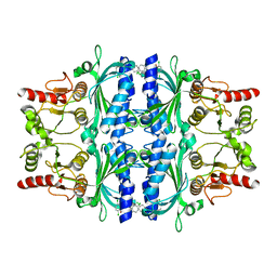
 | | Histone H3 recognition and presentation by the WDR5 module of the MLL1 complex | | 分子名称: | Histone H3 K4-Me3 9-residue peptide, WD-repeat protein 5 | | 著者 | Ruthenburg, A.J, Wang, W.-K, Graybosch, D.M, Li, H, Allis, C.D, Patel, D.J, Verdine, G.L. | | 登録日 | 2006-06-01 | | 公開日 | 2006-07-04 | | 最終更新日 | 2023-08-30 | | 実験手法 | X-RAY DIFFRACTION (1.87 Å) | | 主引用文献 | Histone H3 recognition and presentation by the WDR5 module of the MLL1 complex.
Nat.Struct.Mol.Biol., 13, 2006
|
|
2H68
 | | Histone H3 recognition and presentation by the WDR5 module of the MLL1 complex | | 分子名称: | WD-repeat protein 5 | | 著者 | Ruthenburg, A.J, Wang, W.-K, Graybosch, D.M, Li, H, Allis, C.D, Patel, D.J, Verdine, G.L. | | 登録日 | 2006-05-30 | | 公開日 | 2006-07-04 | | 最終更新日 | 2023-08-30 | | 実験手法 | X-RAY DIFFRACTION (1.79 Å) | | 主引用文献 | Histone H3 recognition and presentation by the WDR5 module of the MLL1 complex.
Nat.Struct.Mol.Biol., 13, 2006
|
|
2H6K
 | | Histone H3 recognition and presentation by the WDR5 module of the MLL1 complex | | 分子名称: | Histone H3 K4-Me 9-residue peptide, WD-repeat protein 5 | | 著者 | Ruthenburg, A.J, Wang, W.-K, Graybosch, D.M, Li, H, Allis, C.D, Patel, D.J, Verdine, G.L. | | 登録日 | 2006-05-31 | | 公開日 | 2006-07-04 | | 最終更新日 | 2023-08-30 | | 実験手法 | X-RAY DIFFRACTION (1.89 Å) | | 主引用文献 | Histone H3 recognition and presentation by the WDR5 module of the MLL1 complex.
Nat.Struct.Mol.Biol., 13, 2006
|
|
2H9L
 | | WDR5delta23 | | 分子名称: | SULFATE ION, WD-repeat protein 5 | | 著者 | Min, J.R, Schuetz, A, Allali-Hassani, A, Martin, F, Loppnau, P, Vedadi, M, Weigelt, J, Sundstrom, M, Edwards, A.M, Arrowsmith, C.H, Bochkarev, A, Plotnikov, A.N, Structural Genomics Consortium (SGC) | | 登録日 | 2006-06-10 | | 公開日 | 2006-08-01 | | 最終更新日 | 2024-02-14 | | 実験手法 | X-RAY DIFFRACTION (1.75 Å) | | 主引用文献 | Structural basis for molecular recognition and presentation of histone H3 By WDR5.
Embo J., 25, 2006
|
|
2GLI
 | | FIVE-FINGER GLI/DNA COMPLEX | | 分子名称: | COBALT (II) ION, DNA (5'-D(*AP*CP*GP*TP*GP*GP*AP*CP*CP*AP*CP*CP*CP*AP*AP*GP*AP*CP*GP*AP*A)-3'), DNA (5'-D(*TP*TP*TP*CP*GP*TP*CP*TP*TP*GP*GP*GP*TP*GP*GP*TP*CP*CP*AP*CP*G)-3'), ... | | 著者 | Pavletich, N.P, Pabo, C.O. | | 登録日 | 1993-11-09 | | 公開日 | 1993-11-09 | | 最終更新日 | 2024-02-14 | | 実験手法 | X-RAY DIFFRACTION (2.6 Å) | | 主引用文献 | Crystal structure of a five-finger GLI-DNA complex: new perspectives on zinc fingers.
Science, 261, 1993
|
|
2H6N
 | | Histone H3 recognition and presentation by the WDR5 module of the MLL1 complex | | 分子名称: | Histone H3 K4-Me2 9-residue peptide, WD-repeat protein 5 | | 著者 | Ruthenburg, A.J, Wang, W.-K, Graybosch, D.M, Li, H, Allis, C.D, Patel, D.J, Verdine, G.L. | | 登録日 | 2006-05-31 | | 公開日 | 2006-07-04 | | 最終更新日 | 2023-08-30 | | 実験手法 | X-RAY DIFFRACTION (1.5 Å) | | 主引用文献 | Histone H3 recognition and presentation by the WDR5 module of the MLL1 complex.
Nat.Struct.Mol.Biol., 13, 2006
|
|
1XCQ
 | | Complex HCV core-Fab 19D9D6-Protein L mutant (D55A,L57H,Y64W) in space group P21 | | 分子名称: | Capsid protein C, Monoclonal antibody 19D9D6 Heavy chain, Monoclonal antibody 19D9D6 Light chain, ... | | 著者 | Menez, R, Housden, N.G, Harrison, S, Jolivet-Reynaud, C, Gore, M.G, Stura, E.A. | | 登録日 | 2004-09-03 | | 公開日 | 2005-05-31 | | 最終更新日 | 2011-07-13 | | 実験手法 | X-RAY DIFFRACTION (3.5 Å) | | 主引用文献 | Different crystal packing in Fab-protein L semi-disordered peptide complex.
Acta Crystallogr.,Sect.D, 61, 2005
|
|
1XCT
 | | Complex HCV core-Fab 19D9D6-Protein L mutant (D55A, L57H, Y64W) in space group P21212 | | 分子名称: | Capsid protein C, Monoclonal antibody 19D9D6 Heavy chain, Monoclonal antibody 19D9D6 Light chain, ... | | 著者 | Menez, R, Housden, N.G, Harrison, S, Jolivet-Reynaud, C, Gore, M.G, Stura, E.A. | | 登録日 | 2004-09-03 | | 公開日 | 2005-05-31 | | 最終更新日 | 2011-07-13 | | 実験手法 | X-RAY DIFFRACTION (3.05 Å) | | 主引用文献 | Different crystal packing in Fab-protein L semi-disordered peptide complex.
Acta Crystallogr.,Sect.D, 61, 2005
|
|
8CR8
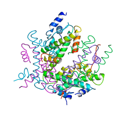
 | | human Interleukin-23 | | 分子名称: | Interleukin-12 subunit beta, Interleukin-23 subunit alpha, TERBIUM(III) ION, ... | | 著者 | Bloch, Y, Savvides, S.N. | | 登録日 | 2023-03-08 | | 公開日 | 2024-02-07 | | 最終更新日 | 2024-05-01 | | 実験手法 | X-RAY DIFFRACTION (2 Å) | | 主引用文献 | Structures of complete extracellular receptor assemblies mediated by IL-12 and IL-23.
Nat.Struct.Mol.Biol., 31, 2024
|
|
2FHY
 | |
8FUQ
 | |
2HIO
 | | HISTONE OCTAMER (CHICKEN), CHROMOSOMAL PROTEIN | | 分子名称: | PROTEIN (HISTONE H2A), PROTEIN (HISTONE H2B), PROTEIN (HISTONE H3), ... | | 著者 | Arents, G, Moudrianakis, E.N. | | 登録日 | 1999-06-15 | | 公開日 | 2000-01-12 | | 最終更新日 | 2023-12-27 | | 実験手法 | X-RAY DIFFRACTION (3.1 Å) | | 主引用文献 | The nucleosomal core histone octamer at 3.1 A resolution: a tripartite protein assembly and a left-handed superhelix.
Proc.Natl.Acad.Sci.USA, 88, 1991
|
|
2HI3
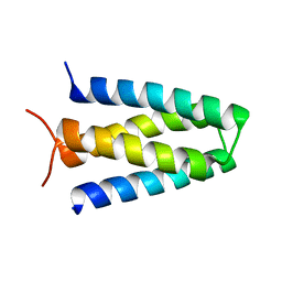
 | | Solution structure of the homeodomain-only protein HOP | | 分子名称: | Homeodomain-only protein | | 著者 | Mackay, J.P, Kook, H, Epstein, J.A, Simpson, R.J, Yung, W.W. | | 登録日 | 2006-06-28 | | 公開日 | 2007-01-02 | | 最終更新日 | 2024-05-29 | | 実験手法 | SOLUTION NMR | | 主引用文献 | Analysis of the structure and function of the transcriptional coregulator HOP
Biochemistry, 45, 2006
|
|
2NOZ
 | | Structure of Q315F human 8-oxoguanine glycosylase distal crosslink to 8-oxoguanine DNA | | 分子名称: | 5'-D(*G*CP*GP*TP*CP*CP*AP*(G42)P*GP*TP*CP*TP*AP*CP*C)-3', 5'-D(*GP*G*TP*AP*GP*AP*CP*CP*TP*GP*GP*AP*CP*GP*C)-3', CALCIUM ION, ... | | 著者 | Radom, C.T, Banerjee, A, Verdine, G.L. | | 登録日 | 2006-10-26 | | 公開日 | 2006-11-21 | | 最終更新日 | 2023-12-27 | | 実験手法 | X-RAY DIFFRACTION (2.43 Å) | | 主引用文献 | Structural characterization of human 8-oxoguanine DNA glycosylase variants bearing active site mutations.
J.Biol.Chem., 282, 2007
|
|
8D5E
 | |
8D5F
 | |
2NOF
 | | Structure of Q315F human 8-oxoguanine glycosylase proximal crosslink to 8-oxoguanine DNA | | 分子名称: | 5'-D(*GP*CP*GP*TP*C*CP*AP*(G42)P*GP*TP*CP*TP*AP*CP*C)-3', 5'-D(*GP*GP*TP*AP*GP*AP*CP*CP*TP*GP*GP*AP*CP*GP*C)-3', CALCIUM ION, ... | | 著者 | Radom, C.T, Banerjee, A, Verdine, G.L. | | 登録日 | 2006-10-25 | | 公開日 | 2006-11-21 | | 最終更新日 | 2023-12-27 | | 実験手法 | X-RAY DIFFRACTION (2.35 Å) | | 主引用文献 | Structural characterization of human 8-oxoguanine DNA glycosylase variants bearing active site mutations.
J.Biol.Chem., 282, 2007
|
|
2NOB
 | | Structure of catalytically inactive H270A human 8-oxoguanine glycosylase crosslinked to 8-oxoguanine DNA | | 分子名称: | 5'-D(*G*CP*GP*TP*CP*CP*AP*(G42)P*GP*TP*CP*TP*AP*CP*C)-3', 5'-D(*T*GP*GP*TP*AP*GP*AP*CP*CP*TP*GP*GP*AP*CP*GP*C)-3', CALCIUM ION, ... | | 著者 | Radom, C.T, Banerjee, A, Verdine, G.L. | | 登録日 | 2006-10-25 | | 公開日 | 2006-11-21 | | 最終更新日 | 2023-12-27 | | 実験手法 | X-RAY DIFFRACTION (2.1 Å) | | 主引用文献 | Structural characterization of human 8-oxoguanine DNA glycosylase variants bearing active site mutations.
J.Biol.Chem., 282, 2007
|
|
2G9A
 | |
2NOI
 | | Structure of G42A human 8-oxoguanine glycosylase crosslinked to undamaged G-containing DNA | | 分子名称: | 5'-D(*GP*CP*GP*TP*C*CP*AP*GP*GP*TP*CP*TP*AP*CP*C)-3', 5'-D(*GP*GP*TP*AP*GP*AP*CP*CP*TP*GP*GP*AP*CP*GP*C)-3', CALCIUM ION, ... | | 著者 | Radom, C.T, Banerjee, A, Verdine, G.L. | | 登録日 | 2006-10-25 | | 公開日 | 2006-11-21 | | 最終更新日 | 2023-12-27 | | 実験手法 | X-RAY DIFFRACTION (2.35 Å) | | 主引用文献 | Structural characterization of human 8-oxoguanine DNA glycosylase variants bearing active site mutations.
J.Biol.Chem., 282, 2007
|
|
2NOE
 | | Structure of catalytically inactive G42A human 8-oxoguanine glycosylase complexed to 8-oxoguanine DNA | | 分子名称: | 5'-D(*G*CP*GP*TP*CP*CP*AP*(G42)P*GP*TP*CP*TP*AP*CP*C)-3', 5'-D(*G*GP*TP*AP*GP*AP*CP*CP*TP*GP*GP*AP*CP*GP*C)-3', CALCIUM ION, ... | | 著者 | Radom, C.T, Banerjee, A, Verdine, G.L. | | 登録日 | 2006-10-25 | | 公開日 | 2006-11-21 | | 最終更新日 | 2023-12-27 | | 実験手法 | X-RAY DIFFRACTION (2.2 Å) | | 主引用文献 | Structural characterization of human 8-oxoguanine DNA glycosylase variants bearing active site mutations.
J.Biol.Chem., 282, 2007
|
|
6GHO
 | |
8D6A
 | |
8UQE
 | | Crystal structure of RNF168 (RING)-UbcH5c fused to H2A-H2B via a 26-residue linker (RING not modeled in density) | | 分子名称: | E3 ubiquitin-protein ligase RNF168,Ubiquitin-conjugating enzyme E2 D3,Histone H2B type 2-E,Histone H2A type 1-B/E | | 著者 | Hu, Q, Botuyan, M.V, Mer, G. | | 登録日 | 2023-10-23 | | 公開日 | 2024-01-17 | | 最終更新日 | 2024-03-20 | | 実験手法 | X-RAY DIFFRACTION (3.562 Å) | | 主引用文献 | Mechanisms of RNF168 nucleosome recognition and ubiquitylation.
Mol.Cell, 84, 2024
|
|


