7Z7P
 
 | |
7Z7Q
 | |
7Z7O
 | |
1TWB
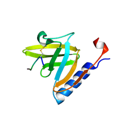 | | SspB disulfide crosslinked to an ssrA degradation tag | | 分子名称: | Stringent starvation protein B homolog, ssrA peptide | | 著者 | Bolon, D.N, Grant, R.A, Baker, T.A, Sauer, R.T. | | 登録日 | 2004-06-30 | | 公開日 | 2004-11-16 | | 最終更新日 | 2024-11-06 | | 実験手法 | X-RAY DIFFRACTION (1.9 Å) | | 主引用文献 | Nucleotide-Dependent Substrate Handoff from the SspB Adaptor to the AAA+ ClpXP Protease.
Mol.Cell, 16, 2004
|
|
7EYF
 | |
5G5Y
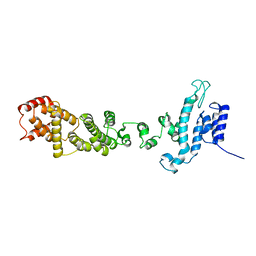
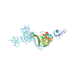
 | | S.pneumoniae ABC-transporter substrate binding protein FusA apo structure | | 分子名称: | ABC TRANSPORTER, SUBSTRATE-BINDING PROTEIN, CALCIUM ION, ... | | 著者 | Culurgioni, S, Harris, G, Singh, A.K, King, S.J, Walsh, M.A. | | 登録日 | 2016-06-10 | | 公開日 | 2017-01-18 | | 最終更新日 | 2024-05-08 | | 実験手法 | X-RAY DIFFRACTION (1.73 Å) | | 主引用文献 | Structural Basis for Regulation and Specificity of Fructooligosaccharide Import in Streptococcus pneumoniae.
Structure, 25, 2017
|
|
7KVV
 | |
7KVT
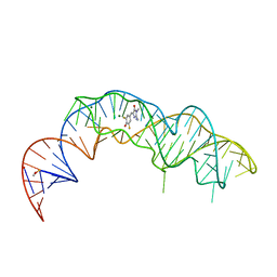
 | | Crystal structure of Squash RNA aptamer in complex with DFHBI-1T with iridium (III) ions | | 分子名称: | (5Z)-5-(3,5-difluoro-4-hydroxybenzylidene)-2-methyl-3-(2,2,2-trifluoroethyl)-3,5-dihydro-4H-imidazol-4-one, IRIDIUM HEXAMMINE ION, MAGNESIUM ION, ... | | 著者 | Truong, L, Ferre-D'Amare, A.R. | | 登録日 | 2020-11-28 | | 公開日 | 2022-01-19 | | 最終更新日 | 2024-05-22 | | 実験手法 | X-RAY DIFFRACTION (2.73 Å) | | 主引用文献 | The fluorescent aptamer Squash extensively repurposes the adenine riboswitch fold.
Nat.Chem.Biol., 18, 2022
|
|
7KVU
 | | Crystal structure of Squash RNA aptamer in complex with DFHBI-1T | | 分子名称: | (5Z)-5-(3,5-difluoro-4-hydroxybenzylidene)-2-methyl-3-(2,2,2-trifluoroethyl)-3,5-dihydro-4H-imidazol-4-one, MAGNESIUM ION, POTASSIUM ION, ... | | 著者 | Truong, L, Ferre-D'Amare, A.R. | | 登録日 | 2020-11-28 | | 公開日 | 2022-01-19 | | 最終更新日 | 2023-10-18 | | 実験手法 | X-RAY DIFFRACTION (2.68 Å) | | 主引用文献 | The fluorescent aptamer Squash extensively repurposes the adenine riboswitch fold.
Nat.Chem.Biol., 18, 2022
|
|
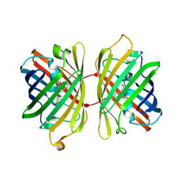
1UIS
 | | The 2.0 crystal structure of eqFP611, a far-red fluorescent protein from the sea anemone Entacmaea quadricolor | | 分子名称: | ACETIC ACID, CALCIUM ION, red fluorescent protein FP611 | | 著者 | Petersen, J, Wilmann, P.G, Beddoe, T, Oakley, A.J, Devenish, R.J, Prescott, M, Rossjohn, J. | | 登録日 | 2003-07-21 | | 公開日 | 2003-10-21 | | 最終更新日 | 2023-12-27 | | 実験手法 | X-RAY DIFFRACTION (2 Å) | | 主引用文献 | The 2.0A crystal structure of eqFP611, a far-red fluorescent protein from the sea anemone Entacmaea quadricolor
J.Biol.Chem., 278, 2003
|
|
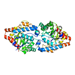
3URA
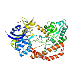
 | | Crystal Structure of PTE mutant H254G/H257W/L303T/K185R/I274N/A80V/S61T | | 分子名称: | COBALT (II) ION, IMIDAZOLE, Parathion hydrolase | | 著者 | Tsai, P, Fox, N.G, Li, Y, Barondeau, D.P, Raushel, F.M. | | 登録日 | 2011-11-21 | | 公開日 | 2012-08-01 | | 最終更新日 | 2023-12-06 | | 実験手法 | X-RAY DIFFRACTION (1.88 Å) | | 主引用文献 | Enzymes for the homeland defense: optimizing phosphotriesterase for the hydrolysis of organophosphate nerve agents.
Biochemistry, 51, 2012
|
|
3URN
 | | Crystal Structure of PTE mutant H254G/H257W/L303T/K185R/I274N/A80V/S61T with cyclohexyl methylphosphonate inhibitor | | 分子名称: | COBALT (II) ION, IMIDAZOLE, Parathion hydrolase, ... | | 著者 | Tsai, P, Fox, N.G, Li, Y, Barondeau, D.P, Raushel, F.M. | | 登録日 | 2011-11-22 | | 公開日 | 2012-08-01 | | 最終更新日 | 2023-12-06 | | 実験手法 | X-RAY DIFFRACTION (1.95 Å) | | 主引用文献 | Enzymes for the homeland defense: optimizing phosphotriesterase for the hydrolysis of organophosphate nerve agents.
Biochemistry, 51, 2012
|
|
3UR2
 | | Crystal Structure of PTE mutant H254G/H257W/L303T/K185R/I274N/A80V | | 分子名称: | 1,2-ETHANEDIOL, COBALT (II) ION, IMIDAZOLE, ... | | 著者 | Tsai, P, Fox, N.G, Li, Y, Barondeau, D.P, Raushel, F.M. | | 登録日 | 2011-11-21 | | 公開日 | 2012-08-01 | | 最終更新日 | 2023-12-06 | | 実験手法 | X-RAY DIFFRACTION (2 Å) | | 主引用文献 | Enzymes for the homeland defense: optimizing phosphotriesterase for the hydrolysis of organophosphate nerve agents.
Biochemistry, 51, 2012
|
|
3UR5
 | | Crystal Structure of PTE mutant K185R/I274N | | 分子名称: | COBALT (II) ION, DIETHYL HYDROGEN PHOSPHATE, Parathion hydrolase | | 著者 | Tsai, P, Fox, N.G, Li, Y, Barondeau, D.P, Raushel, F.M. | | 登録日 | 2011-11-21 | | 公開日 | 2012-08-01 | | 最終更新日 | 2023-12-06 | | 実験手法 | X-RAY DIFFRACTION (1.6 Å) | | 主引用文献 | Enzymes for the homeland defense: optimizing phosphotriesterase for the hydrolysis of organophosphate nerve agents.
Biochemistry, 51, 2012
|
|
3URQ
 | | Crystal Structure of PTE mutant H254G/H257W/L303T/M317L/I106C/F132I/L271I/K185R/I274N/A80V/R67H with cyclohexyl methylphosphonate inhibitor | | 分子名称: | COBALT (II) ION, IMIDAZOLE, Parathion hydrolase, ... | | 著者 | Tsai, P, Fox, N.G, Li, Y, Barondeau, D.P, Raushel, F.M. | | 登録日 | 2011-11-22 | | 公開日 | 2012-08-01 | | 最終更新日 | 2023-12-06 | | 実験手法 | X-RAY DIFFRACTION (2.1 Å) | | 主引用文献 | Enzymes for the homeland defense: optimizing phosphotriesterase for the hydrolysis of organophosphate nerve agents.
Biochemistry, 51, 2012
|
|
3URB
 | | Crystal Structure of PTE mutant H254G/H257W/L303T/M317L/I106C/F132I/L271I/K185R/I274N/A80V/R67H | | 分子名称: | COBALT (II) ION, DIETHYL HYDROGEN PHOSPHATE, IMIDAZOLE, ... | | 著者 | Tsai, P, Fox, N.G, Li, Y, Barondeau, D.P, Raushel, F.M. | | 登録日 | 2011-11-21 | | 公開日 | 2012-08-01 | | 最終更新日 | 2023-12-06 | | 実験手法 | X-RAY DIFFRACTION (1.77 Å) | | 主引用文献 | Enzymes for the homeland defense: optimizing phosphotriesterase for the hydrolysis of organophosphate nerve agents.
Biochemistry, 51, 2012
|
|
4KGE
 | | Crystal structure of near-infrared fluorescent protein with an extended stokes shift, pH 4.5 | | 分子名称: | CHLORIDE ION, TagRFP675, red fluorescent protein | | 著者 | Malashkevich, V.N, Piatkevich, K, Almo, S.C, Verkhusha, V, New York Structural Genomics Research Consortium (NYSGRC) | | 登録日 | 2013-04-29 | | 公開日 | 2013-05-08 | | 最終更新日 | 2023-12-06 | | 実験手法 | X-RAY DIFFRACTION (2.3 Å) | | 主引用文献 | Extended Stokes shift in fluorescent proteins: chromophore-protein interactions in a near-infrared TagRFP675 variant.
Sci Rep, 3, 2013
|
|
4KGF
 | | Crystal structure of near-infrared fluorescent protein with an extended stokes shift, ph 8.0 | | 分子名称: | CHLORIDE ION, TagRFP675, red fluorescent protein | | 著者 | Malashkevich, V.N, Piatkevich, K, Almo, S.C, Verkhusha, V, New York Structural Genomics Research Consortium (NYSGRC) | | 登録日 | 2013-04-29 | | 公開日 | 2013-05-08 | | 最終更新日 | 2023-12-06 | | 実験手法 | X-RAY DIFFRACTION (2.3 Å) | | 主引用文献 | Extended Stokes shift in fluorescent proteins: chromophore-protein interactions in a near-infrared TagRFP675 variant.
Sci Rep, 3, 2013
|
|
3UPM
 | | Crystal Structure of PTE mutant H254Q/H257F/K185R/I274N | | 分子名称: | COBALT (II) ION, Parathion hydrolase | | 著者 | Tsai, P, Fox, N.G, Li, Y, Barondeau, D.P, Raushel, F.M. | | 登録日 | 2011-11-18 | | 公開日 | 2012-08-01 | | 最終更新日 | 2023-12-06 | | 実験手法 | X-RAY DIFFRACTION (1.95 Å) | | 主引用文献 | Enzymes for the homeland defense: optimizing phosphotriesterase for the hydrolysis of organophosphate nerve agents.
Biochemistry, 51, 2012
|
|
8TEC
 | |
8TEE
 | |
3P19
 | | Improved NADPH-dependent Blue Fluorescent Protein | | 分子名称: | NADPH DIHYDRO-NICOTINAMIDE-ADENINE-DINUCLEOTIDE PHOSPHATE, Putative blue fluorescent protein | | 著者 | Kao, T.H, Chen, Y, Pai, C.H, Wang, A.H.J. | | 登録日 | 2010-09-30 | | 公開日 | 2011-07-20 | | 最終更新日 | 2023-11-01 | | 実験手法 | X-RAY DIFFRACTION (2.05 Å) | | 主引用文献 | Structure of a NADPH-dependent blue fluorescent protein revealed the unique role of Gly176 on the fluorescence enhancement.
J.Struct.Biol., 174, 2011
|
|
6B9C
 | | Superfolder Green Fluorescent Protein with 4-nitro-L-phenylalanine at the chromophore (position 66) | | 分子名称: | CARBON DIOXIDE, Green fluorescent protein | | 著者 | Phillips-Piro, C.M, Brewer, S.H, Olenginski, G.M, Piacentini, J. | | 登録日 | 2017-10-10 | | 公開日 | 2018-10-17 | | 最終更新日 | 2024-12-25 | | 実験手法 | X-RAY DIFFRACTION (1.695 Å) | | 主引用文献 | Structural and spectrophotometric investigation of two unnatural amino-acid altered chromophores in the superfolder green fluorescent protein
Acta Crystallogr.,Sect.D, 2021
|
|
5D94
 | | Crystal structure of LC3-LIR peptide complex | | 分子名称: | Microtubule-associated proteins 1A/1B light chain 3B, Peptide from FYVE and coiled-coil domain-containing protein 1 | | 著者 | Takagi, K, Mizushima, T, Johansen, T. | | 登録日 | 2015-08-18 | | 公開日 | 2015-10-21 | | 最終更新日 | 2024-03-20 | | 実験手法 | X-RAY DIFFRACTION (1.53 Å) | | 主引用文献 | FYCO1 Contains a C-terminally Extended, LC3A/B-preferring LC3-interacting Region (LIR) Motif Required for Efficient Maturation of Autophagosomes during Basal Autophagy
J.Biol.Chem., 290, 2015
|
|
3ZU7
 | |


