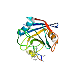
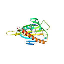
2O2I
 
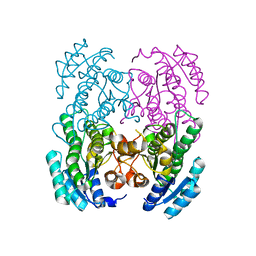
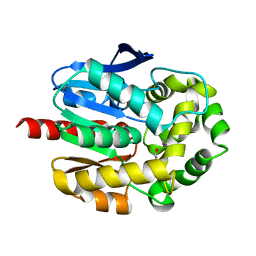 | | Crystal structure of haloalkane dehalogenase Rv2579 from Mycobacterium tuberculosis complexed with 1,3-propandiol | | 分子名称: | 1,3-PROPANDIOL, BROMIDE ION, Haloalkane dehalogenase 3 | | 著者 | Mazumdar, P.A, Hulecki, J, Cherney, M.M, Garen, C.R, James, M.N.G, TB Structural Genomics Consortium (TBSGC) | | 登録日 | 2006-11-29 | | 公開日 | 2007-11-13 | | 最終更新日 | 2023-08-30 | | 実験手法 | X-RAY DIFFRACTION (1.5 Å) | | 主引用文献 | Crystal structure of haloalkane dehalogenase Rv2579 from Mycobacterium tuberculosis complexed with 1,3-propandiol
To be Published
|
|
4DE2
 | |
3SWK
 | |
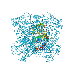
3GZ4
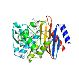
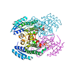
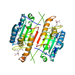
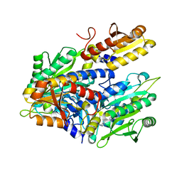
 | | Crystal structure of putative short chain dehydrogenase FROM ESCHERICHIA COLI CFT073 complexed with NADPH | | 分子名称: | Hypothetical oxidoreductase yciK, NADPH DIHYDRO-NICOTINAMIDE-ADENINE-DINUCLEOTIDE PHOSPHATE | | 著者 | Malashkevich, V.N, Toro, R, Morano, C, Sauder, J.M, Burley, S.K, Almo, S.C, New York SGX Research Center for Structural Genomics (NYSGXRC) | | 登録日 | 2009-04-06 | | 公開日 | 2009-04-14 | | 最終更新日 | 2024-02-21 | | 実験手法 | X-RAY DIFFRACTION (2.1 Å) | | 主引用文献 | Crystal structure of putative short chain dehydrogenase
FROM ESCHERICHIA COLI CFT073 complexed with NADPH
To be Published
|
|
4DR5
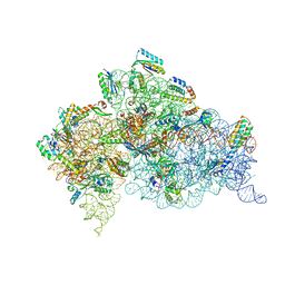 | | Crystal structure of the Thermus thermophilus (HB8) 30S ribosomal subunit with codon, crystallographically disordered cognate transfer RNA anticodon stem-loop and streptomycin bound | | 分子名称: | 16S rRNA, 30S ribosomal protein S10, 30S ribosomal protein S11, ... | | 著者 | Demirci, H, Murphy IV, F, Murphy, E, Gregory, S.T, Dahlberg, A.E, Jogl, G. | | 登録日 | 2012-02-16 | | 公開日 | 2012-11-14 | | 最終更新日 | 2013-01-30 | | 実験手法 | X-RAY DIFFRACTION (3.45 Å) | | 主引用文献 | A structural basis for streptomycin-induced misreading of the genetic code.
Nat Commun, 4, 2013
|
|
3GTY
 | |
4HR2
 | |
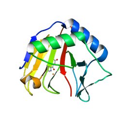
3SVG
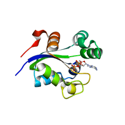
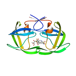
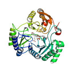
 | | Crystal Structure of the first bromodomain of human BRD4 in complex with a 3,5-dimethylisoxazol ligand | | 分子名称: | (1R)-1-[3-(3,5-dimethyl-1,2-oxazol-4-yl)-5-ethoxyphenyl]ethanol, 1,2-ETHANEDIOL, Bromodomain-containing protein 4 | | 著者 | Filippakopoulos, P, Picaud, S, Felletar, I, Hewings, S.D, von Delft, F, Arrowsmith, C.H, Edwards, A.M, Weigelt, J, Bountra, C, Conway, S.J, Knapp, S, Structural Genomics Consortium (SGC) | | 登録日 | 2011-07-12 | | 公開日 | 2011-08-10 | | 最終更新日 | 2023-09-13 | | 実験手法 | X-RAY DIFFRACTION (1.68 Å) | | 主引用文献 | Crystal Structure of the first bromodomain of human BRD4 in complex with a 3,5-dimethylisoxazol ligand
TO BE PUBLISHED
|
|
4DGC
 | | TRIMCyp cyclophilin domain from Macaca mulatta: cyclosporin A complex | | 分子名称: | TRIMCyp, cyclosporin A | | 著者 | Caines, M.E.C, Bichel, K, Price, A.J, McEwan, W.A, James, L.C. | | 登録日 | 2012-01-25 | | 公開日 | 2012-02-08 | | 最終更新日 | 2023-12-06 | | 実験手法 | X-RAY DIFFRACTION (2.65 Å) | | 主引用文献 | Diverse HIV viruses are targeted by a conformationally dynamic antiviral.
Nat.Struct.Mol.Biol., 19, 2012
|
|
2O4L
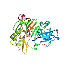
 | | Crystal Structure of HIV-1 Protease (Q7K, I50V) in Complex with Tipranavir | | 分子名称: | CHLORIDE ION, GLYCEROL, N-(3-{(1R)-1-[(6R)-4-HYDROXY-2-OXO-6-PHENETHYL-6-PROPYL-5,6-DIHYDRO-2H-PYRAN-3-YL]PROPYL}PHENYL)-5-(TRIFLUOROMETHYL)-2-PYRIDINESULFONAMIDE, ... | | 著者 | Armstrong, A.A, Muzammil, S, Jakalian, A, Bonneau, P.R, Schmelmer, V, Freire, E, Amzel, L.M. | | 登録日 | 2006-12-04 | | 公開日 | 2006-12-12 | | 最終更新日 | 2023-08-30 | | 実験手法 | X-RAY DIFFRACTION (1.33 Å) | | 主引用文献 | Unique thermodynamic response of tipranavir to human immunodeficiency virus type 1 protease drug resistance mutations.
J.Virol., 81, 2007
|
|
3SVT
 | |
2I51
 | |
3GVC
 | |
3H1P
 | |
4HTR
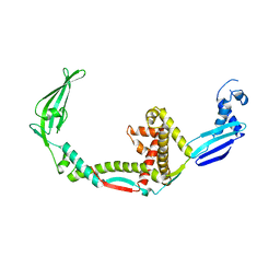
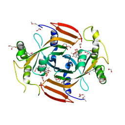
 | | N149W variant of SiRHP bound to sulfite | | 分子名称: | IRON/SULFUR CLUSTER, SIROHEME, SODIUM ION, ... | | 著者 | Smith, K.W, Stroupe, M.E. | | 登録日 | 2012-11-01 | | 公開日 | 2013-01-16 | | 最終更新日 | 2023-12-27 | | 実験手法 | X-RAY DIFFRACTION (1.6 Å) | | 主引用文献 | Mutational analysis of sulfite reductase hemoprotein reveals the mechanism for coordinated electron and proton transfer.
Biochemistry, 51, 2012
|
|
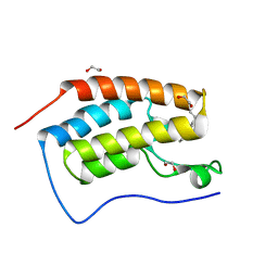
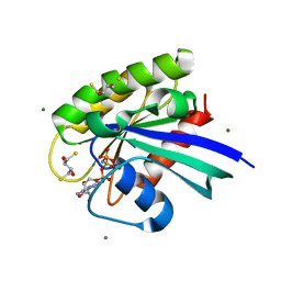
4HU9
 | | E. coli thioredoxin variant with (4S)-FluoroPro76 as single proline residue | | 分子名称: | COPPER (II) ION, Thioredoxin-1 | | 著者 | Scharer, M.A, Rubini, M, Capitani, G, Glockshuber, R. | | 登録日 | 2012-11-02 | | 公開日 | 2013-05-29 | | 最終更新日 | 2017-09-20 | | 実験手法 | X-RAY DIFFRACTION (1.55 Å) | | 主引用文献 | (4R)- and (4S)-Fluoroproline in the Conserved cis-Prolyl Peptide Bond of the Thioredoxin Fold: Tertiary Structure Context Dictates Ring Puckering.
Chembiochem, 14, 2013
|
|
4DJY
 | |
3H3S
 | |
4HWG
 | |
3T3Y
 | |
4DLZ
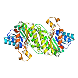
 | | H-Ras Set 2 Ca(OAc)2/DTE, ordered off | | 分子名称: | (2R,3S)-1,4-DIMERCAPTOBUTANE-2,3-DIOL, CALCIUM ION, GTPase HRas, ... | | 著者 | Holzapfel, G, Mattos, C. | | 登録日 | 2012-02-06 | | 公開日 | 2012-08-08 | | 最終更新日 | 2024-02-28 | | 実験手法 | X-RAY DIFFRACTION (1.662 Å) | | 主引用文献 | Shift in the Equilibrium between On and Off States of the Allosteric Switch in Ras-GppNHp Affected by Small Molecules and Bulk Solvent Composition.
Biochemistry, 51, 2012
|
|
3H5G
 | |
4HWP
 | |
3T46
 | |
3T49
 | |


