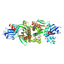
6VQ6
 
 | |
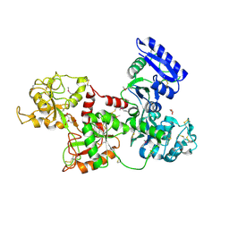
6W9D
 | |
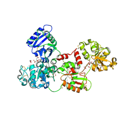
6VQC
 | |
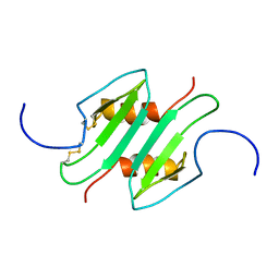
4V4F
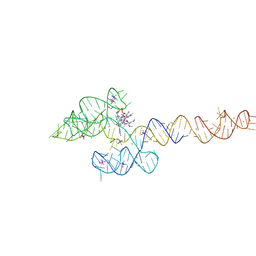
 | | The structure of the trp RNA-binding attenuation protein (TRAP) bound to a RNA molecule containing UAGAU repeats | | 分子名称: | 5'-R(*UP*AP*GP*AP*UP)-3', TRANSCRIPTION ATTENUATION PROTEIN MTRB, TRYPTOPHAN | | 著者 | Hopcroft, N.H, Manfredo, A, Wendt, A.L, Brzozowski, A.M, Gollnick, P, Antson, A.A. | | 登録日 | 2003-12-08 | | 公開日 | 2014-07-09 | | 最終更新日 | 2024-01-10 | | 実験手法 | X-RAY DIFFRACTION (1.9 Å) | | 主引用文献 | The Interaction of RNA with Trap: The Role of Triplet Repeats and Separating Spacer Nucleotides
J.Mol.Biol., 338, 2004
|
|
6VMY
 | | Structure of the B. subtilis cobalamin riboswitch | | 分子名称: | Adenosylcobalamin, B. subtilis cobalamin riboswitch, COBALT HEXAMMINE(III), ... | | 著者 | Chan, C.W, Mondragon, A. | | 登録日 | 2020-01-28 | | 公開日 | 2020-06-10 | | 最終更新日 | 2024-03-06 | | 実験手法 | X-RAY DIFFRACTION (3.25 Å) | | 主引用文献 | Crystal structure of an atypical cobalamin riboswitch reveals RNA structural adaptability as basis for promiscuous ligand binding.
Nucleic Acids Res., 48, 2020
|
|
6VQG
 | |
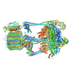
6VWS
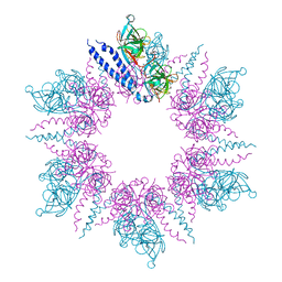
 | | Hexamer of Helical HIV capsid by RASTR method | | 分子名称: | HIV capsid protein | | 著者 | Zhao, H, Iqbal, N, Asturias, F, Kvaratskhelia, M, Vanblerkom, P. | | 登録日 | 2020-02-20 | | 公開日 | 2020-10-21 | | 最終更新日 | 2024-03-06 | | 実験手法 | ELECTRON MICROSCOPY (6.08 Å) | | 主引用文献 | Structural and mechanistic bases for a potent HIV-1 capsid inhibitor.
Science, 370, 2020
|
|
1M1X
 | | CRYSTAL STRUCTURE OF THE EXTRACELLULAR SEGMENT OF INTEGRIN ALPHA VBETA3 BOUND TO MN2+ | | 分子名称: | 2-acetamido-2-deoxy-alpha-D-glucopyranose-(1-4)-2-acetamido-2-deoxy-beta-D-glucopyranose, 2-acetamido-2-deoxy-beta-D-glucopyranose, 2-acetamido-2-deoxy-beta-D-glucopyranose-(1-4)-2-acetamido-2-deoxy-beta-D-glucopyranose, ... | | 著者 | Xiong, J.-P, Stehle, T, Zhang, R, Joachimiak, A, Frech, M, Goodman, S.L, Arnaout, M.A. | | 登録日 | 2002-06-20 | | 公開日 | 2002-08-14 | | 最終更新日 | 2020-07-29 | | 実験手法 | X-RAY DIFFRACTION (3.3 Å) | | 主引用文献 | Crystal structure of the extracellular segment of integrin alpha Vbeta3 in complex with an Arg-Gly-Asp ligand.
Science, 296, 2002
|
|
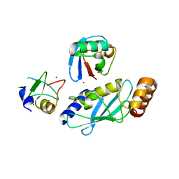
2I0N
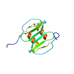
 | | Structure of Dictyostelium discoideum Myosin VII SH3 domain with adjacent proline rich region | | 分子名称: | Class VII unconventional myosin | | 著者 | Wang, Q, Deloia, M.A, Kang, Y, Litchke, C, Titus, M.A, Walters, K.J. | | 登録日 | 2006-08-10 | | 公開日 | 2007-01-09 | | 最終更新日 | 2024-05-29 | | 実験手法 | SOLUTION NMR | | 主引用文献 | The SH3 domain of a M7 interacts with its C-terminal proline-rich region.
Protein Sci., 16, 2007
|
|
1NYF
 | |
1LNY
 | | Crystal structure of the recombinant mouse-muscle adenylosuccinate synthetase complexed with 6-phosphoryl-IMP, GDP and Mg | | 分子名称: | 6-O-PHOSPHORYL INOSINE MONOPHOSPHATE, Adenylosuccinate Synthetase, GUANOSINE-5'-DIPHOSPHATE, ... | | 著者 | Iancu, C.V, Borza, T, Fromm, H.J, Honzatko, R.B. | | 登録日 | 2002-05-04 | | 公開日 | 2002-08-28 | | 最終更新日 | 2023-10-25 | | 実験手法 | X-RAY DIFFRACTION (2.2 Å) | | 主引用文献 | IMP, GTP, and 6-phosphoryl-IMP complexes of recombinant mouse muscle adenylosuccinate synthetase.
J.Biol.Chem., 277, 2002
|
|
1NLP
 | | STRUCTURE OF SIGNAL TRANSDUCTION PROTEIN, NMR, MINIMIZED AVERAGE STRUCTURE | | 分子名称: | C-SRC, NL2 (MN8-MN1-PLPPLP) | | 著者 | Feng, S, Kapoor, T.M, Shirai, F, Combs, A.P, Schreiber, S.L. | | 登録日 | 1996-08-04 | | 公開日 | 1997-01-27 | | 最終更新日 | 2023-11-15 | | 実験手法 | SOLUTION NMR | | 主引用文献 | Molecular basis for the binding of SH3 ligands with non-peptide elements identified by combinatorial synthesis.
Chem.Biol., 3, 1996
|
|
1NLO
 | | STRUCTURE OF SIGNAL TRANSDUCTION PROTEIN, NMR, MINIMIZED AVERAGE STRUCTURE | | 分子名称: | C-SRC, NL1 (MN7-MN2-MN1-PLPPLP) | | 著者 | Feng, S, Kapoor, T.M, Shirai, F, Combs, A.P, Schreiber, S.L. | | 登録日 | 1996-08-04 | | 公開日 | 1997-01-27 | | 最終更新日 | 2023-11-15 | | 実験手法 | SOLUTION NMR | | 主引用文献 | Molecular basis for the binding of SH3 ligands with non-peptide elements identified by combinatorial synthesis.
Chem.Biol., 3, 1996
|
|
1NYG
 | |
2IAK
 | |
2I7P
 | | Crystal structure of human PANK3 in complex with AcCoA | | 分子名称: | ACETYL COENZYME *A, Pantothenate kinase 3 | | 著者 | Hong, B.S, Wang, L, Shen, L, Tempel, W, Loppnau, P, Finerty, P, Arrowsmith, C.H, Edwards, A.M, Sundstrom, M, Weigelt, J, Bochkarev, A, Park, H.W, Structural Genomics Consortium (SGC) | | 登録日 | 2006-08-31 | | 公開日 | 2006-12-26 | | 最終更新日 | 2024-02-21 | | 実験手法 | X-RAY DIFFRACTION (2.05 Å) | | 主引用文献 | Crystal structures of human pantothenate kinases. Insights into allosteric regulation and mutations linked to a neurodegeneration disorder.
J.Biol.Chem., 282, 2007
|
|
2FV8
 | | The crystal structure of RhoB in the GDP-bound state | | 分子名称: | GUANOSINE-5'-DIPHOSPHATE, Rho-related GTP-binding protein RhoB | | 著者 | Turnbull, A.P, Soundararajan, M, Smee, C, Johansson, C, Schoch, G, Gorrec, F, Bray, J, Papagrigoriou, E, von Delft, F, Weigelt, J, Edwards, A, Arrowsmith, C, Sundstrom, M, Doyle, D, Structural Genomics Consortium (SGC) | | 登録日 | 2006-01-30 | | 公開日 | 2006-02-28 | | 最終更新日 | 2024-04-03 | | 実験手法 | X-RAY DIFFRACTION (1.9 Å) | | 主引用文献 | The crystal structure of RhoB in the GDP-bound state
To be Published
|
|
2I7N
 | | Crystal structure of human PANK1 alpha: the catalytic core domain in complex with AcCoA | | 分子名称: | ACETYL COENZYME *A, Pantothenate kinase 1 | | 著者 | Hong, B.S, Wang, L, Tempel, W, Loppnau, P, Allali-Hassani, A, Arrowsmith, C.H, Edwards, A.M, Sundstrom, M, Weigelt, J, Bochkarev, A, Park, H.W. | | 登録日 | 2006-08-31 | | 公開日 | 2006-12-26 | | 最終更新日 | 2024-02-21 | | 実験手法 | X-RAY DIFFRACTION (1.9 Å) | | 主引用文献 | Crystal structures of human pantothenate kinases. Insights into allosteric regulation and mutations linked to a neurodegeneration disorder.
J.Biol.Chem., 282, 2007
|
|
2HAU
 | | Apo-Human Serum Transferrin (Non-Glycosylated) | | 分子名称: | CITRIC ACID, GLYCEROL, Serotransferrin | | 著者 | Wally, J, Everse, S.J. | | 登録日 | 2006-06-13 | | 公開日 | 2006-06-27 | | 最終更新日 | 2021-10-20 | | 実験手法 | X-RAY DIFFRACTION (2.7 Å) | | 主引用文献 | The Crystal Structure of Iron-free Human Serum Transferrin Provides Insight into Inter-lobe Communication and Receptor Binding.
J.Biol.Chem., 281, 2006
|
|
2HAV
 | | Apo-Human Serum Transferrin (Glycosylated) | | 分子名称: | CITRIC ACID, GLYCEROL, Serotransferrin | | 著者 | Wally, J, Everse, S.J. | | 登録日 | 2006-06-13 | | 公開日 | 2006-06-27 | | 最終更新日 | 2023-08-30 | | 実験手法 | X-RAY DIFFRACTION (2.7 Å) | | 主引用文献 | The Crystal Structure of Iron-free Human Serum Transferrin Provides Insight into Inter-lobe Communication and Receptor Binding.
J.Biol.Chem., 281, 2006
|
|
1MSG
 | |
2J5U
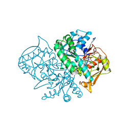
 | | MreC Lysteria monocytogenes | | 分子名称: | MREC PROTEIN | | 著者 | van den Ent, F, Leaver, M, Bendezu, F, Errington, J, de Boer, P, Lowe, J. | | 登録日 | 2006-09-19 | | 公開日 | 2006-12-11 | | 最終更新日 | 2024-05-08 | | 実験手法 | X-RAY DIFFRACTION (2.5 Å) | | 主引用文献 | Dimeric Structure of the Cell Shape Protein Mrec and its Functional Implications.
Mol.Microbiol., 62, 2006
|
|
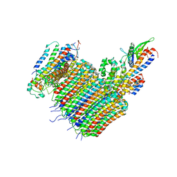
1N11
 | | D34 REGION OF HUMAN ANKYRIN-R AND LINKER | | 分子名称: | Ankyrin, BROMIDE ION, CHLORIDE ION | | 著者 | Michaely, P, Tomchick, D.R, Machius, M, Anderson, R.G.W. | | 登録日 | 2002-10-16 | | 公開日 | 2002-12-11 | | 最終更新日 | 2024-02-14 | | 実験手法 | X-RAY DIFFRACTION (2.7 Å) | | 主引用文献 | Crystal structure of a 12 ANK repeat stack from human ankyrinR
Embo J., 21, 2002
|
|
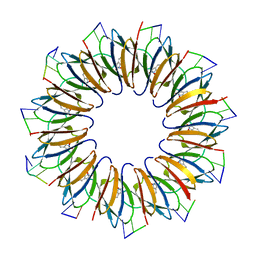
2J7Z
 | | Crystal Structure of recombinant Human Stromal Cell-Derived Factor- 1alpha | | 分子名称: | STROMAL CELL-DERIVED FACTOR 1 ALPHA | | 著者 | Ryu, E.K, Kim, T.G, Kwon, T.H, Jung, I.D, Ryu, D.W, Park, Y.-M, Ahn, K, Ban, C. | | 登録日 | 2006-10-18 | | 公開日 | 2006-10-23 | | 最終更新日 | 2023-12-13 | | 実験手法 | X-RAY DIFFRACTION (1.95 Å) | | 主引用文献 | Crystal Structure of Recombinant Human Stromal Cell-Derived Factor-1Alpha.
Proteins, 67, 2007
|
|
1MEZ
 | | Structure of the Recombinant Mouse-Muscle Adenylosuccinate Synthetase Complexed with SAMP, GDP, SO4(2-), and Mg(2+) | | 分子名称: | 2-[9-(3,4-DIHYDROXY-5-PHOSPHONOOXYMETHYL-TETRAHYDRO-FURAN-2-YL)-9H-PURIN-6-YLAMINO]-SUCCINIC ACID, Adenylosuccinate Synthetase, GUANOSINE-5'-DIPHOSPHATE, ... | | 著者 | Iancu, C.V, Borza, T, Fromm, H.J, Honzatko, R.B. | | 登録日 | 2002-08-09 | | 公開日 | 2002-10-30 | | 最終更新日 | 2024-03-13 | | 実験手法 | X-RAY DIFFRACTION (2.4 Å) | | 主引用文献 | Feedback inhibition and product complexes of recombinant mouse muscle adenylosuccinate synthetase.
J.Biol.Chem., 277, 2002
|
|


