3MJH
 
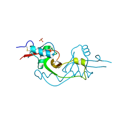
 | | Crystal Structure of Human Rab5A in complex with the C2H2 Zinc Finger of EEA1 | | 分子名称: | Early endosome antigen 1, GUANOSINE-5'-TRIPHOSPHATE, MAGNESIUM ION, ... | | 著者 | Mishra, A.K, Eathiraj, S, Lambright, D.G. | | 登録日 | 2010-04-12 | | 公開日 | 2010-05-05 | | 最終更新日 | 2024-02-21 | | 実験手法 | X-RAY DIFFRACTION (2.03 Å) | | 主引用文献 | Structural basis for Rab GTPase recognition and endosome tethering by the C2H2 zinc finger of Early Endosomal Autoantigen 1 (EEA1).
Proc.Natl.Acad.Sci.USA, 107, 2010
|
|
3MOU
 | |
6Q46
 | |
6Q4D
 | | CDK2 in complex with FragLite31 | | 分子名称: | 2-(4-bromanyl-2-methoxy-phenyl)ethanoic acid, Cyclin-dependent kinase 2 | | 著者 | Wood, D.J, Martin, M.P, Noble, M.E.M. | | 登録日 | 2018-12-05 | | 公開日 | 2019-03-20 | | 最終更新日 | 2024-01-24 | | 実験手法 | X-RAY DIFFRACTION (1.07 Å) | | 主引用文献 | FragLites-Minimal, Halogenated Fragments Displaying Pharmacophore Doublets. An Efficient Approach to Druggability Assessment and Hit Generation.
J.Med.Chem., 62, 2019
|
|
4YT3
 | | CYP106A2 | | 分子名称: | ACETATE ION, Cytochrome P450(MEG), PROTOPORPHYRIN IX CONTAINING FE | | 著者 | janocha, S, carius, y, bernhardt, r, lancaster, c.r.d. | | 登録日 | 2015-03-17 | | 公開日 | 2016-02-24 | | 最終更新日 | 2024-01-10 | | 実験手法 | X-RAY DIFFRACTION (1.8 Å) | | 主引用文献 | Crystal Structure of CYP106A2 in Substrate-Free and Substrate-Bound Form.
Chembiochem, 17, 2016
|
|
6Q6C
 | | Pore-modulating toxins exploit inherent slow inactivation to block K+ channels | | 分子名称: | 1,2-ETHANEDIOL, Kunitz-type conkunitzin-S1, NITRATE ION, ... | | 著者 | Karbat, I, Gueta, H, Fine, S, Szanto, T, Hamer-Rogotner, S, Dym, O, Frolow, F, Gordon, D, Panyi, G, Gurevitz, M, Reuveny, E. | | 登録日 | 2018-12-10 | | 公開日 | 2019-08-21 | | 最終更新日 | 2024-01-24 | | 実験手法 | X-RAY DIFFRACTION (1.3 Å) | | 主引用文献 | Pore-modulating toxins exploit inherent slow inactivation to block K+channels.
Proc.Natl.Acad.Sci.USA, 116, 2019
|
|
6Q6U
 | |
4YGG
 | |
3M0U
 | |
8W9J
 | |
6Q82
 | | Crystal structure of the biportin Pdr6 in complex with RanGTP | | 分子名称: | GTP-binding nuclear protein Ran, GUANOSINE-5'-TRIPHOSPHATE, Importin beta-like protein KAP122, ... | | 著者 | Aksu, M, Vera-Rodriguez, A, Trakhanov, S, Gorlich, D. | | 登録日 | 2018-12-14 | | 公開日 | 2019-05-01 | | 最終更新日 | 2019-06-12 | | 実験手法 | X-RAY DIFFRACTION (2.994 Å) | | 主引用文献 | Structural basis for the nuclear import and export functions of the biportin Pdr6/Kap122.
J.Cell Biol., 218, 2019
|
|
4YV5
 | | Crystal Structure of Myotoxin II from Bothrops moojeni complexed to Suramin | | 分子名称: | 2-{2-[2-(2-{2-[2-(2-ETHOXY-ETHOXY)-ETHOXY]-ETHOXY}-ETHOXY)-ETHOXY]-ETHOXY}-ETHANOL, 8,8'-[CARBONYLBIS[IMINO-3,1-PHENYLENECARBONYLIMINO(4-METHYL-3,1-PHENYLENE)CARBONYLIMINO]]BIS-1,3,5-NAPHTHALENETRISULFON IC ACID, Basic phospholipase A2 homolog 2, ... | | 著者 | Salvador, G.H.M, Fontes, M.R.M. | | 登録日 | 2015-03-19 | | 公開日 | 2015-10-14 | | 最終更新日 | 2023-09-27 | | 実験手法 | X-RAY DIFFRACTION (1.9 Å) | | 主引用文献 | Structural and functional evidence for membrane docking and disruption sites on phospholipase A2-like proteins revealed by complexation with the inhibitor suramin.
Acta Crystallogr. D Biol. Crystallogr., 71, 2015
|
|
8XEY
 | | The Crystal Structure of C-terminal kinase domain of RSK2 from Biortus | | 分子名称: | 1,2-ETHANEDIOL, DI(HYDROXYETHYL)ETHER, Ribosomal protein S6 kinase alpha-3 | | 著者 | Wang, F, Cheng, W, Lv, Z, Meng, Q, Zhang, B. | | 登録日 | 2023-12-13 | | 公開日 | 2024-03-06 | | 実験手法 | X-RAY DIFFRACTION (2.65 Å) | | 主引用文献 | The Crystal Structure of C-terminal kinase domain of RSK2 from Biortus
To Be Published
|
|
8XFY
 | | The Crystal Structure of RSK2 from Biortus. | | 分子名称: | 2,6-bis(fluoranyl)-4-[4-(4-morpholin-4-ylphenyl)pyridin-3-yl]phenol, Ribosomal protein S6 kinase alpha-3 | | 著者 | Wang, F, Cheng, W, Lv, Z, Lin, D, Pan, W. | | 登録日 | 2023-12-14 | | 公開日 | 2024-03-06 | | 実験手法 | X-RAY DIFFRACTION (3.2 Å) | | 主引用文献 | The Crystal Structure of RSK2 from Biortus.
To Be Published
|
|
4YB1
 | | 20A Mutant c-di-GMP Vc2 Riboswitch bound with 3',3'-cGAMP | | 分子名称: | 2-amino-9-[(2R,3R,3aS,5R,7aR,9R,10R,10aS,12R,14aR)-9-(6-amino-9H-purin-9-yl)-3,5,10,12-tetrahydroxy-5,12-dioxidooctahydro-2H,7H-difuro[3,2-d:3',2'-j][1,3,7,9,2,8]tetraoxadiphosphacyclododecin-2-yl]-1,9-dihydro-6H-purin-6-one, MAGNESIUM ION, RNA (91-MER), ... | | 著者 | Ren, A.M, Patel, D.J, Rajashankar, R.K. | | 登録日 | 2015-02-18 | | 公開日 | 2015-04-15 | | 最終更新日 | 2024-02-28 | | 実験手法 | X-RAY DIFFRACTION (2.081 Å) | | 主引用文献 | Structural Basis for Molecular Discrimination by a 3',3'-cGAMP Sensing Riboswitch.
Cell Rep, 11, 2015
|
|
3M42
 | | Crystal structure of MAPKAP kinase 2 (MK2) complexed with a tetracyclic ATP site inhibitor | | 分子名称: | 2-[5-(2-methoxyethoxy)pyridin-3-yl]-8,9,10,11-tetrahydro-7H-pyrido[3',4':4,5]pyrrolo[2,3-f]isoquinolin-7-one, MAGNESIUM ION, MAP kinase-activated protein kinase 2 | | 著者 | Scheufler, C, Revesz, L, Be, C, Izaac, A, Huppertz, C, Schlapbach, A, Kroemer, M. | | 登録日 | 2010-03-10 | | 公開日 | 2011-03-23 | | 最終更新日 | 2024-02-21 | | 実験手法 | X-RAY DIFFRACTION (2.68 Å) | | 主引用文献 | Novel pyrrolo[2,3-f]isoquinoline based MAPKAP-K2 (MK2) inhibitors with potent in vitro and in vivo activity
To be Published
|
|
6QHA
 | |
4YB9
 | | Crystal structure of the Bovine Fructose transporter GLUT5 in an open inward-facing conformation | | 分子名称: | Solute carrier family 2, facilitated glucose transporter member 5 | | 著者 | Verdon, G, Kang, H.J, Iwata, S, Drew, D. | | 登録日 | 2015-02-18 | | 公開日 | 2015-10-14 | | 最終更新日 | 2024-05-01 | | 実験手法 | X-RAY DIFFRACTION (3.2 Å) | | 主引用文献 | Structure and mechanism of the mammalian fructose transporter GLUT5.
Nature, 526, 2015
|
|
6QIH
 | |
4YM8
 | |
4YBQ
 | | Rat GLUT5 with Fv in the outward-open form | | 分子名称: | Solute carrier family 2, facilitated glucose transporter member 5, antibody Fv fragment heavy chain, ... | | 著者 | Nomura, N, Shimamura, T, Iwata, S. | | 登録日 | 2015-02-19 | | 公開日 | 2015-10-07 | | 最終更新日 | 2023-11-08 | | 実験手法 | X-RAY DIFFRACTION (3.27 Å) | | 主引用文献 | Structure and mechanism of the mammalian fructose transporter GLUT5
Nature, 526, 2015
|
|
8XOV
 | | The Crystal Structure of N-terminal kinase domain of human RSK-1 from Biortus. | | 分子名称: | 1,2-ETHANEDIOL, PHOSPHOAMINOPHOSPHONIC ACID-ADENYLATE ESTER, Ribosomal protein S6 kinase alpha-1 | | 著者 | Wang, F, Cheng, W, Lv, Z, Meng, Q, Zhang, B. | | 登録日 | 2024-01-02 | | 公開日 | 2024-03-06 | | 実験手法 | X-RAY DIFFRACTION (2.55 Å) | | 主引用文献 | The Crystal Structure of N-terminal kinase domain of human RSK-1 from Biortus.
To Be Published
|
|
6QHC
 | |
4YD7
 | |
3M9Z
 | | Crystal Structure of extracellular domain of mouse NKR-P1A | | 分子名称: | Killer cell lectin-like receptor subfamily B member 1A, PHOSPHATE ION | | 著者 | Kolenko, P, Rozbesky, D, Bezouska, K, Hasek, J, Dohnalek, J. | | 登録日 | 2010-03-23 | | 公開日 | 2011-04-06 | | 最終更新日 | 2023-09-06 | | 実験手法 | X-RAY DIFFRACTION (1.7 Å) | | 主引用文献 | Molecular architecture of mouse activating NKR-P1 receptors.
J.Struct.Biol., 175, 2011
|
|


