5LYE
 
 | |
9CPM
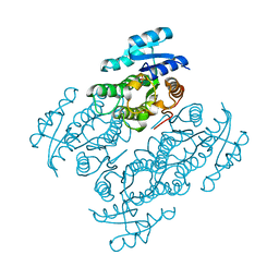
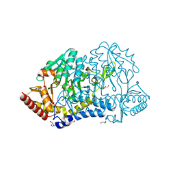
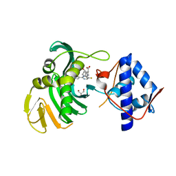
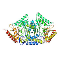
 | | Thermus thermophilus HB27 laccase (Tth-Lac) mutant with partial deletion of beta-hairpin sequence | | 分子名称: | COPPER (II) ION, GLYCEROL, Laccase | | 著者 | Lee, J, Miranda-Zaragoza, B, Rodriguez-Almazan, C, Strynadka, N.C.J. | | 登録日 | 2024-07-18 | | 公開日 | 2025-02-05 | | 実験手法 | X-RAY DIFFRACTION (1.6 Å) | | 主引用文献 | Structure-Function Relationship of the beta-Hairpin of Thermus thermophilus HB27 Laccase.
Int J Mol Sci, 26, 2025
|
|
6LPK
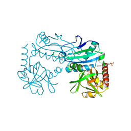
 | | A2AR crystallized in EROCOC17+4, LCP-SFX at 293 K | | 分子名称: | 4-{2-[(7-amino-2-furan-2-yl[1,2,4]triazolo[1,5-a][1,3,5]triazin-5-yl)amino]ethyl}phenol, Adenosine receptor A2a,Soluble cytochrome b562,Adenosine receptor A2a, CHOLESTEROL, ... | | 著者 | Ihara, K, Hato, M, Nakane, T, Yamashita, K, Kimura-Someya, T, Hosaka, T, Ishizuka-Katsura, Y, Tanaka, R, Tanaka, T, Sugahara, M, Hirata, K, Yamamoto, M, Nureki, O, Tono, K, Nango, E, Iwata, S, Shirouzu, M. | | 登録日 | 2020-01-10 | | 公開日 | 2020-11-25 | | 最終更新日 | 2024-10-16 | | 実験手法 | X-RAY DIFFRACTION (1.8 Å) | | 主引用文献 | Isoprenoid-chained lipid EROCOC 17+4 : a new matrix for membrane protein crystallization and a crystal delivery medium in serial femtosecond crystallography.
Sci Rep, 10, 2020
|
|
6H2Z
 | | The crystal structure of human carbonic anhydrase II in complex with 4-(4-phenylpiperidine-1-carbonyl)benzenesulfonamide. | | 分子名称: | 4-(4-phenylpiperidin-1-yl)carbonylbenzenesulfonamide, Carbonic anhydrase 2, GLYCEROL, ... | | 著者 | Buemi, M.R, Di Fiore, A, De Luca, L, Ferro, S, Mancuso, F, Monti, S.M, Buonanno, M, Angeli, A, Russo, E, De Sarro, G, Supuran, C.T, De Simone, G, Gitto, R. | | 登録日 | 2018-07-17 | | 公開日 | 2018-12-19 | | 最終更新日 | 2024-01-17 | | 実験手法 | X-RAY DIFFRACTION (1.94 Å) | | 主引用文献 | Exploring structural properties of potent human carbonic anhydrase inhibitors bearing a 4-(cycloalkylamino-1-carbonyl)benzenesulfonamide moiety.
Eur J Med Chem, 163, 2018
|
|
6H33
 | | The crystal structure of human carbonic anhydrase II in complex with 4-(4-phenyl)-4-hydroxy-1-piperidine-1-carbonyl)benzenesulfonamide. | | 分子名称: | 4-(4-oxidanyl-4-phenyl-piperidin-1-yl)carbonylbenzenesulfonamide, Carbonic anhydrase 2, GLYCEROL, ... | | 著者 | Buemi, M.R, Di Fiore, A, De Luca, L, Ferro, S, Mancuso, F, Monti, S.M, Buonanno, M, Angeli, A, Russo, E, De Sarro, G, Supuran, C.T, De Simone, G, Gitto, R. | | 登録日 | 2018-07-17 | | 公開日 | 2018-12-19 | | 最終更新日 | 2024-01-17 | | 実験手法 | X-RAY DIFFRACTION (1.58 Å) | | 主引用文献 | Exploring structural properties of potent human carbonic anhydrase inhibitors bearing a 4-(cycloalkylamino-1-carbonyl)benzenesulfonamide moiety.
Eur J Med Chem, 163, 2018
|
|
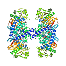
5JO9
 | | Structural characterization of the thermostable Bradyrhizobium japonicum d-sorbitol dehydrogenase | | 分子名称: | PHOSPHATE ION, Ribitol 2-dehydrogenase, sorbitol | | 著者 | Fredslund, F, Otten, H, Navarro Poulsen, J.-C, Lo Leggio, L. | | 登録日 | 2016-05-02 | | 公開日 | 2016-11-09 | | 最終更新日 | 2024-01-10 | | 実験手法 | X-RAY DIFFRACTION (2.894 Å) | | 主引用文献 | Structural characterization of the thermostable Bradyrhizobium japonicumD-sorbitol dehydrogenase.
Acta Crystallogr F Struct Biol Commun, 72, 2016
|
|
1GK7
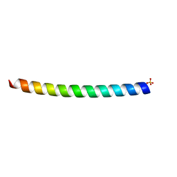 | | HUMAN VIMENTIN COIL 1A FRAGMENT (1A) | | 分子名称: | SULFATE ION, VIMENTIN | | 著者 | Strelkov, S.V, Herrmann, H, Geisler, N, Zimbelmann, R, Aebi, U, Burkhard, P. | | 登録日 | 2001-08-08 | | 公開日 | 2002-03-15 | | 最終更新日 | 2023-12-13 | | 実験手法 | X-RAY DIFFRACTION (1.4 Å) | | 主引用文献 | Conserved Segments 1A and 2B of the Intermediate Filament Dimer: Their Atomic Structures and Role in Filament Assembly.
Embo J., 21, 2002
|
|
5J78
 | | Crystal structure of an Acetylating Aldehyde Dehydrogenase from Geobacillus thermoglucosidasius | | 分子名称: | ACETATE ION, Acetaldehyde dehydrogenase (Acetylating), GLYCEROL, ... | | 著者 | Crennell, S.J, Extance, J.P, Danson, M.J. | | 登録日 | 2016-04-06 | | 公開日 | 2016-09-07 | | 最終更新日 | 2024-11-20 | | 実験手法 | X-RAY DIFFRACTION (2.1 Å) | | 主引用文献 | Structure of an acetylating aldehyde dehydrogenase from the thermophilic ethanologen Geobacillus thermoglucosidasius.
Protein Sci., 25, 2016
|
|
6UX5
 | |
6MBB
 | |
5J7I
 | |
6IN7
 | |
6UCK
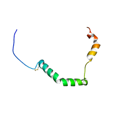 | | proIAPP in DPC Micelles - Two-Conformer Ensemble Refinement, Bent Conformer | | 分子名称: | Islet amyloid polypeptide | | 著者 | DeLisle, C.F, Malooley, A.L, Banerjee, I, Lorieau, J.L. | | 登録日 | 2019-09-16 | | 公開日 | 2020-02-26 | | 最終更新日 | 2024-11-20 | | 実験手法 | SOLUTION NMR | | 主引用文献 | Pro-islet amyloid polypeptide in micelles contains a helical prohormone segment.
Febs J., 287, 2020
|
|
6UCJ
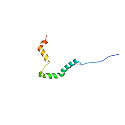 | | proIAPP in DPC Micelles - Two-Conformer Ensemble Refinement, Open Conformer | | 分子名称: | Islet amyloid polypeptide | | 著者 | DeLisle, C.F, Malooley, A.L, Banerjee, I, Lorieau, J.L. | | 登録日 | 2019-09-16 | | 公開日 | 2020-02-26 | | 最終更新日 | 2024-10-30 | | 実験手法 | SOLUTION NMR | | 主引用文献 | Pro-islet amyloid polypeptide in micelles contains a helical prohormone segment.
Febs J., 287, 2020
|
|
6KFY
 | |
8YRU
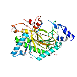
 | | Crystal structure of D-amino acid transaminase from Haliscomenobacter hydrossis (apo form) after 15 sec of soaking with phenylhydrazine | | 分子名称: | ACETATE ION, Aminotransferase class IV, [6-methyl-5-oxidanyl-4-[(2-phenylhydrazinyl)methyl]pyridin-3-yl]methyl dihydrogen phosphate | | 著者 | Matyuta, I.O, Bakunova, A.K, Minyaev, M.E, Popov, V.O, Boyko, K.M. | | 登録日 | 2024-03-21 | | 公開日 | 2024-04-17 | | 最終更新日 | 2024-10-30 | | 実験手法 | X-RAY DIFFRACTION (2 Å) | | 主引用文献 | Incorporation of pyridoxal-5'-phosphate into the apoenzyme: A structural study of D-amino acid transaminase from Haliscomenobacter hydrossis.
Biochim Biophys Acta Proteins Proteom, 1873, 2024
|
|
1H33
 | | Oxidised SoxAX complex from Rhodovulum sulfidophilum | | 分子名称: | CYTOCHROME C, DIHEME CYTOCHROME C, HEME C | | 著者 | Bamford, V.A, Bruno, S, Rasmussen, T, Appia-Ayme, C, Cheesman, M.R, Berks, B.C, Hemmings, A.M. | | 登録日 | 2002-08-21 | | 公開日 | 2002-11-07 | | 最終更新日 | 2024-11-13 | | 実験手法 | X-RAY DIFFRACTION (1.75 Å) | | 主引用文献 | Structural Basis for the Oxidation of Thiosulfate by a Sulfur Cycle Enzyme
Embo J., 21, 2002
|
|
5NPU
 | | Inferred ancestral pyruvate decarboxylase | | 分子名称: | ANC27, DI(HYDROXYETHYL)ETHER, MAGNESIUM ION, ... | | 著者 | Buddrus, L, Crennell, S.J, Leak, D.J, Danson, M.J, Andrews, E.S.V, Arcus, V.L. | | 登録日 | 2017-04-19 | | 公開日 | 2018-03-07 | | 最終更新日 | 2024-01-17 | | 実験手法 | X-RAY DIFFRACTION (3.5 Å) | | 主引用文献 | Crystal structure of an inferred ancestral bacterial pyruvate decarboxylase.
Acta Crystallogr F Struct Biol Commun, 74, 2018
|
|
6S5V
 | | Crystal structure of the Cap-Midlink region of the H5N1 Influenza A virus polymerase in complex with a Cap-domain binding analogue | | 分子名称: | (1~{S},2~{S},3~{S},6~{R})-2-[[2-[5,7-bis(fluoranyl)-1~{H}-indol-3-yl]-5-fluoranyl-pyrimidin-4-yl]amino]-3,6-dimethyl-cyclohexane-1-carboxylic acid, GLYCEROL, POTASSIUM ION, ... | | 著者 | Keown, J.R, Fodor, E, Grimes, J.M. | | 登録日 | 2019-07-02 | | 公開日 | 2019-11-06 | | 最終更新日 | 2024-05-15 | | 実験手法 | X-RAY DIFFRACTION (1.35 Å) | | 主引用文献 | Design, Synthesis, and Biological Evaluation of Novel Indoles Targeting the Influenza PB2 Cap Binding Region.
J.Med.Chem., 62, 2019
|
|
6MBC
 | |
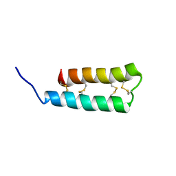
6MBD
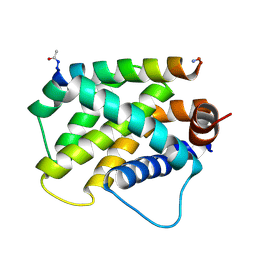
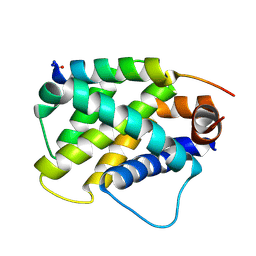
 | | Human Mcl-1 in complex with the designed peptide dM1 | | 分子名称: | Induced myeloid leukemia cell differentiation protein Mcl-1, ZINC ION, dM1 | | 著者 | Jenson, J.M, Keating, A.E. | | 登録日 | 2018-08-29 | | 公開日 | 2019-03-06 | | 最終更新日 | 2024-10-23 | | 実験手法 | X-RAY DIFFRACTION (1.95 Å) | | 主引用文献 | Tertiary Structural Motif Sequence Statistics Enable Facile Prediction and Design of Peptides that Bind Anti-apoptotic Bfl-1 and Mcl-1.
Structure, 27, 2019
|
|
5O7Y
 | | Thebaine 6-O-demethylase (T6ODM) from Papaver somniferum in complex with succinate | | 分子名称: | 1,2-ETHANEDIOL, DI(HYDROXYETHYL)ETHER, NICKEL (II) ION, ... | | 著者 | Kluza, A, Niedzialkowska, E, Kurpiewska, K, Porebski, P.J, Borowski, T. | | 登録日 | 2017-06-10 | | 公開日 | 2018-02-14 | | 最終更新日 | 2024-01-17 | | 実験手法 | X-RAY DIFFRACTION (1.97 Å) | | 主引用文献 | Crystal structure of thebaine 6-O-demethylase from the morphine biosynthesis pathway.
J. Struct. Biol., 202, 2018
|
|
6TOR
 | | human O-phosphoethanolamine phospho-lyase | | 分子名称: | 4'-DEOXY-4'-AMINOPYRIDOXAL-5'-PHOSPHATE, Ethanolamine-phosphate phospho-lyase, GLYCEROL | | 著者 | Vettraino, C, Donini, S, Parisini, E. | | 登録日 | 2019-12-11 | | 公開日 | 2020-04-15 | | 最終更新日 | 2024-01-24 | | 実験手法 | X-RAY DIFFRACTION (2.05 Å) | | 主引用文献 | Structural characterization of human O-phosphoethanolamine phospho-lyase.
Acta Crystallogr.,Sect.F, 76, 2020
|
|
1H0Y
 | | Structure of Alba: an archaeal chromatin protein modulated by acetylation | | 分子名称: | DNA BINDING PROTEIN SSO10B, SULFATE ION | | 著者 | Wardleworth, B.N, Russell, R.J.M, Bell, S.D, Taylor, G.L, White, M.F. | | 登録日 | 2002-07-01 | | 公開日 | 2002-09-05 | | 最終更新日 | 2023-12-13 | | 実験手法 | X-RAY DIFFRACTION (2.8 Å) | | 主引用文献 | Structure of Alba: An Archaeal Chromatin Protein Modulated by Acetylation
Embo J., 21, 2002
|
|
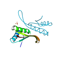
1H0M
 | | Three-dimensional structure of the quorum sensing protein TraR bound to its autoinducer and to its target DNA | | 分子名称: | 3-OXO-OCTANOIC ACID (2-OXO-TETRAHYDRO-FURAN-3-YL)-AMIDE, 5'-D(*AP*TP*GP*TP*GP*CP*AP*GP*AP*TP *CP*TP*GP*CP*AP*CP*AP*T)-3', Transcriptional activator protein TraR | | 著者 | Vannini, A, Volpari, C, Gargioli, C, Muraglia, E, Cortese, R, De Francesco, R, Neddermann, P, Di Marco, S. | | 登録日 | 2002-06-25 | | 公開日 | 2002-08-29 | | 最終更新日 | 2024-11-13 | | 実験手法 | X-RAY DIFFRACTION (3 Å) | | 主引用文献 | The Crystal Structure of the Quorum Sensing Protein Trar Bound to its Autoinducer and Target DNA
Embo J., 21, 2002
|
|


