1TNM
 
 | |
2R15
 | |
2NCM
 | |
1WWB
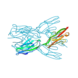
 | | LIGAND BINDING DOMAIN OF HUMAN TRKB RECEPTOR | | 分子名称: | PROTEIN (Brain Derived Neurotrophic Factor Receptor TrkB) | | 著者 | Wiesmann, C, Ultsch, M.H, Bass, S.H, De Vos, A.M. | | 登録日 | 1999-05-03 | | 公開日 | 1999-07-07 | | 最終更新日 | 2023-08-23 | | 実験手法 | X-RAY DIFFRACTION (2.1 Å) | | 主引用文献 | Crystal structures of the neurotrophin-binding domain of TrkA, TrkB and TrkC.
J.Mol.Biol., 290, 1999
|
|
1WVZ
 | |
1X44
 | | Solution structure of the third ig-like domain of Myosin-dinding protein C, slow-type | | 分子名称: | Myosin-binding protein C, slow-type | | 著者 | Qin, X.-R, Kurosaki, C, Hayashi, F, Yoshida, M, Yokoyama, S, RIKEN Structural Genomics/Proteomics Initiative (RSGI) | | 登録日 | 2005-05-13 | | 公開日 | 2005-11-13 | | 最終更新日 | 2024-05-29 | | 実験手法 | SOLUTION NMR | | 主引用文献 | Solution structure of the third ig-like domain of Myosin-dinding protein C, slow-type
to be published
|
|
1WWC
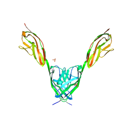
 | | NT3 BINDING DOMAIN OF HUMAN TRKC RECEPTOR | | 分子名称: | PROTEIN (NT-3 GROWTH FACTOR RECEPTOR TRKC) | | 著者 | Ultsch, M.H, Wiesmann, C, Simmons, L.C, Henrich, J, Yang, M, Reilly, D, Bass, S.H, De Vos, A.M. | | 登録日 | 1999-04-30 | | 公開日 | 1999-07-07 | | 最終更新日 | 2023-12-27 | | 実験手法 | X-RAY DIFFRACTION (1.9 Å) | | 主引用文献 | Crystal structures of the neurotrophin-binding domain of TrkA, TrkB and TrkC.
J.Mol.Biol., 290, 1999
|
|
1WIU
 | |
1WIT
 | |
6H4L
 | | Structure of Titin M4 trigonal form | | 分子名称: | CHLORIDE ION, Titin, ZINC ION | | 著者 | Sauer, F, Wilmanns, M. | | 登録日 | 2018-07-21 | | 公開日 | 2019-08-07 | | 最終更新日 | 2020-02-19 | | 実験手法 | X-RAY DIFFRACTION (1.6 Å) | | 主引用文献 | Structural diversity in the atomic resolution 3D fingerprint of the titin M-band segment.
Plos One, 14, 2019
|
|
6FWX
 | | Chimeric titin Z1Z2-Z1Z2 tandem (Z1212) functionalized with a GRGDS exogenous peptide from fibronectin | | 分子名称: | 1,2-ETHANEDIOL, Titin,Titin,Titin | | 著者 | Mayans, O, Fleming, J, Hill, C. | | 登録日 | 2018-03-07 | | 公開日 | 2019-03-27 | | 最終更新日 | 2024-01-17 | | 実験手法 | X-RAY DIFFRACTION (3 Å) | | 主引用文献 | Self-Assembling Proteins as High-Performance Substrates for Embryonic Stem Cell Self-Renewal.
Adv. Mater. Weinheim, 31, 2019
|
|
2A38
 | | Crystal structure of the N-Terminus of titin | | 分子名称: | CADMIUM ION, Titin | | 著者 | Marino, M, Muhle-Goll, C, Svergun, D, Demirel, M, Mayans, O. | | 登録日 | 2005-06-24 | | 公開日 | 2006-06-24 | | 最終更新日 | 2023-10-25 | | 実験手法 | X-RAY DIFFRACTION (2 Å) | | 主引用文献 | The Ig doublet Z1Z2: a model system for the hybrid analysis of conformational dynamics in Ig tandems from titin
Structure, 14, 2006
|
|
4OF7
 | | Crystal Structure of SYG-1 D1, Crystal Form 2 | | 分子名称: | 2-acetamido-2-deoxy-beta-D-glucopyranose, Protein SYG-1, isoform b, ... | | 著者 | Ozkan, E, Garcia, K.C. | | 登録日 | 2014-01-14 | | 公開日 | 2014-02-19 | | 最終更新日 | 2023-09-20 | | 実験手法 | X-RAY DIFFRACTION (2.1 Å) | | 主引用文献 | Extracellular Architecture of the SYG-1/SYG-2 Adhesion Complex Instructs Synaptogenesis.
Cell(Cambridge,Mass.), 156, 2014
|
|
3PUC
 | |
3PXH
 | | Tandem Ig domains of tyrosine phosphatase LAR | | 分子名称: | Receptor-type tyrosine-protein phosphatase F, SULFATE ION | | 著者 | Biersmith, B.H, Bouyain, S. | | 登録日 | 2010-12-09 | | 公開日 | 2011-03-23 | | 最終更新日 | 2023-09-13 | | 実験手法 | X-RAY DIFFRACTION (2.0009 Å) | | 主引用文献 | The Immunoglobulin-like Domains 1 and 2 of the Protein Tyrosine Phosphatase LAR Adopt an Unusual Horseshoe-like Conformation.
J.Mol.Biol., 408, 2011
|
|
3PXJ
 | | Tandem Ig repeats of Dlar | | 分子名称: | Tyrosine-protein phosphatase Lar | | 著者 | Biersmith, B.H, Bouyain, S. | | 登録日 | 2010-12-10 | | 公開日 | 2011-03-23 | | 最終更新日 | 2023-09-13 | | 実験手法 | X-RAY DIFFRACTION (2.3003 Å) | | 主引用文献 | The Immunoglobulin-like Domains 1 and 2 of the Protein Tyrosine Phosphatase LAR Adopt an Unusual Horseshoe-like Conformation.
J.Mol.Biol., 408, 2011
|
|
4OF6
 | | Crystal Structure of SYG-1 D1, Crystal form 1 | | 分子名称: | 1,2-ETHANEDIOL, Protein SYG-1, isoform b, ... | | 著者 | Ozkan, E, Garcia, K.C. | | 登録日 | 2014-01-14 | | 公開日 | 2014-02-19 | | 最終更新日 | 2023-09-20 | | 実験手法 | X-RAY DIFFRACTION (1.696 Å) | | 主引用文献 | Extracellular Architecture of the SYG-1/SYG-2 Adhesion Complex Instructs Synaptogenesis.
Cell(Cambridge,Mass.), 156, 2014
|
|
3Q5O
 | |
2K1M
 | |
2J8O
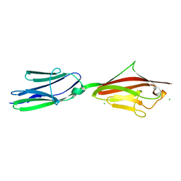
 | | Structure of the immunoglobulin tandem repeat of titin A168-A169 | | 分子名称: | GLYCEROL, TITIN | | 著者 | Mueller, S, Lange, S, Kursula, I, Gautel, M, Wilmanns, M. | | 登録日 | 2006-10-26 | | 公開日 | 2007-08-21 | | 最終更新日 | 2023-12-13 | | 実験手法 | X-RAY DIFFRACTION (2.49 Å) | | 主引用文献 | Rigid Conformation of an Immunoglobulin Domain Tandem Repeat in the A-Band of the Elastic Muscle Protein Titin
J.Mol.Biol., 371, 2007
|
|
2J8H
 | | Structure of the immunoglobulin tandem repeat A168-A169 of titin | | 分子名称: | GLYCEROL, TITIN | | 著者 | Mueller, S, Lange, S, Kursula, I, Gautel, M, Wilmanns, M. | | 登録日 | 2006-10-25 | | 公開日 | 2007-08-21 | | 最終更新日 | 2024-05-08 | | 実験手法 | X-RAY DIFFRACTION (1.99 Å) | | 主引用文献 | Rigid Conformation of an Immunoglobulin Domain Tandem Repeat in the A-Band of the Elastic Muscle Protein Titin
J.Mol.Biol., 371, 2007
|
|
2IEP
 | |
2ILL
 | | Anomalous substructure of Titin-A168169 | | 分子名称: | CHLORIDE ION, Titin | | 著者 | Mueller-Dieckmann, C, Weiss, M.S. | | 登録日 | 2006-10-03 | | 公開日 | 2007-02-20 | | 最終更新日 | 2024-03-13 | | 実験手法 | X-RAY DIFFRACTION (2.2 Å) | | 主引用文献 | On the routine use of soft X-rays in macromolecular crystallography. Part IV. Efficient determination of anomalous substructures in biomacromolecules using longer X-ray wavelengths
ACTA CRYSTALLOGR.,SECT.D, 63, 2007
|
|
2KKQ
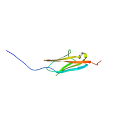 | | Solution NMR Structure of the Ig-like C2-type 2 Domain of Human Myotilin. Northeast Structural Genomics Target HR3158. | | 分子名称: | Myotilin | | 著者 | Rossi, P, Shastry, R, Ciccosanti, C, Hamilton, K, Xiao, R, Acton, T.B, Swapna, G.V.T, Nair, R, Everett, J.K, Rost, B, Montelione, G.T, Northeast Structural Genomics Consortium (NESG) | | 登録日 | 2009-06-29 | | 公開日 | 2009-07-07 | | 最終更新日 | 2024-05-01 | | 実験手法 | SOLUTION NMR | | 主引用文献 | Solution NMR Structure of the Ig-like C2-type 2 Domain of Human Myotilin. Northeast Structural Genomics Target HR3158.
To be Published
|
|
2KDG
 | | Solution Structure of the 1st Ig domain of Myotilin | | 分子名称: | Myotilin | | 著者 | Heikkinen, O, Kilpelainen, I, Permi, P, Koskela, H, Ylanne, J, Carpen, O. | | 登録日 | 2009-01-08 | | 公開日 | 2009-07-21 | | 最終更新日 | 2024-05-29 | | 実験手法 | SOLUTION NMR | | 主引用文献 | Solution structure of the first immunoglobulin domain of human myotilin
J.Biomol.Nmr, 44, 2009
|
|


