Loading
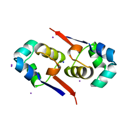
PDBj
メニューPDBj@FacebookPDBj@TwitterPDBj@YouTubewwPDB FoundationwwPDB
RCSB PDBPDBeBMRBAdv. SearchSearch help

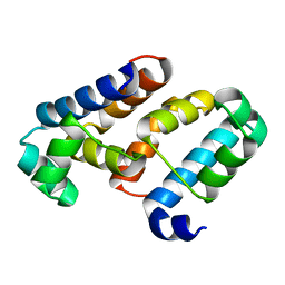
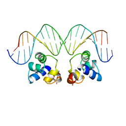
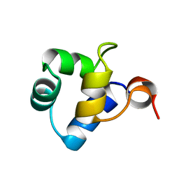
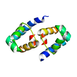
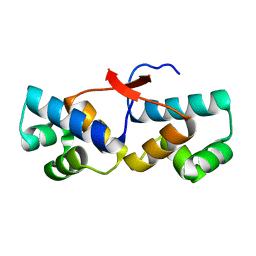
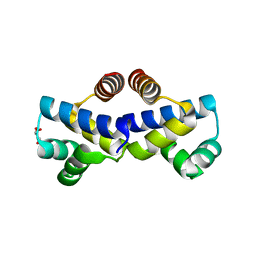
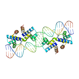
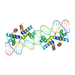
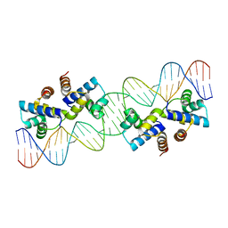
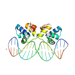
3JXD
DownloadVisualize
BU of 3jxd by Molmil
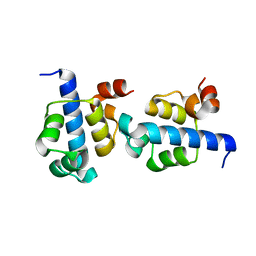
Crystal structure of the P22 c2 repressor protein in complex with synthetic operator 9C in the presence of Rb+
分子名称: 5'-D(*CP*AP*TP*TP*TP*AP*AP*GP*AP*CP*GP*TP*CP*TP*TP*AP*AP*AP*TP*G)-3', RUBIDIUM ION, Repressor protein C2
著者Watkins, D, Koudelka, G.B, Williams, L.D.
登録日2009-09-18
公開日2010-01-19
最終更新日2023-09-06
実験手法X-RAY DIFFRACTION (2.1 Å)
主引用文献Sequence Recognition of DNA by Protein-Induced Conformational Transitions.
J.Mol.Biol., 396, 2010
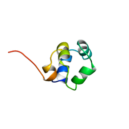
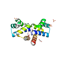
1UTX
DownloadVisualize
BU of 1utx by Molmil
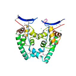
Regulation of Cytolysin Expression by Enterococcus faecalis: Role of CylR2
分子名称: CYLR2, IODIDE ION, SODIUM ION
著者Razeto, A, Rumpel, S, Pillar, C.M, Gilmore, M.S, Becker, S, Zweckstetter, M.
登録日2003-12-12
公開日2004-09-16
最終更新日2024-05-08
実験手法X-RAY DIFFRACTION (1.9 Å)
主引用文献Structure and DNA-Binding Properties of the Cytolysin Regulator CylR2 from Enterococcus Faecalis
Embo J., 23, 2004
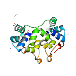
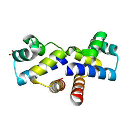
3KZ3
DownloadVisualize
BU of 3kz3 by Molmil
A structure of a lambda repressor fragment mutant
分子名称: Repressor protein CI
著者Gruebele, M, Liu, F, Gao, Y.
登録日2009-12-07
公開日2010-02-23
最終更新日2024-02-21
実験手法X-RAY DIFFRACTION (1.64 Å)
主引用文献A survey of lambda repressor fragments from two-state to downhill folding.
J.Mol.Biol., 397, 2010
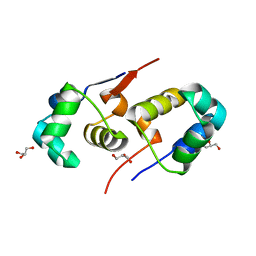
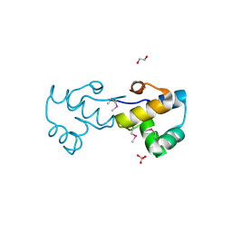
3KXA
DownloadVisualize
BU of 3kxa by Molmil
Crystal Structure of NGO0477 from Neisseria gonorrhoeae
分子名称: ASPARAGINE, CHLORIDE ION, Putative uncharacterized protein, ...
著者Ren, J, Sainsbury, S, Nettleship, J.E, Owens, R.J, Oxford Protein Production Facility (OPPF)
登録日2009-12-02
公開日2010-01-19
最終更新日2011-07-13
実験手法X-RAY DIFFRACTION (2.8 Å)
主引用文献The crystal structure of NGO0477 from Neisseria gonorrhoeae reveals a novel protein fold incorporating a helix-turn-helix motif.
Proteins, 78, 2010
6B9T
DownloadVisualize
BU of 6b9t by Molmil
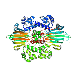
Crystal structure of MPnS with substrate 2-hydroxyethylphosphonate (2-HEP) and Fe(II) bound
分子名称: (2-hydroxyethyl)phosphonic acid, FE (III) ION, FORMIC ACID, ...
著者Born, D.A, Drennan, C.L.
登録日2017-10-11
公開日2017-12-20
最終更新日2024-03-13
実験手法X-RAY DIFFRACTION (2.35 Å)
主引用文献Structural basis for methylphosphonate biosynthesis.
Science, 358, 2017
6B9S
DownloadVisualize
BU of 6b9s by Molmil
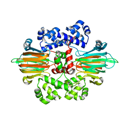
MPnS crystallized in the absence of substrate
分子名称: FE (III) ION, Methylphosphonate synthase
著者Born, D.A, Drennan, C.L.
登録日2017-10-11
公開日2017-12-20
最終更新日2024-03-13
実験手法X-RAY DIFFRACTION (2.373 Å)
主引用文献Structural basis for methylphosphonate biosynthesis.
Science, 358, 2017
3EUS
DownloadVisualize
BU of 3eus by Molmil
The crystal structure of the DNA binding protein from Silicibacter pomeroyi
分子名称: DNA-binding protein
著者Zhang, R, Li, H, Freeman, L, Joachimiak, A, Midwest Center for Structural Genomics (MCSG)
登録日2008-10-10
公開日2008-11-25
最終更新日2023-12-27
実験手法X-RAY DIFFRACTION (1.8 Å)
主引用文献The crystal structure of the DNA binding protein from Silicibacter pomeroyi
To be Published
1RPE
DownloadVisualize
BU of 1rpe by Molmil
THE PHAGE 434 OR2/R1-69 COMPLEX AT 2.5 ANGSTROMS RESOLUTION
分子名称: DNA (5'-D(*AP*CP*AP*AP*AP*CP*AP*AP*GP*AP*TP*AP*CP*AP*TP*TP*G P*TP*AP*T)-3'), DNA (5'-D(*TP*AP*TP*AP*CP*AP*AP*TP*GP*TP*AP*TP*CP*TP*TP*GP*T P*TP*TP*G)-3'), PROTEIN (434 REPRESSOR)
著者Shimon, L.J.W, Harrison, S.C.
登録日1993-03-24
公開日1994-01-31
最終更新日2024-02-14
実験手法X-RAY DIFFRACTION (2.5 Å)
主引用文献The phage 434 OR2/R1-69 complex at 2.5 A resolution.
J.Mol.Biol., 232, 1993
1R63
DownloadVisualize
BU of 1r63 by Molmil
STRUCTURAL ROLE OF A BURIED SALT BRIDGE IN THE 434 REPRESSOR DNA-BINDING DOMAIN, NMR, 20 STRUCTURES
分子名称: REPRESSOR PROTEIN FROM BACTERIOPHAGE 434
著者Pervushin, K.V, Billeter, M, Siegal, G, Wuthrich, K.
登録日1996-11-08
公開日1997-06-16
最終更新日2024-05-22
実験手法SOLUTION NMR
主引用文献Structural role of a buried salt bridge in the 434 repressor DNA-binding domain.
J.Mol.Biol., 264, 1996
1Y7Y
DownloadVisualize
BU of 1y7y by Molmil
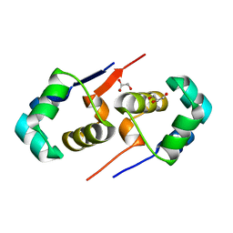
High-resolution crystal structure of the restriction-modification controller protein C.AhdI from Aeromonas hydrophila
分子名称: C.AhdI
著者McGeehan, J.E, Streeter, S.D, Papapanagiotou, I, Fox, G.C, Kneale, G.G.
登録日2004-12-10
公開日2005-02-22
最終更新日2024-04-03
実験手法X-RAY DIFFRACTION (1.69 Å)
主引用文献High-resolution crystal structure of the restriction-modification controller protein C.AhdI from Aeromonas hydrophila.
J.Mol.Biol., 346, 2005
4PU7
DownloadVisualize
BU of 4pu7 by Molmil
Shewanella oneidensis Toxin Antitoxin System Antitoxin Protein HipB Resolution 1.85
分子名称: Toxin-antitoxin system antidote transcriptional repressor Xre family
著者Wen, Y, Behiels, E, Felix, J, Elegheert, J, Vergauwen, B, Devreese, B, Savvides, S.
登録日2014-03-12
公開日2014-08-06
最終更新日2023-09-20
実験手法X-RAY DIFFRACTION (1.85 Å)
主引用文献The bacterial antitoxin HipB establishes a ternary complex with operator DNA and phosphorylated toxin HipA to regulate bacterial persistence.
Nucleic Acids Res., 42, 2014
1ZUG
DownloadVisualize
BU of 1zug by Molmil
STRUCTURE OF PHAGE 434 CRO PROTEIN, NMR, 20 STRUCTURES
分子名称: PHAGE 434 CRO PROTEIN
著者Padmanabhan, S, Jimenez, M.A, Gonzalez, C, Sanz, J.M, Gimenez-Gallego, G, Rico, M.
登録日1997-03-14
公開日1997-07-07
最終更新日2024-05-22
実験手法SOLUTION NMR
主引用文献Three-dimensional solution structure and stability of phage 434 Cro protein.
Biochemistry, 36, 1997
2XCJ
DownloadVisualize
BU of 2xcj by Molmil
Crystal structure of P2 C, the immunity repressor of temperate E. coli phage P2
分子名称: C PROTEIN, FORMIC ACID, GLYCEROL, ...
著者Massad, T, Skaar, K, Hogbom, M, Stenmark, P.
登録日2010-04-23
公開日2010-07-28
最終更新日2024-05-08
実験手法X-RAY DIFFRACTION (1.8 Å)
主引用文献Crystal Structure of the P2 C-Repressor: A Binder of Non-Palindromic Direct DNA Repeats.
Nucleic Acids Res., 38, 2010
2XI8
DownloadVisualize
BU of 2xi8 by Molmil
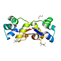
High resolution structure of native CylR2
分子名称: GLYCEROL, PUTATIVE TRANSCRIPTION REGULATOR
著者Gruene, T, Cho, M.-K, Karyagina, I, Kim, H.-Y, Grosse, C, Giller, K, Zweckstetter, M, Becker, S.
登録日2010-06-28
公開日2011-02-09
最終更新日2023-12-20
実験手法X-RAY DIFFRACTION (1.21 Å)
主引用文献Integrated Analysis of the Conformation of a Protein-Linked Spin Label by Crystallography, Epr and NMR Spectroscopy.
J.Biomol.NMR, 49, 2011
2XIU
DownloadVisualize
BU of 2xiu by Molmil
High resolution structure of MTSL-tagged CylR2.
分子名称: CYLR2, GLYCEROL, S-[(1-oxyl-2,2,5,5-tetramethyl-2,5-dihydro-1H-pyrrol-3-yl)methyl] methanesulfonothioate
著者Gruene, T, Cho, M.-K, Karyagina, I, Kim, H.-Y, Grosse, C, Giller, K, Zweckstetter, M, Becker, S.
登録日2010-07-01
公開日2011-02-09
最終更新日2023-12-20
実験手法X-RAY DIFFRACTION (1.5 Å)
主引用文献Integrated Analysis of the Conformation of a Protein-Linked Spin Label by Crystallography, Epr and NMR Spectroscopy.
J.Biomol.NMR, 49, 2011
4F8D
DownloadVisualize
BU of 4f8d by Molmil
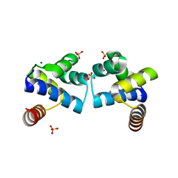
Crystal Structure of an R46A mutant of the Restriction-Modification Controller Protein C.Esp1396I (Monoclinic Form)
分子名称: Regulatory protein, SODIUM ION, SULFATE ION
著者Martin, R.N.A, McGeehan, J.E, Kneale, G.G.
登録日2012-05-17
公開日2013-04-10
最終更新日2024-02-28
実験手法X-RAY DIFFRACTION (1.5 Å)
主引用文献Structural and Mutagenic Analysis of the RM Controller Protein C.Esp1396I.
Plos One, 9, 2014
2XJ3
DownloadVisualize
BU of 2xj3 by Molmil
High resolution structure of the T55C mutant of CylR2.
分子名称: CYLR2 SYNONYM CYTOLYSIN REPRESSOR 2, GLYCEROL
著者Gruene, T, Cho, M.K, Karyagina, I, Kim, H.Y, Grosse, C, Giller, K, Zweckstetter, M, Becker, S.
登録日2010-07-02
公開日2011-02-09
最終更新日2023-12-20
実験手法X-RAY DIFFRACTION (1.23 Å)
主引用文献Integrated Analysis of the Conformation of a Protein-Linked Spin Label by Crystallography, Epr and NMR Spectroscopy.
J.Biomol.NMR, 49, 2011
4FN3
DownloadVisualize
BU of 4fn3 by Molmil
Crystal Structure of an S52A mutant of the Restriction-Modification Controller Protein C.Esp1396I
分子名称: Regulatory protein, SULFATE ION
著者Martin, R.N.A, McGeehan, J.E, Kneale, G.G.
登録日2012-06-19
公開日2013-05-29
最終更新日2024-02-28
実験手法X-RAY DIFFRACTION (1.79 Å)
主引用文献Structural and Mutagenic Analysis of the RM Controller Protein C.Esp1396I.
Plos One, 9, 2014
4FBI
DownloadVisualize
BU of 4fbi by Molmil
Crystal Structure of an R46A mutant of the Restriction-Modification Controller Protein C.Esp1396I (Trigonal Form)
分子名称: GLYCEROL, Regulatory protein
著者Martin, R.N.A, McGeehan, J.E, Kneale, G.G.
登録日2012-05-23
公開日2013-04-10
最終更新日2024-02-28
実験手法X-RAY DIFFRACTION (1.5 Å)
主引用文献Structural and Mutagenic Analysis of the RM Controller Protein C.Esp1396I.
Plos One, 9, 2014
4I6R
DownloadVisualize
BU of 4i6r by Molmil
High Resolution Crystal Structure of the Wild-Type Restriction-Modification Controller Protein C.Esp1396I (Triclinic form)
分子名称: GLYCEROL, Regulatory protein, SULFATE ION
著者Martin, R.N.A, McGeehan, J.E, Kneale, G.G.
登録日2012-11-30
公開日2013-11-13
最終更新日2023-09-20
実験手法X-RAY DIFFRACTION (1.38 Å)
主引用文献Structural and Mutagenic Analysis of the RM Controller Protein C.Esp1396I.
Plos One, 9, 2014
4IA8
DownloadVisualize
BU of 4ia8 by Molmil
Crystal Structure of a Y37A mutant of the Restriction-Modification Controller Protein C.Esp1396I
分子名称: Regulatory protein, SULFATE ION
著者Martin, R.N.A, McGeehan, J.E, Kneale, G.G.
登録日2012-12-06
公開日2013-12-11
最終更新日2023-09-20
実験手法X-RAY DIFFRACTION (1.85 Å)
主引用文献Structural and Mutagenic Analysis of the RM Controller Protein C.Esp1396I.
Plos One, 9, 2014
3BS3
DownloadVisualize
BU of 3bs3 by Molmil
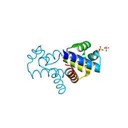
Crystal structure of a putative DNA-binding protein from Bacteroides fragilis
分子名称: 1,2-ETHANEDIOL, Putative DNA-binding protein, SULFATE ION
著者Cuff, M.E, Bigelow, L, Clancy, S, Joachimiak, A, Midwest Center for Structural Genomics (MCSG)
登録日2007-12-21
公開日2008-01-15
最終更新日2017-10-25
実験手法X-RAY DIFFRACTION (1.65 Å)
主引用文献The structure of a putative DNA-binding protein from Bacteroides fragilis.
TO BE PUBLISHED
4X4E
DownloadVisualize
BU of 4x4e by Molmil
RADIATION DAMAGE TO THE NUCLEOPROTEIN COMPLEX C.Esp1396I: DOSE (DWD) 14.4 MGy
分子名称: 35-MER DNA, Regulatory protein
著者Bury, C.S, McGeehan, J.E, Garman, E.F.
登録日2014-12-02
公開日2015-03-11
最終更新日2024-01-10
実験手法X-RAY DIFFRACTION (2.8 Å)
主引用文献Radiation damage to nucleoprotein complexes in macromolecular crystallography.
J.Synchrotron Radiat., 22, 2015
4X4I
DownloadVisualize
BU of 4x4i by Molmil
RADIATION DAMAGE TO THE NUCLEOPROTEIN COMPLEX C.Esp1396I: DOSE (DWD) 44.6 MGy
分子名称: 35-MER DNA, Regulatory protein
著者Bury, C.S, McGeehan, J.E, Garman, E.F.
登録日2014-12-02
公開日2015-03-11
最終更新日2024-01-10
実験手法X-RAY DIFFRACTION (2.8 Å)
主引用文献Radiation damage to nucleoprotein complexes in macromolecular crystallography.
J.Synchrotron Radiat., 22, 2015
4X4G
DownloadVisualize
BU of 4x4g by Molmil
RADIATION DAMAGE TO THE NUCLEOPROTEIN COMPLEX C.Esp1396I: DOSE (DWD) 26.8 MGy
分子名称: 35-MER DNA, Regulatory protein
著者Bury, C.S, McGeehan, J.E, Garman, E.F.
登録日2014-12-02
公開日2015-03-11
最終更新日2024-01-10
実験手法X-RAY DIFFRACTION (2.8 Å)
主引用文献Radiation damage to nucleoprotein complexes in macromolecular crystallography.
J.Synchrotron Radiat., 22, 2015

223532

件を2024-08-07に公開中

PDB statisticsPDBj update infoContact PDBjnumon