Loading
PDBj
メニューPDBj@FacebookPDBj@TwitterPDBj@YouTubewwPDB FoundationwwPDB
RCSB PDBPDBeBMRBAdv. SearchSearch help

4F16
DownloadVisualize
BU of 4f16 by Molmil
Crystal Structure of the alpha spectrin SH3 domain at pH 5
分子名称: Spectrin alpha chain, brain
著者Camara-Artigas, A, Gavira, J.A.
登録日2012-05-06
公開日2013-05-08
最終更新日2023-09-13
実験手法X-RAY DIFFRACTION (1.93 Å)
主引用文献Crystal Structure of the alpha spectrin SH3 domain at pH 5
To be Published
4F17
DownloadVisualize
BU of 4f17 by Molmil
Crystal Structure of the alpha spectrin SH3 domain at pH 9
分子名称: Spectrin alpha chain, brain
著者Camara-Artigas, A, Gavira, J.A.
登録日2012-05-06
公開日2013-05-08
最終更新日2023-09-13
実験手法X-RAY DIFFRACTION (1.55 Å)
主引用文献Crystal Structure of the alpha spectrin SH3 domain at pH 9
To be Published
5ZAU
DownloadVisualize
BU of 5zau by Molmil
Complex of the human FYN SH3 and monobody binder
分子名称: Monobody Binder, Tyrosine-protein kinase Fyn
著者Reddy, P.P, Gulyani, A, Das, R.
登録日2018-02-09
公開日2019-09-11
最終更新日2024-05-15
実験手法SOLUTION NMR
主引用文献A Fyn biosensor reveals pulsatile, spatially localized kinase activity and signaling crosstalk in live mammalian cells.
Elife, 9, 2020
6A9C
DownloadVisualize
BU of 6a9c by Molmil
Crystal Structure c-terminal SH3 domain of Myosin IB from Entamoeba histolytica bound to EhFP10(GEF) peptide.
分子名称: Peptide from Rho guanine nucleotide exchange factor, SULFATE ION, Unconventional myosin IB
著者Gautam, G, Gourinath, S.
登録日2018-07-13
公開日2019-06-12
最終更新日2023-11-22
実験手法X-RAY DIFFRACTION (1.98 Å)
主引用文献EhFP10: A FYVE family GEF interacts with myosin IB to regulate cytoskeletal dynamics during endocytosis in Entamoeba histolytica.
Plos Pathog., 15, 2019
6XVO
DownloadVisualize
BU of 6xvo by Molmil
Crystal structure of the intertwined dimer of the c-Src SH3 domain without ATCUN motif
分子名称: DI(HYDROXYETHYL)ETHER, Proto-oncogene tyrosine-protein kinase Src, TRIETHYLENE GLYCOL
著者Camara-Artigas, A, Plaza-Garrido, M.
登録日2020-01-22
公開日2020-04-22
最終更新日2024-01-24
実験手法X-RAY DIFFRACTION (1.7 Å)
主引用文献The effect of an engineered ATCUN motif on the structure and biophysical properties of the SH3 domain of c-Src tyrosine kinase.
J.Biol.Inorg.Chem., 25, 2020
6XX2
DownloadVisualize
BU of 6xx2 by Molmil
Crystal structure of the c-Src SH3 domain H122R-Q128K mutant in complex with Cu(II) at pH 7.5 co-crystallized with methyl beta-cyclodextrin
分子名称: COPPER (II) ION, Cyclic 2,3-di-O-methyl-alpha-D-glucopyranose-(1-4)-2-O-methyl-alpha-D-glucopyranose-(1-4)-2,6-di-O-methyl-alpha-D-glucopyranose-(1-4)-2-O-methyl-alpha-D-glucopyranose-(1-4)-alpha-D-glucopyranose-(1-4)-alpha-D-glucopyranose-(1-4)-3-O-methyl-alpha-D-glucopyranose, Proto-oncogene tyrosine-protein kinase Src
著者Camara-Artigas, A.
登録日2020-01-26
公開日2020-04-22
最終更新日2024-01-24
実験手法X-RAY DIFFRACTION (1.25 Å)
主引用文献The effect of an engineered ATCUN motif on the structure and biophysical properties of the SH3 domain of c-Src tyrosine kinase.
J.Biol.Inorg.Chem., 25, 2020
6ATV
DownloadVisualize
BU of 6atv by Molmil
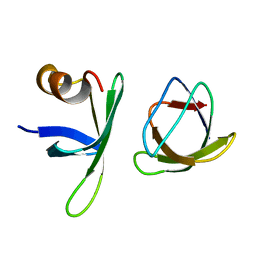
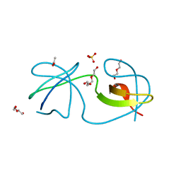
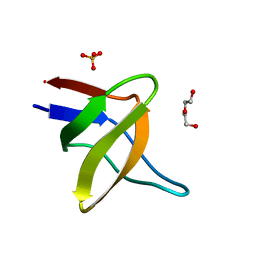
The molecular mechanisms by which NS1 of the 1918 Spanish influenza A virus hijack host protein-protein interactions
分子名称: Adapter molecule crk, proline-rich motif in IAV-NS1
著者Shen, Q, Zeng, D, Zhao, B, Li, P, Cho, J.H.
登録日2017-08-29
公開日2018-08-08
最終更新日2023-10-04
実験手法X-RAY DIFFRACTION (1.751 Å)
主引用文献Molecular Mechanisms of Tight Binding through Fuzzy Interactions.
Biophys. J., 114, 2018
6XX4
DownloadVisualize
BU of 6xx4 by Molmil
Crystal structure of the c-Src SH3 domain H122R-Q128E mutant in complex with Ni(II) at pH 7.5 co-crystallized with methyl beta-cyclodextrin
分子名称: Cyclic 2,3-di-O-methyl-alpha-D-glucopyranose-(1-4)-2-O-methyl-alpha-D-glucopyranose-(1-4)-2,6-di-O-methyl-alpha-D-glucopyranose-(1-4)-2-O-methyl-alpha-D-glucopyranose-(1-4)-alpha-D-glucopyranose-(1-4)-alpha-D-glucopyranose-(1-4)-3-O-methyl-alpha-D-glucopyranose, NICKEL (II) ION, Proto-oncogene tyrosine-protein kinase Src
著者Camara-Artigas, A.
登録日2020-01-26
公開日2020-04-22
最終更新日2024-01-24
実験手法X-RAY DIFFRACTION (1.05 Å)
主引用文献The effect of an engineered ATCUN motif on the structure and biophysical properties of the SH3 domain of c-Src tyrosine kinase.
J.Biol.Inorg.Chem., 25, 2020
6XVN
DownloadVisualize
BU of 6xvn by Molmil
Crystal structure of c-Src SH3 domain without ATCUN motif: monomer 1
分子名称: Proto-oncogene tyrosine-protein kinase Src
著者Camara-Artigas, A, Plaza-Garrido, M.
登録日2020-01-22
公開日2020-04-22
最終更新日2024-01-24
実験手法X-RAY DIFFRACTION (1.7 Å)
主引用文献The effect of an engineered ATCUN motif on the structure and biophysical properties of the SH3 domain of c-Src tyrosine kinase.
J.Biol.Inorg.Chem., 25, 2020
6XVM
DownloadVisualize
BU of 6xvm by Molmil
Crystal structure of c-Src SH3 domain without ATCUN motif: monomer 2
分子名称: GLYCEROL, Proto-oncogene tyrosine-protein kinase Src
著者Camara-Artigas, A, Plaza-Garrido, M.
登録日2020-01-22
公開日2020-04-22
最終更新日2024-01-24
実験手法X-RAY DIFFRACTION (0.9 Å)
主引用文献The effect of an engineered ATCUN motif on the structure and biophysical properties of the SH3 domain of c-Src tyrosine kinase.
J.Biol.Inorg.Chem., 25, 2020
6XX3
DownloadVisualize
BU of 6xx3 by Molmil
Crystal structure of the c-Src SH3 domain H122R-Q128E mutant in complex with Cu(II) at pH 6.5 co-crystallized with methyl beta-cyclodextrin
分子名称: COPPER (II) ION, Cyclic 2,3-di-O-methyl-alpha-D-glucopyranose-(1-4)-2-O-methyl-alpha-D-glucopyranose-(1-4)-2,6-di-O-methyl-alpha-D-glucopyranose-(1-4)-2-O-methyl-alpha-D-glucopyranose-(1-4)-alpha-D-glucopyranose-(1-4)-alpha-D-glucopyranose-(1-4)-3-O-methyl-alpha-D-glucopyranose, Proto-oncogene tyrosine-protein kinase Src
著者Camara-Artigas, A.
登録日2020-01-26
公開日2020-04-22
最終更新日2024-01-24
実験手法X-RAY DIFFRACTION (1.36 Å)
主引用文献The effect of an engineered ATCUN motif on the structure and biophysical properties of the SH3 domain of c-Src tyrosine kinase.
J.Biol.Inorg.Chem., 25, 2020
6XX5
DownloadVisualize
BU of 6xx5 by Molmil
Crystal structure of the c-Src SH3 domain H122R-Q128K mutant in complex with Ni(II) at pH 7.5 co-crystallized with methyl beta-cyclodextrin
分子名称: Cyclic 2,3-di-O-methyl-alpha-D-glucopyranose-(1-4)-2-O-methyl-alpha-D-glucopyranose-(1-4)-2,6-di-O-methyl-alpha-D-glucopyranose-(1-4)-2-O-methyl-alpha-D-glucopyranose-(1-4)-alpha-D-glucopyranose-(1-4)-alpha-D-glucopyranose-(1-4)-3-O-methyl-alpha-D-glucopyranose, NICKEL (II) ION, Proto-oncogene tyrosine-protein kinase Src
著者Camara-Artigas, A.
登録日2020-01-26
公開日2020-05-27
最終更新日2024-01-24
実験手法X-RAY DIFFRACTION (1.3 Å)
主引用文献The effect of an engineered ATCUN motif on the structure and biophysical properties of the SH3 domain of c-Src tyrosine kinase.
J.Biol.Inorg.Chem., 25, 2020
6C4S
DownloadVisualize
BU of 6c4s by Molmil
Human cSrc SH3 Domain in complex with Choline Kinase fragment 60-69
分子名称: Proto-oncogene tyrosine-protein kinase Src,cSrc SH3 Domain, ZINC ION
著者Kall, S.L, Lavie, A.
登録日2018-01-12
公開日2019-01-23
最終更新日2024-03-13
実験手法X-RAY DIFFRACTION (1.5 Å)
主引用文献Molecular basis for the interaction between human choline kinase alpha and the SH3 domain of the c-Src tyrosine kinase.
Sci Rep, 9, 2019
5HCK
DownloadVisualize
BU of 5hck by Molmil
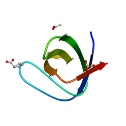
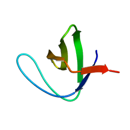
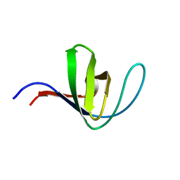
HUMAN HCK SH3 DOMAIN, NMR, MINIMIZED AVERAGE STRUCTURE
分子名称: HEMATOPOIETIC CELL KINASE
著者Horita, D.A, Baldisseri, D.M, Zhang, W, Altieri, A.S, Smithgall, T.E, Gmeiner, W.H, Byrd, R.A.
登録日1998-03-09
公開日1998-06-17
最終更新日2024-05-01
実験手法SOLUTION NMR
主引用文献Solution structure of the human Hck SH3 domain and identification of its ligand binding site.
J.Mol.Biol., 278, 1998
5I11
DownloadVisualize
BU of 5i11 by Molmil
Crystal structure of the intertwined form of the Src tyrosine kinase SH3 domain T114S-Q128R mutant
分子名称: ACETATE ION, DI(HYDROXYETHYL)ETHER, Proto-oncogene tyrosine-protein kinase Src, ...
著者Camara-Artigas, A.
登録日2016-02-05
公開日2016-03-09
最終更新日2024-01-10
実験手法X-RAY DIFFRACTION (1.95 Å)
主引用文献Crystal structure of the intertwined form of the Src tyrosine kinase SH3 domain T114S-Q128R mutant
To Be Published
5IHK
DownloadVisualize
BU of 5ihk by Molmil
Crystal Structure of the alpha spectrin SH3 domain mutant N47A
分子名称: ACETATE ION, PROLINE, SULFATE ION, ...
著者Camara-Artigas, A.
登録日2016-02-29
公開日2017-03-29
最終更新日2024-01-10
実験手法X-RAY DIFFRACTION (1.35 Å)
主引用文献Crystal Structure of the alpha spectrin SH3 domain mutant N47A
to be published
5IHN
DownloadVisualize
BU of 5ihn by Molmil
Crystal Structure of the alpha spectrin SH3 domain mutant N47G
分子名称: FORMIC ACID, SODIUM ION, Spectrin alpha chain, ...
著者Camara-Artigas, A.
登録日2016-02-29
公開日2017-03-29
最終更新日2024-01-10
実験手法X-RAY DIFFRACTION (1.5 Å)
主引用文献Crystal Structure of the alpha spectrin SH3 domain mutant N47G
to be published
5IH2
DownloadVisualize
BU of 5ih2 by Molmil
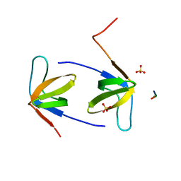
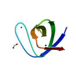
Structure, thermodynamics, and the role of conformational dynamics in the interactions between the N-terminal SH3 domain of CrkII and proline-rich motifs in cAbl
分子名称: 1-ETHOXY-2-(2-ETHOXYETHOXY)ETHANE, Adapter molecule crk, DI(HYDROXYETHYL)ETHER, ...
著者Bhatt, V.S, Zeng, D, Krieger, I, Sacchettini, J.C, Cho, J.-H.
登録日2016-02-28
公開日2016-06-29
最終更新日2023-09-27
実験手法X-RAY DIFFRACTION (1.8 Å)
主引用文献Binding Mechanism of the N-Terminal SH3 Domain of CrkII and Proline-Rich Motifs in cAbl.
Biophys.J., 110, 2016
5IHI
DownloadVisualize
BU of 5ihi by Molmil
Crystal Structure of the alpha spectrin SH3 domain double mutant V46G-D48G
分子名称: DI(HYDROXYETHYL)ETHER, SULFATE ION, Spectrin alpha chain, ...
著者Camara-Artigas, A.
登録日2016-02-29
公開日2017-03-29
最終更新日2024-01-10
実験手法X-RAY DIFFRACTION (1.45 Å)
主引用文献Crystal Structure of the alpha spectrin SH3 domain double mutant V46G-D48G
to be published
3M0P
DownloadVisualize
BU of 3m0p by Molmil
Crystal Structure of the R21D mutant of alpha-spectrin SH3 domain. Crystal obtained in ammonium sulphate at pH 4.
分子名称: SULFATE ION, Spectrin alpha chain, brain
著者Camara-Artigas, A, Gavira, J.A.
登録日2010-03-03
公開日2011-01-19
最終更新日2023-09-06
実験手法X-RAY DIFFRACTION (2.6 Å)
主引用文献Understanding the polymorphic behaviour of a mutant of the alpha-spectrin SH3 domain by means of two 1.1 A structures
Acta Crystallogr.,Sect.D, 2011
3M0S
DownloadVisualize
BU of 3m0s by Molmil
Crystal Structure of the R21D mutant of alpha-spectrin SH3 domain. Crystal obtained in ammonium sulphate at pH 7
分子名称: SULFATE ION, Spectrin alpha chain, brain
著者Camara-Artigas, A, Gavira, J.A.
登録日2010-03-03
公開日2011-01-19
最終更新日2023-09-06
実験手法X-RAY DIFFRACTION (1.6 Å)
主引用文献Understanding the polymorphic behaviour of a mutant of the alpha-spectrin SH3 domain by means of two 1.1 A structures
Acta Crystallogr.,Sect.D, 2011
3M0U
DownloadVisualize
BU of 3m0u by Molmil
Crystal Structure of the R21D mutant of alpha-spectrin SH3 domain. Hexagonal crystal obtained in sodium formate at pH 6.5.
分子名称: FORMIC ACID, Spectrin alpha chain, brain
著者Gavira, J.A, Camara-Artigas, A.
登録日2010-03-03
公開日2011-01-19
最終更新日2023-09-06
実験手法X-RAY DIFFRACTION (1.1 Å)
主引用文献Understanding the polymorphic behaviour of a mutant of the alpha-spectrin SH3 domain by means of two 1.1 A structures
Acta Crystallogr.,Sect.D, 2011
3M0Q
DownloadVisualize
BU of 3m0q by Molmil
Crystal Structure of the R21D mutant of alpha-spectrin SH3 domain. Crystal obtained in ammonium sulphate at pH 5.
分子名称: SULFATE ION, Spectrin alpha chain, brain
著者Camara-Artigas, A, Gavira, J.A.
登録日2010-03-03
公開日2011-01-19
最終更新日2023-09-06
実験手法X-RAY DIFFRACTION (1.75 Å)
主引用文献Understanding the polymorphic behaviour of a mutant of the alpha-spectrin SH3 domain by means of two 1.1 A structures
Acta Crystallogr.,Sect.D, 2011
3M0R
DownloadVisualize
BU of 3m0r by Molmil
Crystal Structure of the R21D mutant of alpha-spectrin SH3 domain. Crystal obtained in ammonium sulphate at pH 6.
分子名称: SULFATE ION, Spectrin alpha chain, brain
著者Gavira, J.A, Camara-Artigas, A.
登録日2010-03-03
公開日2011-01-19
最終更新日2023-09-06
実験手法X-RAY DIFFRACTION (1.1 Å)
主引用文献Understanding the polymorphic behaviour of a mutant of the alpha-spectrin SH3 domain by means of two 1.1 A structures
Acta Crystallogr.,Sect.D, 2011
3NGP
DownloadVisualize
BU of 3ngp by Molmil
High resolution structure of alpha-spectrin SH3 domain mutant with a redesigned core
分子名称: Spectrin alpha chain, brain
著者Camara-Artigas, A, Gavira, J.A, Martin-Garcia, J.M.
登録日2010-06-12
公開日2010-09-08
最終更新日2023-09-06
実験手法X-RAY DIFFRACTION (1.084 Å)
主引用文献High-resolution structure of an alpha-spectrin SH3-domain mutant with a redesigned hydrophobic core.
Acta Crystallogr.,Sect.F, 66, 2010

222415

件を2024-07-10に公開中

PDB statisticsPDBj update infoContact PDBjnumon