1NF3
 
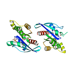
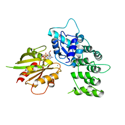
 | | Structure of Cdc42 in a complex with the GTPase-binding domain of the cell polarity protein, Par6 | | 分子名称: | G25K GTP-binding protein, placental isoform, MAGNESIUM ION, ... | | 著者 | Garrard, S.M, Capaldo, C.T, Gao, L, Rosen, M.K, Macara, I.G, Tomchick, D.R. | | 登録日 | 2002-12-12 | | 公開日 | 2003-03-04 | | 最終更新日 | 2023-08-16 | | 実験手法 | X-RAY DIFFRACTION (2.1 Å) | | 主引用文献 | Structure of Cdc42 in a complex with the GTPase-binding domain of the cell polarity protein, Par6
Embo J., 22, 2003
|
|
1B4S
 | |
7PKN
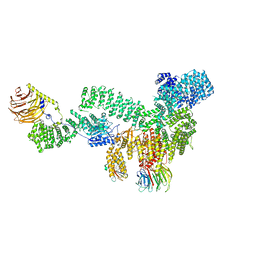
 | | Structure of the human CCAN deltaCT complex | | 分子名称: | Centromere protein H, Centromere protein I, Centromere protein K, ... | | 著者 | Muir, K.W, Yatskevich, S, Bellini, D, Barford, D. | | 登録日 | 2021-08-25 | | 公開日 | 2022-04-27 | | 最終更新日 | 2022-06-01 | | 実験手法 | ELECTRON MICROSCOPY (3.2 Å) | | 主引用文献 | Structure of the human inner kinetochore bound to a centromeric CENP-A nucleosome.
Science, 376, 2022
|
|
1BUX
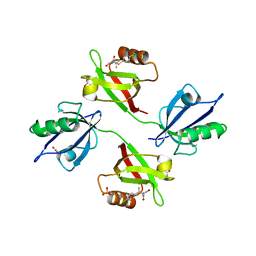
 | | 3'-PHOSPHORYLATED NUCLEOTIDES BINDING TO NUCLEOSIDE DIPHOSPHATE KINASE | | 分子名称: | 3'-PHOSPHATE-ADENOSINE-5'-PHOSPHATE SULFATE, NUCLEOSIDE DIPHOSPHATE KINASE | | 著者 | Xu, Y, Schneider, B, Deville-Bonne, D, Veron, M, Janin, J. | | 登録日 | 1998-09-07 | | 公開日 | 1999-04-27 | | 最終更新日 | 2024-02-07 | | 実験手法 | X-RAY DIFFRACTION (2.8 Å) | | 主引用文献 | 3'-Phosphorylated nucleotides are tight binding inhibitors of nucleoside diphosphate kinase activity.
J.Biol.Chem., 273, 1998
|
|
1AGI
 | |
7QOO
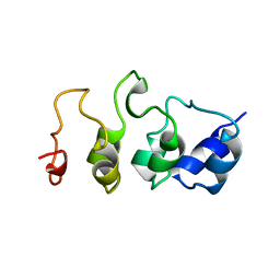
 | | Structure of the human inner kinetochore CCAN complex | | 分子名称: | Centromere protein C, Centromere protein H, Centromere protein I, ... | | 著者 | Vetter, I.R, Pesenti, M, Raisch, T. | | 登録日 | 2021-12-24 | | 公開日 | 2022-06-08 | | 最終更新日 | 2022-06-15 | | 実験手法 | ELECTRON MICROSCOPY (4.6 Å) | | 主引用文献 | Structure of the human inner kinetochore CCAN complex and its significance for human centromere organization.
Mol.Cell, 82, 2022
|
|
6R3V
 | | Crystal Structure of RhoA-GDP-Pi in Complex with RhoGAP | | 分子名称: | 2,3-DIHYDROXY-1,4-DITHIOBUTANE, 2-(N-MORPHOLINO)-ETHANESULFONIC ACID, GUANOSINE-5'-DIPHOSPHATE, ... | | 著者 | Jin, Y. | | 登録日 | 2019-03-21 | | 公開日 | 2019-05-08 | | 最終更新日 | 2024-01-24 | | 実験手法 | X-RAY DIFFRACTION (1.75 Å) | | 主引用文献 | A GAP-GTPase-GDP-PiIntermediate Crystal Structure Analyzed by DFT Shows GTP Hydrolysis Involves Serial Proton Transfers.
Chemistry, 25, 2019
|
|
1CD9
 | | 2:2 COMPLEX OF G-CSF WITH ITS RECEPTOR | | 分子名称: | 2-acetamido-2-deoxy-beta-D-glucopyranose, PROTEIN (G-CSF RECEPTOR), PROTEIN (GRANULOCYTE COLONY-STIMULATING FACTOR) | | 著者 | Aritomi, M, Kunishima, N, Okamoto, T, Kuroki, R, Ota, Y, Morikawa, K. | | 登録日 | 1999-03-08 | | 公開日 | 2000-03-08 | | 最終更新日 | 2023-12-27 | | 実験手法 | X-RAY DIFFRACTION (2.8 Å) | | 主引用文献 | Atomic structure of the GCSF-receptor complex showing a new cytokine-receptor recognition scheme.
Nature, 401, 1999
|
|
1AIN
 | |
1B99
 | | 3'-FLUORO-URIDINE DIPHOSPHATE BINDING TO NUCLEOSIDE DIPHOSPHATE KINASE | | 分子名称: | 2',3'-DIDEOXY-3'-FLUORO-URIDIDINE-5'-DIPHOSPHATE, PROTEIN (NUCLEOSIDE DIPHOSPHATE KINASE), PYROPHOSPHATE 2- | | 著者 | Janin, J, Xu, Y. | | 登録日 | 1999-02-22 | | 公開日 | 1999-06-28 | | 最終更新日 | 2023-08-09 | | 実験手法 | X-RAY DIFFRACTION (2.7 Å) | | 主引用文献 | Catalytic mechanism of nucleoside diphosphate kinase investigated using nucleotide analogues, viscosity effects, and X-ray crystallography.
Biochemistry, 38, 1999
|
|
3GCG
 | | crystal structure of MAP and CDC42 complex | | 分子名称: | Cell division control protein 42 homolog, L0028 (Mitochondria associated protein) | | 著者 | Chai, J, Huang, Z, Feng, Y, Wu, X. | | 登録日 | 2009-02-22 | | 公開日 | 2009-07-21 | | 最終更新日 | 2024-03-20 | | 実験手法 | X-RAY DIFFRACTION (2.3 Å) | | 主引用文献 | Structural insights into host GTPase isoform selection by a family of bacterial GEF mimics
Nat.Struct.Mol.Biol., 16, 2009
|
|
8ETD
 | |
8ERA
 | | RMC-5552 in complex with mTORC1 and FKBP12 | | 分子名称: | (3S,5R,6R,7E,9R,10R,12R,14S,15E,17E,19E,21S,23S,26R,27R,30R,34aS)-5,9,27-trihydroxy-3-{(2R)-1-[(1S,3R,4R)-4-hydroxy-3-methoxycyclohexyl]propan-2-yl}-10,21-dimethoxy-6,8,12,14,20,26-hexamethyl-5,6,9,10,12,13,14,21,22,23,24,25,26,27,32,33,34,34a-octadecahydro-3H-23,27-epoxypyrido[2,1-c][1,4]oxazacyclohentriacontine-1,11,28,29(4H,31H)-tetrone, 1-[6-{[(3M)-4-amino-3-(2-amino-1,3-benzoxazol-5-yl)-1H-pyrazolo[3,4-d]pyrimidin-1-yl]methyl}-3,4-dihydroisoquinolin-2(1H)-yl]-3-hydroxypropan-1-one, Peptidyl-prolyl cis-trans isomerase FKBP1A, ... | | 著者 | Tomlinson, A.C.A, Yano, J.K. | | 登録日 | 2022-10-11 | | 公開日 | 2022-12-28 | | 最終更新日 | 2024-06-19 | | 実験手法 | ELECTRON MICROSCOPY (2.86 Å) | | 主引用文献 | Discovery of RMC-5552, a Selective Bi-Steric Inhibitor of mTORC1, for the Treatment of mTORC1-Activated Tumors.
J.Med.Chem., 66, 2023
|
|
6L1U
 | | Cryo-EM structure of phosphorylated Tyr39 alpha-synuclein amyloid fibril | | 分子名称: | Alpha-synuclein | | 著者 | Liu, C, Li, Y.M, Zhao, K, Lim, Y.J, Liu, Z.Y. | | 登録日 | 2019-09-30 | | 公開日 | 2020-08-12 | | 最終更新日 | 2020-09-02 | | 実験手法 | ELECTRON MICROSCOPY (3.37 Å) | | 主引用文献 | Parkinson's disease-related phosphorylation at Tyr39 rearranges alpha-synuclein amyloid fibril structure revealed by cryo-EM.
Proc.Natl.Acad.Sci.USA, 117, 2020
|
|
6R9H
 | |
1MYO
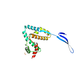
 | | SOLUTION STRUCTURE OF MYOTROPHIN, NMR, 44 STRUCTURES | | 分子名称: | MYOTROPHIN | | 著者 | Yang, Y, Nanduri, S, Sen, S, Qin, J. | | 登録日 | 1998-08-17 | | 公開日 | 1999-08-17 | | 最終更新日 | 2024-05-22 | | 実験手法 | SOLUTION NMR | | 主引用文献 | The structural basis of ankyrin-like repeat function as revealed by the solution structure of myotrophin.
Structure, 6, 1998
|
|
6KZ1
 | | Complex structure of Whirlin and Myosin XVa | | 分子名称: | Myosin XVa, Whirlin | | 著者 | Lin, L, Wang, M, Shi, Y, Zhu, J, Zhang, R. | | 登録日 | 2019-09-22 | | 公開日 | 2020-09-23 | | 最終更新日 | 2023-11-22 | | 実験手法 | X-RAY DIFFRACTION (1.694 Å) | | 主引用文献 | Phase separation-mediated condensation of Whirlin-Myo15-Eps8 stereocilia tip complex.
Cell Rep, 34, 2021
|
|
3LVR
 | | The crystal structure of ASAP3 in complex with Arf6 in transition state soaked with Calcium | | 分子名称: | ALUMINUM FLUORIDE, Arf-GAP with SH3 domain, ANK repeat and PH domain-containing protein 3, ... | | 著者 | Ismail, S.A, Vetter, I.R, Sot, B, Wittinghofer, A. | | 登録日 | 2010-02-22 | | 公開日 | 2010-06-09 | | 最終更新日 | 2023-11-01 | | 実験手法 | X-RAY DIFFRACTION (3.38 Å) | | 主引用文献 | The structure of an Arf-ArfGAP complex reveals a Ca2+ regulatory mechanism
Cell(Cambridge,Mass.), 141, 2010
|
|
3LYQ
 | | Crystal structure of IpgB2 from Shigella flexneri | | 分子名称: | CITRATE ANION, IpgB2, MU-OXO-DIIRON | | 著者 | Klink, B.U, Barden, S, Heidler, T.V, Borchers, C, Ladwein, M, Stradal, T.E.B, Rottner, K, Heinz, D.W. | | 登録日 | 2010-02-28 | | 公開日 | 2010-03-31 | | 最終更新日 | 2024-03-20 | | 実験手法 | X-RAY DIFFRACTION (2.3 Å) | | 主引用文献 | Structure of Shigella IPGB2 in complex with human RhoA: Implications for the mechanism of bacterial GEF-mimicry
J.Biol.Chem., 285, 2010
|
|
6M67
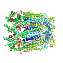
 | | The Cryo-EM Structure of Human Pannexin 1 with D376E/D379E Mutation | | 分子名称: | Pannexin-1 | | 著者 | Jin, Q, Bo, Z, Xiang, Z, Xiaokang, Z, Ye, S. | | 登録日 | 2020-03-13 | | 公開日 | 2020-04-15 | | 最終更新日 | 2020-05-13 | | 実験手法 | ELECTRON MICROSCOPY (3.6 Å) | | 主引用文献 | Cryo-EM structures of human pannexin 1 channel.
Cell Res., 30, 2020
|
|
6LRQ
 | |
6LTO
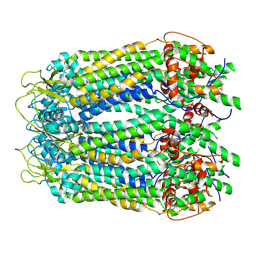 | | cryo-EM structure of full length human Pannexin1 | | 分子名称: | Pannexin-1 | | 著者 | Mou, L.Q, Ke, M, Xiao, Q.J, Wu, J.P, Deng, D. | | 登録日 | 2020-01-23 | | 公開日 | 2020-05-13 | | 最終更新日 | 2020-05-27 | | 実験手法 | ELECTRON MICROSCOPY (3.1 Å) | | 主引用文献 | Structural basis for gating mechanism of Pannexin 1 channel.
Cell Res., 30, 2020
|
|
6M68
 | | The Cryo-EM Structure of Human Pannexin 1 in the Presence of CBX | | 分子名称: | Pannexin-1 | | 著者 | Jin, Q, Bo, Z, Xiang, Z, Xiaokang, Z, Ye, S. | | 登録日 | 2020-03-13 | | 公開日 | 2020-04-15 | | 最終更新日 | 2020-05-13 | | 実験手法 | ELECTRON MICROSCOPY (4.6 Å) | | 主引用文献 | Cryo-EM structures of human pannexin 1 channel.
Cell Res., 30, 2020
|
|
6M66
 | | The Cryo-EM Structure of Human Pannexin 1 | | 分子名称: | Pannexin-1 | | 著者 | Jin, Q, Bo, Z, Xiang, Z, Xiaokang, Z, Ye, S. | | 登録日 | 2020-03-13 | | 公開日 | 2020-04-15 | | 最終更新日 | 2020-05-13 | | 実験手法 | ELECTRON MICROSCOPY (4.1 Å) | | 主引用文献 | Cryo-EM structures of human pannexin 1 channel.
Cell Res., 30, 2020
|
|
4L1B
 | | Crystal Structure of p110alpha complexed with niSH2 of p85alpha | | 分子名称: | Phosphatidylinositol 3-kinase regulatory subunit alpha, Phosphatidylinositol 4,5-bisphosphate 3-kinase catalytic subunit alpha isoform, SULFATE ION | | 著者 | Zhang, J, Zhao, Y.L, Chen, Y.Y, Huang, M, Jiang, F. | | 登録日 | 2013-06-03 | | 公開日 | 2014-01-01 | | 最終更新日 | 2023-09-20 | | 実験手法 | X-RAY DIFFRACTION (2.586 Å) | | 主引用文献 | Crystal Structures of PI3K alpha Complexed with PI103 and Its Derivatives: New Directions for Inhibitors Design.
ACS Med Chem Lett, 5, 2014
|
|


