1MAP
 
 | |
9G7Q
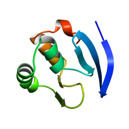
 | | Structure of the StayRose dimer | | 分子名称: | 1,2-ETHANEDIOL, CHLORIDE ION, StayRose | | 著者 | Crow, A. | | 登録日 | 2024-07-22 | | 公開日 | 2025-05-21 | | 実験手法 | X-RAY DIFFRACTION (1.65 Å) | | 主引用文献 | StayRose: a photostable StayGold derivative red-shifted by genetic code expansion
Biorxiv, 2024
|
|
1MB1
 | | MBP1 FROM SACCHAROMYCES CEREVISIAE | | 分子名称: | MLU1-BOX BINDING PROTEIN | | 著者 | Taylor, I.A, Smerdon, S.J. | | 登録日 | 1997-07-23 | | 公開日 | 1998-07-29 | | 最終更新日 | 2024-02-14 | | 実験手法 | X-RAY DIFFRACTION (2.1 Å) | | 主引用文献 | The X-ray structure of the DNA-binding domain from the Saccharomyces cerevisiae cell-cycle transcription factor Mbp1 at 2.1 A resolution.
J.Mol.Biol., 272, 1997
|
|
7RE8
 | |
9GSN
 | |
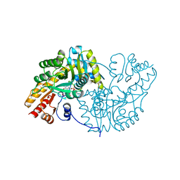
1MB9
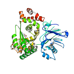 | | BETA-LACTAM SYNTHETASE COMPLEXED WITH ATP | | 分子名称: | ADENOSINE MONOPHOSPHATE, ADENOSINE-5'-TRIPHOSPHATE, BETA-LACTAM SYNTHETASE, ... | | 著者 | Miller, M.T, Bachmann, B.O, Townsend, C.A, Rosenzweig, A.C. | | 登録日 | 2002-08-02 | | 公開日 | 2002-10-23 | | 最終更新日 | 2024-02-14 | | 実験手法 | X-RAY DIFFRACTION (2.11 Å) | | 主引用文献 | The catalytic cycle of beta -lactam synthetase observed by x-ray crystallographic snapshots
Proc.Natl.Acad.Sci.USA, 99, 2002
|
|
7RHD
 | | darkmRuby M94T/F96Y mutant at pH 7.5 | | 分子名称: | 1,2-ETHANEDIOL, darkmRuby M94T/F96Y mutant | | 著者 | Huang, M, Ng, H.L, Zhang, S, Deng, M, Chu, J. | | 登録日 | 2021-07-16 | | 公開日 | 2022-07-27 | | 最終更新日 | 2023-11-15 | | 実験手法 | X-RAY DIFFRACTION (1.9 Å) | | 主引用文献 | A Long-range Interaction Affects Brightness and pH Stability of a Dark Fluorescent Protein
To Be Published
|
|
9FZW
 | | Structure of Urethanase UMG-SP2 | | 分子名称: | GLYCEROL, SULFATE ION, Urethanase UMG-SP2 | | 著者 | Singh, P, Lennartz, F, Bornscheuer, U.T, Wei, R, Weber, G. | | 登録日 | 2024-07-06 | | 公開日 | 2025-05-21 | | 実験手法 | X-RAY DIFFRACTION (2.59 Å) | | 主引用文献 | Structure of Urethanase UMG-SP2
To Be Published
|
|
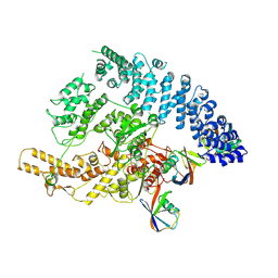
1MC0
 | | Regulatory Segment of Mouse 3',5'-Cyclic Nucleotide Phosphodiesterase 2A, Containing the GAF A and GAF B Domains | | 分子名称: | 3',5'-cyclic nucleotide phosphodiesterase 2A, CYCLIC GUANOSINE MONOPHOSPHATE | | 著者 | Martinez, S, Wu, A, Glavas, N, Tang, X, Turley, S, Hol, W, Beavo, J. | | 登録日 | 2002-08-04 | | 公開日 | 2002-10-02 | | 最終更新日 | 2024-11-13 | | 実験手法 | X-RAY DIFFRACTION (2.86 Å) | | 主引用文献 | The two GAF domains in phosphodiesterase 2A have distinct roles in dimerization and in cGMP binding.
Proc.Natl.Acad.Sci.USA, 99, 2002
|
|
9GKM
 | |
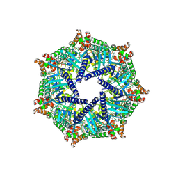
1M90
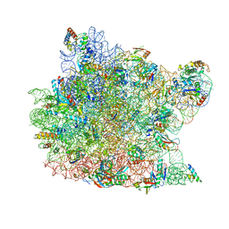 | | Co-crystal structure of CCA-Phe-caproic acid-biotin and sparsomycin bound to the 50S ribosomal subunit | | 分子名称: | 23S RRNA, 5S RRNA, 6-AMINOHEXANOIC ACID, ... | | 著者 | Hansen, J.L, Schmeing, T.M, Moore, P.B, Steitz, T.A. | | 登録日 | 2002-07-26 | | 公開日 | 2002-09-06 | | 最終更新日 | 2023-11-15 | | 実験手法 | X-RAY DIFFRACTION (2.8 Å) | | 主引用文献 | Structural insights into peptide bond formation.
Proc.Natl.Acad.Sci.USA, 99, 2002
|
|
9GOJ
 | |
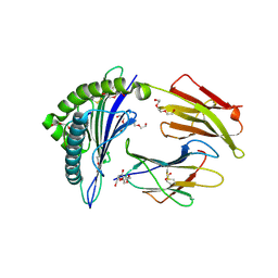
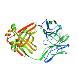
1MGR
 | | Crystal structure of RNase Sa3,cytotoxic microbial ribonuclease | | 分子名称: | Guanyl-specific ribonuclease Sa3, SULFATE ION | | 著者 | Sevcik, J, Urbanikova, L, Leland, P.A, Raines, R.T. | | 登録日 | 2002-08-16 | | 公開日 | 2003-02-04 | | 最終更新日 | 2024-10-30 | | 実験手法 | X-RAY DIFFRACTION (1.7 Å) | | 主引用文献 | Links X-ray Structure of Two Crystalline Forms of a Streptomycete Ribonuclease with Cytotoxic Activity
J.Biol.Chem., 277, 2002
|
|
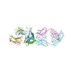
1MGW
 | | Crystal structure of RNase Sa3, cytotoxic microbial ribonuclease | | 分子名称: | Guanyl-specific ribonuclease Sa3, LITHIUM ION | | 著者 | Sevcik, J, Urbanikova, L, Leland, P.A, Raines, R.T. | | 登録日 | 2002-08-16 | | 公開日 | 2003-02-04 | | 最終更新日 | 2024-11-20 | | 実験手法 | X-RAY DIFFRACTION (2 Å) | | 主引用文献 | Links X-ray Structure of Two Crystalline Forms of a Streptomycete Ribonuclease with Cytotoxic Activity
J.Biol.Chem., 277, 2002
|
|
9GS7
 | | Cryo-EM structure of human SLC35B1-E33A variant with AMP-PNP | | 分子名称: | Fv-MBP, PHOSPHOAMINOPHOSPHONIC ACID-ADENYLATE ESTER, Solute carrier family 35 member B1 | | 著者 | Gulati, A, Ahn, D, Suades, A, Drew, D. | | 登録日 | 2024-09-13 | | 公開日 | 2025-05-21 | | 最終更新日 | 2025-08-20 | | 実験手法 | ELECTRON MICROSCOPY (3.15 Å) | | 主引用文献 | Stepwise ATP translocation into the endoplasmic reticulum by human SLC35B1.
Nature, 643, 2025
|
|
1MH4
 | |
9FY9
 | | Cryo-EM structure of the type 1 chaperone-usher pilus FimD-tip complex (FimDHGFC) - Conformer 1 | | 分子名称: | Chaperone protein FimC, Outer membrane usher protein FimD, Protein FimF, ... | | 著者 | Bachmann, P, Afanasyev, P, Boehringer, D, Glockshuber, R. | | 登録日 | 2024-07-03 | | 公開日 | 2025-06-04 | | 実験手法 | ELECTRON MICROSCOPY (3.3 Å) | | 主引用文献 | Cryo-EM structure of the type 1 chaperone-usher pilus FimD-tip complex (FimDHGFC) - Conformer 1
To Be Published
|
|
9G7J
 | |
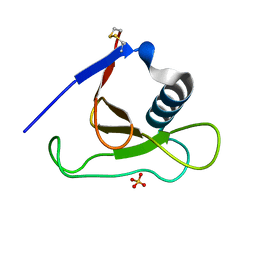
1MCC
 | | PRINCIPLES AND PITFALLS IN DESIGNING SITE DIRECTED PEPTIDE LIGANDS | | 分子名称: | IMMUNOGLOBULIN LAMBDA DIMER MCG (LIGHT CHAIN), PEPTIDE N-ACETYL-L-GLN-D-PHE-L-HIS-D-PRO-NH2 | | 著者 | Edmundson, A.B, Harris, D.L, Fan, Z.-C, Guddat, L.W. | | 登録日 | 1993-02-25 | | 公開日 | 1994-01-31 | | 最終更新日 | 2024-10-23 | | 実験手法 | X-RAY DIFFRACTION (2.7 Å) | | 主引用文献 | Principles and pitfalls in designing site-directed peptide ligands.
Proteins, 16, 1993
|
|
7RE7
 | |
9FXS
 | |
7RK4
 | |
9GIM
 | |
1MHH
 | |
2P65
 | | Crystal Structure of the first nucleotide binding domain of chaperone ClpB1, putative, (Pv089580) from Plasmodium Vivax | | 分子名称: | Hypothetical protein PF08_0063 | | 著者 | Wernimont, A.K, Lew, J, Kozieradzki, I, Lin, Y.H, Hassanali, A, Zhao, Y, Arrowsmith, C.H, Edwards, A.M, Weigelt, J, Sundstrom, M, Bochkarev, A, Hui, R, Artz, J.D, Structural Genomics Consortium (SGC) | | 登録日 | 2007-03-16 | | 公開日 | 2007-04-03 | | 最終更新日 | 2023-08-30 | | 実験手法 | X-RAY DIFFRACTION (1.7 Å) | | 主引用文献 | Crystal Structure of the first nucleotide binding domain of chaperone ClpB1, putative, (Pv089580) from Plasmodium Vivax
To be Published
|
|


