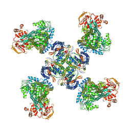
6ZMJ
 
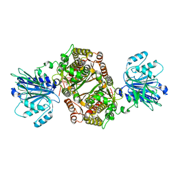 | | Crystal structure of human GFAT-1 R203H | | 分子名称: | GLUCOSE-6-PHOSPHATE, GLUTAMIC ACID, Glutamine--fructose-6-phosphate aminotransferase [isomerizing] 1 | | 著者 | Ruegenberg, S, Mayr, F, Miethe, S, Atanassov, I, Baumann, U, Denzel, M.S. | | 登録日 | 2020-07-02 | | 公開日 | 2020-08-05 | | 最終更新日 | 2024-01-31 | | 実験手法 | X-RAY DIFFRACTION (2.774 Å) | | 主引用文献 | Protein kinase A controls the hexosamine pathway by tuning the feedback inhibition of GFAT-1.
Nat Commun, 12, 2021
|
|
8F8K
 | | The structure of Rv2173 from M. tuberculosis with IPP bound | | 分子名称: | (2E,6E)-farnesyl diphosphate synthase, 3-METHYLBUT-3-ENYL TRIHYDROGEN DIPHOSPHATE, ACETATE ION, ... | | 著者 | Johnston, J.M, Allison, T.M, Titterington, J, Beasley, C.P.H. | | 登録日 | 2022-11-22 | | 公開日 | 2023-11-29 | | 実験手法 | X-RAY DIFFRACTION (2.2 Å) | | 主引用文献 | The structure of Rv2173 from M. tuberculosis in APO-, IPP-, and DMAP-bound forms.
To be Published
|
|
8F8F
 | | The structure of Rv2173 from M. tuberculosis (APO form) | | 分子名称: | (2E,6E)-farnesyl diphosphate synthase, GLYCEROL, MAGNESIUM ION | | 著者 | Johnston, J.M, Allison, T.M, Titterington, J. | | 登録日 | 2022-11-22 | | 公開日 | 2023-11-29 | | 実験手法 | X-RAY DIFFRACTION (2 Å) | | 主引用文献 | The structure of Rv2173 from M. tuberculosis in APO-, IPP-, and DMAP-bound forms.
To be Published
|
|
8F8L
 | | The structure of Rv2173 from M. tuberculosis with DMAP bound | | 分子名称: | (2E,6E)-farnesyl diphosphate synthase, CALCIUM ION, DIMETHYLALLYL DIPHOSPHATE, ... | | 著者 | Johnston, J.M, Allison, T.M, Titterington, J. | | 登録日 | 2022-11-22 | | 公開日 | 2023-11-29 | | 実験手法 | X-RAY DIFFRACTION (2.2 Å) | | 主引用文献 | The structure of Rv2173 from M. tuberculosis in APO-, IPP-, and DMAP-bound forms.
To be Published
|
|
5WUA
 | | Structure of a Pancreatic ATP-sensitive Potassium Channel | | 分子名称: | ATP-sensitive inward rectifier potassium channel 11,superfolder GFP, SUR1 | | 著者 | Li, N, Wu, J.-X, Chen, L, Gao, N. | | 登録日 | 2016-12-16 | | 公開日 | 2017-01-25 | | 実験手法 | ELECTRON MICROSCOPY (5.6 Å) | | 主引用文献 | Structure of a Pancreatic ATP-Sensitive Potassium Channel
Cell, 168, 2017
|
|
3AI4
 | | Crystal structure of yeast enhanced green fluorescent protein - mouse polymerase iota ubiquitin binding motif fusion protein | | 分子名称: | SULFATE ION, yeast enhanced green fluorescent protein,DNA polymerase iota | | 著者 | Suzuki, N, Wakatsuki, S, Kawasaki, M. | | 登録日 | 2010-05-10 | | 公開日 | 2010-09-29 | | 最終更新日 | 2023-11-15 | | 実験手法 | X-RAY DIFFRACTION (1.6 Å) | | 主引用文献 | Crystallization of small proteins assisted by green fluorescent protein
Acta Crystallogr.,Sect.D, 66, 2010
|
|
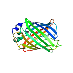
2C9J
 | | Structure of the fluorescent protein cmFP512 at 1.35A from Cerianthus membranaceus | | 分子名称: | GREEN FLUORESCENT PROTEIN FP512 | | 著者 | Renzi, F, Nienhaus, K, Wiedenmann, J, Vallone, B, Nienhaus, G.U. | | 登録日 | 2005-12-12 | | 公開日 | 2006-10-30 | | 最終更新日 | 2023-11-15 | | 実験手法 | X-RAY DIFFRACTION (1.35 Å) | | 主引用文献 | Exploring Chromophore-Protein Interactions in Fluorescent Protein Cmfp512 from Cerianthus Membranaceus: X-Ray Structure Analysis and Optical Spectroscopy.
Biochemistry, 45, 2006
|
|
1S6Z
 | | Enhanced Green Fluorescent Protein Containing the Y66L Substitution | | 分子名称: | CHLORIDE ION, green fluorescent protein | | 著者 | Rosenow, M.A, Huffman, H.A, Phail, M.E, Wachter, R.M. | | 登録日 | 2004-01-28 | | 公開日 | 2004-05-04 | | 最終更新日 | 2023-11-15 | | 実験手法 | X-RAY DIFFRACTION (1.5 Å) | | 主引用文献 | The Crystal Structure of the Y66L Variant of Green Fluorescent Protein Supports a Cyclization-Oxidation-Dehydration Mechanism for Chromophore Maturation(,).
Biochemistry, 43, 2004
|
|
2C9I
 | | Structure of the fluorescent protein asFP499 from Anemonia sulcata | | 分子名称: | GREEN FLUORESCENT PROTEIN ASFP499 | | 著者 | Renzi, F, Nienhaus, K, Wiedenmann, J, Vallone, B, Nienhaus, G.U. | | 登録日 | 2005-12-12 | | 公開日 | 2007-01-16 | | 最終更新日 | 2023-11-15 | | 実験手法 | X-RAY DIFFRACTION (1.82 Å) | | 主引用文献 | Chromophore-Protein Interactions in the Anthozoan Green Fluorescent Protein Asfp499
Biophys.J., 91, 2006
|
|
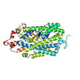
1QYQ
 | | Crystal Structure of the cyclized S65G Y66G GFP variant | | 分子名称: | green-fluorescent protein | | 著者 | Barondeau, D.P, Putnam, C.D, Kassmann, C.J, Tainer, J.A, Getzoff, E.D. | | 登録日 | 2003-09-11 | | 公開日 | 2003-09-30 | | 最終更新日 | 2023-11-15 | | 実験手法 | X-RAY DIFFRACTION (1.8 Å) | | 主引用文献 | Mechanism and energetics of green fluorescent protein chromophore synthesis revealed by trapped intermediate structures.
Proc.Natl.Acad.Sci.Usa, 100, 2003
|
|
5JZK
 | | The Structure of Ultra Stable Green Fluorescent Protein | | 分子名称: | 1,2-ETHANEDIOL, CHLORIDE ION, GLYCEROL, ... | | 著者 | Yong, K.J, Gunn, N.J, Scott, D.J, Griffin, M.D.W. | | 登録日 | 2016-05-17 | | 公開日 | 2017-12-06 | | 最終更新日 | 2023-11-15 | | 実験手法 | X-RAY DIFFRACTION (1.9 Å) | | 主引用文献 | A Novel Ultra-Stable, Monomeric Green Fluorescent Protein For Direct Volumetric Imaging of Whole Organs Using CLARITY.
Sci Rep, 8, 2018
|
|
5JZL
 | | The Structure of Monomeric Ultra Stable Green Fluorescent Protein | | 分子名称: | CHLORIDE ION, Green fluorescent protein, SODIUM ION | | 著者 | Gunn, N.J, Yong, K.J, Scott, D.J, Griffin, M.D.W. | | 登録日 | 2016-05-17 | | 公開日 | 2017-12-06 | | 最終更新日 | 2023-11-15 | | 実験手法 | X-RAY DIFFRACTION (1.8 Å) | | 主引用文献 | A Novel Ultra-Stable, Monomeric Green Fluorescent Protein For Direct Volumetric Imaging of Whole Organs Using CLARITY.
Sci Rep, 8, 2018
|
|
2G6X
 | |
2G6Y
 | |
6LZ2
 | | Crystal structure of a thermostable green fluorescent protein (TGP) with a synthetic nanobody (Sb44) | | 分子名称: | 2-AMINO-2-HYDROXYMETHYL-PROPANE-1,3-DIOL, ACETATE ION, GLYCEROL, ... | | 著者 | Cai, H, Yao, H, Li, T, Hutter, C, Tang, Y, Li, Y, Seeger, M, Li, D. | | 登録日 | 2020-02-17 | | 公開日 | 2020-12-23 | | 最終更新日 | 2023-11-29 | | 実験手法 | X-RAY DIFFRACTION (2.03 Å) | | 主引用文献 | An improved fluorescent tag and its nanobodies for membrane protein expression, stability assay, and purification.
Commun Biol, 3, 2020
|
|
8OVN
 | | X-ray structure of the SF-iGluSnFR-S72A | | 分子名称: | CITRIC ACID, Putative periplasmic binding transport protein,Green fluorescent protein | | 著者 | Tarnawski, M, Hellweg, L, Bergner, A, Hiblot, J, Leippe, P, Johnsson, K. | | 登録日 | 2023-04-26 | | 公開日 | 2023-05-17 | | 実験手法 | X-RAY DIFFRACTION (2.6 Å) | | 主引用文献 | X-ray structure of the SF-iGluSnFR-S72A
To Be Published
|
|
8OVO
 | | X-ray structure of the SF-iGluSnFR-S72A in complex with L-aspartate | | 分子名称: | ASPARTIC ACID, Putative periplasmic binding transport protein,Green fluorescent protein | | 著者 | Tarnawski, M, Hellweg, L, Bergner, A, Hiblot, J, Leippe, P, Johnsson, K. | | 登録日 | 2023-04-26 | | 公開日 | 2023-05-17 | | 実験手法 | X-RAY DIFFRACTION (1.7 Å) | | 主引用文献 | X-ray structure of the SF-iGluSnFR-S72A in complex with L-aspartate
To Be Published
|
|
8OVP
 | | X-ray structure of the iAspSnFR in complex with L-aspartate | | 分子名称: | ACETATE ION, ASPARTIC ACID, MAGNESIUM ION, ... | | 著者 | Tarnawski, M, Hellweg, L, Bergner, A, Hiblot, J, Leippe, P, Johnsson, K. | | 登録日 | 2023-04-26 | | 公開日 | 2023-05-17 | | 実験手法 | X-RAY DIFFRACTION (1.7 Å) | | 主引用文献 | X-ray structure of the SF-iAspSnFR in complex with L-aspartate
To Be Published
|
|
2Z1O
 | | Crystal structure of a photoswitchable GFP-like protein Dronpa in the bright-state | | 分子名称: | Fluorescent protein Dronpa | | 著者 | Kikuchi, A, Jeyakanthan, J, Taka, J, Shiro, Y, Mizuno, H, Miyawaki, A. | | 登録日 | 2007-05-10 | | 公開日 | 2008-05-13 | | 最終更新日 | 2023-11-15 | | 実験手法 | X-RAY DIFFRACTION (1.75 Å) | | 主引用文献 | Light-dependent regulation of structural flexibility in a photochromic fluorescent protein
Proc.Natl.Acad.Sci.Usa, 105, 2008
|
|
2Z6Z
 | | Crystal structure of a photoswitchable GFP-like protein Dronpa in the bright-state | | 分子名称: | Fluorescent protein Dronpa | | 著者 | Kikuchi, A, Jeyakanthan, J, Taka, J, Shiro, Y, Mizuno, H, Miyawaki, A. | | 登録日 | 2007-08-09 | | 公開日 | 2008-07-22 | | 最終更新日 | 2023-11-15 | | 実験手法 | X-RAY DIFFRACTION (1.8 Å) | | 主引用文献 | Light-dependent regulation of structural flexibility in a photochromic fluorescent protein.
Proc.Natl.Acad.Sci.Usa, 105, 2008
|
|
2Z6X
 | | Crystal structure of 22G, the wild-type protein of the photoswitchable GFP-like protein Dronpa | | 分子名称: | photochromic protein Dronpa | | 著者 | Kikuchi, A, Jeyakanthan, J, Taka, J, Shiro, Y, Mizuno, H, Miyawaki, A. | | 登録日 | 2007-08-09 | | 公開日 | 2008-07-22 | | 最終更新日 | 2023-11-15 | | 実験手法 | X-RAY DIFFRACTION (2.3 Å) | | 主引用文献 | Light-dependent regulation of structural flexibility in a photochromic fluorescent protein.
Proc.Natl.Acad.Sci.Usa, 105, 2008
|
|
2Z6Y
 | | Crystal structure of a photoswitchable GFP-like protein Dronpa in the bright-state | | 分子名称: | Fluorescent protein Dronpa | | 著者 | Kikuchi, A, Jeyakanthan, J, Taka, J, Shiro, Y, Mizuno, H, Miyawaki, A. | | 登録日 | 2007-08-09 | | 公開日 | 2008-07-22 | | 最終更新日 | 2023-11-15 | | 実験手法 | X-RAY DIFFRACTION (2 Å) | | 主引用文献 | Light-dependent regulation of structural flexibility in a photochromic fluorescent protein.
Proc.Natl.Acad.Sci.Usa, 105, 2008
|
|
2IB5
 | |
2IB6
 | | Structural characterization of a blue chromoprotein and its yellow mutant from the sea anemone cnidopus japonicus | | 分子名称: | PHOSPHATE ION, Yellow mutant chromo protein | | 著者 | Chan, M.C.Y, Bosanac, I, Ho, D, Prive, G, Ikura, M. | | 登録日 | 2006-09-10 | | 公開日 | 2006-10-10 | | 最終更新日 | 2023-11-15 | | 実験手法 | X-RAY DIFFRACTION (2 Å) | | 主引用文献 | Structural Characterization of a Blue Chromoprotein and Its Yellow Mutant from the Sea Anemone Cnidopus Japonicus
J.Biol.Chem., 281, 2006
|
|
8XB3
 | |


