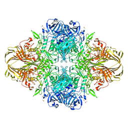
4DV5
 
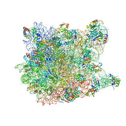
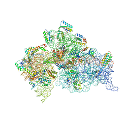 | | Crystal structure of the Thermus thermophilus 30S ribosomal subunit with a 16S rRNA mutation, A914G, bound with streptomycin | | 分子名称: | 16S rRNA, MAGNESIUM ION, STREPTOMYCIN, ... | | 著者 | Demirci, H, Murphy IV, F, Murphy, E, Gregory, S.T, Dahlberg, A.E, Jogl, G. | | 登録日 | 2012-02-22 | | 公開日 | 2013-02-27 | | 実験手法 | X-RAY DIFFRACTION (3.683 Å) | | 主引用文献 | A structural basis for streptomycin resistance
To be Published
|
|
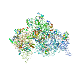
4DV2
 | | Crystal structure of the Thermus thermophilus 30S ribosomal subunit with a 16S rRNA mutation, C912A | | 分子名称: | 16S rRNA, MAGNESIUM ION, ZINC ION, ... | | 著者 | Demirci, H, Murphy IV, F, Murphy, E, Gregory, S.T, Dahlberg, A.E, Jogl, G. | | 登録日 | 2012-02-22 | | 公開日 | 2013-02-27 | | 実験手法 | X-RAY DIFFRACTION (3.646 Å) | | 主引用文献 | A structural basis for streptomycin resistance
To be Published
|
|
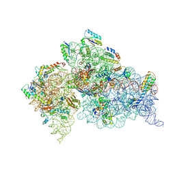
4DV7
 | | Crystal structure of the Thermus thermophilus 30S ribosomal subunit with a 16S rRNA mutation, A915G, bound with streptomycin | | 分子名称: | 16S rRNA, MAGNESIUM ION, STREPTOMYCIN, ... | | 著者 | Demirci, H, Murphy IV, F, Murphy, E, Gregory, S.T, Dahlberg, A.E, Jogl, G. | | 登録日 | 2012-02-22 | | 公開日 | 2013-02-27 | | 実験手法 | X-RAY DIFFRACTION (3.294 Å) | | 主引用文献 | A structural basis for streptomycin resistance
To be Published
|
|
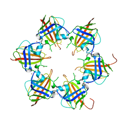
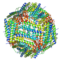
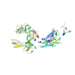
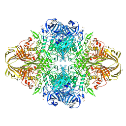
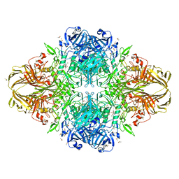
1RVV
 | | SYNTHASE/RIBOFLAVIN SYNTHASE COMPLEX OF BACILLUS SUBTILIS | | 分子名称: | 5-NITRO-6-RIBITYL-AMINO-2,4(1H,3H)-PYRIMIDINEDIONE, PHOSPHATE ION, RIBOFLAVIN SYNTHASE | | 著者 | Ritsert, K, Huber, R, Turk, D, Ladenstein, R, Schmidt-Baese, K, Bacher, A. | | 登録日 | 1995-10-25 | | 公開日 | 1996-12-07 | | 最終更新日 | 2024-02-14 | | 実験手法 | X-RAY DIFFRACTION (2.4 Å) | | 主引用文献 | Studies on the lumazine synthase/riboflavin synthase complex of Bacillus subtilis: crystal structure analysis of reconstituted, icosahedral beta-subunit capsids with bound substrate analogue inhibitor at 2.4 A resolution.
J.Mol.Biol., 253, 1995
|
|
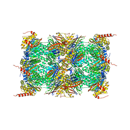
3OTO
 | | Crystal Structure of the 30S ribosomal subunit from a KsgA mutant of Thermus thermophilus (HB8) | | 分子名称: | 16S rRNA, 30S RIBOSOMAL PROTEIN S10, 30S RIBOSOMAL PROTEIN S11, ... | | 著者 | Demirci, H, Murphy IV, F, Belardinelli, R, Kelley, A.C, Ramakrishnan, V, Gregory, S.T, Dahlberg, A.E, Jogl, G. | | 登録日 | 2010-09-13 | | 公開日 | 2010-09-29 | | 最終更新日 | 2023-09-06 | | 実験手法 | X-RAY DIFFRACTION (3.69 Å) | | 主引用文献 | Modification of 16S ribosomal RNA by the KsgA methyltransferase restructures the 30S subunit to optimize ribosome function.
Rna, 16, 2010
|
|
3RCC
 | |
1PX3
 | | E. COLI (LACZ) BETA-GALACTOSIDASE (G794A) | | 分子名称: | DIMETHYL SULFOXIDE, MAGNESIUM ION, SODIUM ION, ... | | 著者 | Juers, D.H, Hakda, S, Matthews, B.W, Huber, R.E. | | 登録日 | 2003-07-02 | | 公開日 | 2004-06-15 | | 最終更新日 | 2023-08-16 | | 実験手法 | X-RAY DIFFRACTION (1.6 Å) | | 主引用文献 | Structural Basis for the Altered Activity of Gly794 Variants of Escherichia coli Beta-Galactosidase
Biochemistry, 42, 2003
|
|
3RBC
 | | Bullfrog M ferritin with iron(III) bound to the ferroxidase site | | 分子名称: | FE (III) ION, Ferritin, middle subunit | | 著者 | Bertini, I, Lalli, D, Mangani, S, Pozzi, C, Rosa, C, Turano, P. | | 登録日 | 2011-03-29 | | 公開日 | 2012-04-04 | | 最終更新日 | 2023-09-13 | | 実験手法 | X-RAY DIFFRACTION (2.7 Å) | | 主引用文献 | Structural insights into the ferroxidase site of ferritins from higher eukaryotes.
J.Am.Chem.Soc., 134, 2012
|
|
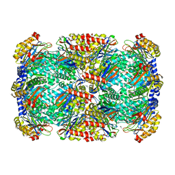
4R67
 | | Human constitutive 20S proteasome in complex with carfilzomib | | 分子名称: | N-{(2S)-2-[(morpholin-4-ylacetyl)amino]-4-phenylbutanoyl}-L-leucyl-N-[(2R,3S,4S)-1,3-dihydroxy-2,6-dimethylheptan-4-yl]-L-phenylalaninamide, Proteasome subunit alpha type-1, Proteasome subunit alpha type-2, ... | | 著者 | Sacchettini, J.C, Harshbarger, W.H. | | 登録日 | 2014-08-22 | | 公開日 | 2015-02-11 | | 最終更新日 | 2015-02-18 | | 実験手法 | X-RAY DIFFRACTION (2.89 Å) | | 主引用文献 | Crystal Structure of the Human 20S Proteasome in Complex with Carfilzomib.
Structure, 23, 2015
|
|
3R90
 | | Crystal structure of Malignant T cell-amplified sequence 1 protein | | 分子名称: | GLYCEROL, Malignant T cell-amplified sequence 1, SULFATE ION, ... | | 著者 | Hong, B, Dimov, S, Tempel, W, Tong, Y, Wernimont, A.K, Arrowsmith, C.H, Edwards, A.M, Bountra, C, Weigelt, J, Park, H, Structural Genomics Consortium (SGC) | | 登録日 | 2011-03-24 | | 公開日 | 2011-04-13 | | 最終更新日 | 2024-02-21 | | 実験手法 | X-RAY DIFFRACTION (1.7 Å) | | 主引用文献 | Crystal structure of Malignant T cell-amplified sequence 1 protein
to be published
|
|
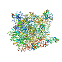
3RJ1
 | | Architecture of the Mediator Head module | | 分子名称: | Mediator of RNA polymerase II transcription subunit 11, Mediator of RNA polymerase II transcription subunit 17, Mediator of RNA polymerase II transcription subunit 18, ... | | 著者 | Imasaki, T, Calero, G, Cai, G, Tsai, K.L, Yamada, K, Cardelli, F, Erdjument-Bromage, H, Tempst, P, Berger, I, Kornberg, G.L, Asturias, F.J, Kornberg, R.D, Takagi, Y. | | 登録日 | 2011-04-14 | | 公開日 | 2011-07-27 | | 最終更新日 | 2017-08-02 | | 実験手法 | X-RAY DIFFRACTION (4.3 Å) | | 主引用文献 | Architecture of the Mediator head module.
Nature, 475, 2011
|
|
4R0D
 | | Crystal structure of a eukaryotic group II intron lariat | | 分子名称: | GROUP IIB INTRON LARIAT, IRIDIUM HEXAMMINE ION, LIGATED EXONS, ... | | 著者 | Robart, A.R, Chan, R.T, Peters, J.K, Rajashankar, K.R, Toor, N. | | 登録日 | 2014-07-30 | | 公開日 | 2014-10-01 | | 最終更新日 | 2024-02-28 | | 実験手法 | X-RAY DIFFRACTION (3.676 Å) | | 主引用文献 | Crystal structure of a eukaryotic group II intron lariat.
Nature, 514, 2014
|
|
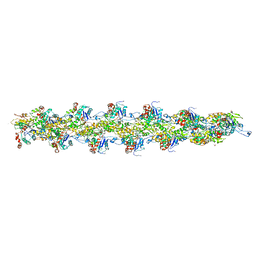
1SMY
 | | Structural basis for transcription regulation by alarmone ppGpp | | 分子名称: | DNA-directed RNA polymerase alpha chain, DNA-directed RNA polymerase beta chain, DNA-directed RNA polymerase beta' chain, ... | | 著者 | Artsimovitch, I, Patlan, V, Sekine, S, Vassylyeva, M.N, Hosaka, T, Ochi, K, Yokoyama, S, Vassylyev, D.G, RIKEN Structural Genomics/Proteomics Initiative (RSGI) | | 登録日 | 2004-03-10 | | 公開日 | 2004-05-18 | | 最終更新日 | 2023-10-25 | | 実験手法 | X-RAY DIFFRACTION (2.7 Å) | | 主引用文献 | Structural basis for transcription regulation by alarmone ppGpp
Cell(Cambridge,Mass.), 117, 2004
|
|
3HHQ
 | | Crystal structure of apo dUT1p from Saccharomyces cerevisiae | | 分子名称: | 1,2-ETHANEDIOL, CHLORIDE ION, DI(HYDROXYETHYL)ETHER, ... | | 著者 | Singer, A.U, Evdokimova, E, Kudritska, M, Dong, A, Edwards, A.M, Yakunin, A.F, Savchenko, A. | | 登録日 | 2009-05-15 | | 公開日 | 2009-06-16 | | 最終更新日 | 2023-09-06 | | 実験手法 | X-RAY DIFFRACTION (2 Å) | | 主引用文献 | Structure and activity of the Saccharomyces cerevisiae dUTP pyrophosphatase DUT1, an essential housekeeping enzyme.
Biochem.J., 437, 2011
|
|
3HFA
 | |
3G6E
 | | Co-crystal structure of Homoharringtonine bound to the large ribosomal subunit | | 分子名称: | (3beta)-O~3~-[(2R)-2,6-dihydroxy-2-(2-methoxy-2-oxoethyl)-6-methylheptanoyl]cephalotaxine, 23S ribosomal RNA, 50S ribosomal protein L10E, ... | | 著者 | Gurel, G, Blaha, G, Moore, P.B, Steitz, T.A. | | 登録日 | 2009-02-06 | | 公開日 | 2009-04-28 | | 最終更新日 | 2023-09-06 | | 実験手法 | X-RAY DIFFRACTION (2.7 Å) | | 主引用文献 | U2504 determines the species specificity of the A-site cleft antibiotics: the structures of tiamulin, homoharringtonine, and bruceantin bound to the ribosome.
J.Mol.Biol., 389, 2009
|
|
3I3D
 | | E. COLI (lacZ) BETA-GALACTOSIDASE (M542A) IN COMPLEX WITH IPTG | | 分子名称: | 1-methylethyl 1-thio-beta-D-galactopyranoside, Beta-galactosidase, DIMETHYL SULFOXIDE, ... | | 著者 | Dugdale, M.L, Dymianiw, D, Minhas, B, Huber, R.E. | | 登録日 | 2009-06-30 | | 公開日 | 2010-05-12 | | 最終更新日 | 2023-09-06 | | 実験手法 | X-RAY DIFFRACTION (2.2 Å) | | 主引用文献 | Role of Met-542 as a guide for the conformational changes of Phe-601 that occur during the reaction of β-galactosidase (Escherichia coli).
Biochem.Cell Biol., 88, 2010
|
|
3I55
 | | Co-crystal structure of Mycalamide A Bound to the Large Ribosomal Subunit | | 分子名称: | 23S ribosomal RNA, 50S ribosomal protein L10E, 50S ribosomal protein L10e, ... | | 著者 | Gurel, G, Blaha, G, Steitz, T.A, Moore, P.B. | | 登録日 | 2009-07-03 | | 公開日 | 2010-03-09 | | 最終更新日 | 2024-02-21 | | 実験手法 | X-RAY DIFFRACTION (3.11 Å) | | 主引用文献 | Structures of triacetyloleandomycin and mycalamide A bind to the large ribosomal subunit of Haloarcula marismortui.
Antimicrob.Agents Chemother., 53, 2009
|
|
3G37
 | | Cryo-EM structure of actin filament in the presence of phosphate | | 分子名称: | ADENOSINE-5'-DIPHOSPHATE, Actin, alpha skeletal muscle, ... | | 著者 | Wakabayshi, T, Murakami, K, Yasunaga, T, Noguchi, T.Q, Uyeda, T.Q. | | 登録日 | 2009-02-02 | | 公開日 | 2010-11-03 | | 最終更新日 | 2019-12-18 | | 実験手法 | ELECTRON MICROSCOPY (6 Å) | | 主引用文献 | Structural basis for actin assembly, activation of ATP hydrolysis, and delayed phosphate release
Cell(Cambridge,Mass.), 143, 2010
|
|
3G71
 | | Co-crystal structure of Bruceantin bound to the large ribosomal subunit | | 分子名称: | 23S ribosomal RNA, 50S ribosomal protein L10E, 50S ribosomal protein L10e, ... | | 著者 | Gurel, G, Blaha, G, Moore, P.B, Steitz, T.A. | | 登録日 | 2009-02-09 | | 公開日 | 2009-04-28 | | 最終更新日 | 2023-09-06 | | 実験手法 | X-RAY DIFFRACTION (2.85 Å) | | 主引用文献 | U2504 determines the species specificity of the A-site cleft antibiotics: the structures of tiamulin, homoharringtonine, and bruceantin bound to the ribosome.
J.Mol.Biol., 389, 2009
|
|
3I3B
 | | E.coli (lacz) Beta-Galactosidase (M542A) in Complex with D-Galactopyranosyl-1-on | | 分子名称: | Beta-galactosidase, D-galactonolactone, DIMETHYL SULFOXIDE, ... | | 著者 | Dugdale, M.L, Dymianiw, D, Minhas, B, Huber, R.E. | | 登録日 | 2009-06-30 | | 公開日 | 2010-05-12 | | 最終更新日 | 2023-09-06 | | 実験手法 | X-RAY DIFFRACTION (2.2 Å) | | 主引用文献 | Role of Met-542 as a guide for the conformational changes of Phe-601 that occur during the reaction of β-galactosidase (Escherichia coli).
Biochem.Cell Biol., 88, 2010
|
|
3I56
 | | Co-crystal structure of Triacetyloleandomcyin Bound to the Large Ribosomal Subunit | | 分子名称: | 23S ribosomal RNA, 50S ribosomal protein L10E, 50S ribosomal protein L10e, ... | | 著者 | Gurel, G, Blaha, G, Steitz, T.A, Moore, P.B. | | 登録日 | 2009-07-03 | | 公開日 | 2010-03-09 | | 最終更新日 | 2024-02-21 | | 実験手法 | X-RAY DIFFRACTION (2.9 Å) | | 主引用文献 | Structures of triacetyloleandomycin and mycalamide A bind to the large ribosomal subunit of Haloarcula marismortui.
Antimicrob.Agents Chemother., 53, 2009
|
|
6ZW6
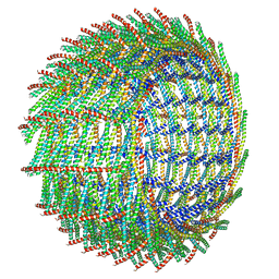 | | C16 symmetry: Bacterial Vipp1 and PspA are members of the ancient ESCRT-III membrane-remodeling superfamily. | | 分子名称: | vipp1 | | 著者 | Liu, J, Tassinari, M, Souza, D.P, Naskar, S, Noel, J.K, Bohuszewicz, O, Buck, M, Williams, T.A, Baum, B, Low, H.H. | | 登録日 | 2020-07-27 | | 公開日 | 2021-08-04 | | 最終更新日 | 2022-05-04 | | 実験手法 | ELECTRON MICROSCOPY (7.4 Å) | | 主引用文献 | Bacterial Vipp1 and PspA are members of the ancient ESCRT-III membrane-remodeling superfamily.
Cell, 184, 2021
|
|
6ZW7
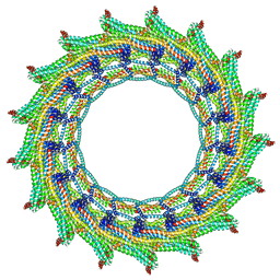 | | C17 symmetry: Bacterial Vipp1 and PspA are members of the ancient ESCRT-III membrane-remodeling superfamily. | | 分子名称: | vipp1 | | 著者 | Liu, J, Tassinari, M, Souza, D.P, Naskar, S, Noel, J.K, Bohuszewicz, O, Buck, M, Williams, T.A, Baum, B, Low, H.H. | | 登録日 | 2020-07-27 | | 公開日 | 2021-08-04 | | 最終更新日 | 2024-07-10 | | 実験手法 | ELECTRON MICROSCOPY (9.4 Å) | | 主引用文献 | Bacterial Vipp1 and PspA are members of the ancient ESCRT-III membrane-remodeling superfamily.
Cell, 184, 2021
|
|
3G4S
 | | Co-crystal structure of Tiamulin bound to the large ribosomal subunit | | 分子名称: | 23S ribosomal RNA, 50S ribosomal protein L10, 50S ribosomal protein L10e, ... | | 著者 | Gurel, G, Blaha, G, Moore, P.B, Steitz, T.A. | | 登録日 | 2009-02-04 | | 公開日 | 2009-04-28 | | 最終更新日 | 2023-09-06 | | 実験手法 | X-RAY DIFFRACTION (3.2 Å) | | 主引用文献 | U2504 determines the species specificity of the A-site cleft antibiotics: the structures of tiamulin, homoharringtonine, and bruceantin bound to the ribosome.
J.Mol.Biol., 389, 2009
|
|


